Anti-Adenosine Receptor A2a antibody (ab3461)
 - Anti-Adenosine Receptor A2a antibody (ab3461)")
Key features and details
- Rabbit polyclonal to Adenosine Receptor A2a
- Suitable for: ICC, WB, IHC-P
- Reacts with: Mouse, Human
- Isotype: IgG
Get better batch-to-batch reproducibility with a recombinant antibody


- Research with confidence – consistent and reproducible results with every batch
- Long-term and scalable supply – powered by recombinant technology for fast production
- Success from the first experiment – confirmed specificity through extensive validation
- Ethical standards compliant – production is animal-free
Overview
-
Product name
Anti-Adenosine Receptor A2a antibody
See all Adenosine Receptor A2a primary antibodies -
Description
Rabbit polyclonal to Adenosine Receptor A2a -
Host species
Rabbit -
Specificity
Detects adenosine receptor A2a. This antibody does not detect other AR subtypes. -
Tested applications
Suitable for: ICC, WB, IHC-Pmore details -
Species reactivity
Reacts with: Mouse, Human
Predicted to work with: Horse
-
Immunogen
Synthetic peptide corresponding to Dog Adenosine Receptor A2a aa 373-391.
Sequence:ESHGDMGLPDVELLSHELK
-
Positive control
- IHC-P: Human testis and placenta tissues. ICC: U251 and SKNSH cells. WB: HepG2 and HeLa cell lysates; Human placenta tissue lysate; Mouse liver tissue lysate.
-
General notes
The Life Science industry has been in the grips of a reproducibility crisis for a number of years. Abcam is leading the way in addressing this with our range of recombinant monoclonal antibodies and knockout edited cell lines for gold-standard validation. Please check that this product meets your needs before purchasing.
If you have any questions, special requirements or concerns, please send us an inquiry and/or contact our Support team ahead of purchase. Recommended alternatives for this product can be found below, along with publications, customer reviews and Q&As
Properties
-
Form
Liquid -
Storage instructions
Shipped at 4°C. Store at +4°C short term (1-2 weeks). Upon delivery aliquot. Store at -20°C or -80°C. Avoid freeze / thaw cycle. -
Storage buffer
Preservative: 0.05% Sodium azide
Constituents: 0.1% BSA, 99% PBS -
 Concentration information loading...
 -
Purity
Immunogen affinity purified -
Clonality
Polyclonal -
Isotype
IgG -
Research areas
Associated products
-
Compatible Secondaries
-
Isotype control
-
Recombinant Protein
Applications
The Abpromise guarantee
Our Abpromise guarantee covers the use of ab3461 in the following tested applications.
The application notes include recommended starting dilutions; optimal dilutions/concentrations should be determined by the end user.
| Application | Abreviews | Notes |
|---|---|---|
| ICC |
Use at an assay dependent concentration.
|
|
| WB | (1) |
1/1000.
|
| IHC-P |
1/20 - 1/200.
|
| Notes |
|---|
|
ICC
Use at an assay dependent concentration. |
|
WB
1/1000. |
|
IHC-P
1/20 - 1/200. |
Target
-
Function
Receptor for adenosine. The activity of this receptor is mediated by G proteins which activate adenylyl cyclase. -
Sequence similarities
Belongs to the G-protein coupled receptor 1 family. -
Domain
The cytoplasmic C-terminal domain is necessary for targeting the non-ubiquitinated form of this protein to the cell surface. -
Post-translational
modificationsUbiquitinated. Deubiquitinated by USP4; leading to stabilization and expression at the cell surface. -
Cellular localization
Cell membrane. - Information by UniProt
-
Database links
- Entrez Gene: 135 Human
- Entrez Gene: 11540 Mouse
- Omim: 102776 Human
- SwissProt: Q6TLI7 Horse
- SwissProt: P29274 Human
- SwissProt: Q60613 Mouse
- Unigene: 197029 Human
- Unigene: 333734 Mouse
-
Alternative names
- A2AAR antibody
- A2aR antibody
- AA2AR_HUMAN antibody
see all
Images
-
Immunohistochemistry (Formalin/PFA-fixed paraffin-embedded sections) - Anti-Adenosine Receptor A2a antibody (ab3461)
ab3461 labelling Adenosine Receptor A2a in the cytoplasm and membrane of Mouse testis tissue (right) compared with a negative control (left) by Immunohistochemisty (formalin/PFA-fixed paraffin-embedded sections). To expose target proteins, antigen retrieval method was performed using 10mM sodium citrate (pH 6.0) microwaved for 8-15 min. Following antigen retrieval, tissues were blocked in 3% H2O2-methanol for 15 min at room temperature. Tissue sections were incubated with the primary antibody (1:20 in 3% BSA-PBS) overnight at 4°C. A HRP-conjugated anti-rabbit IgG was as the secondary antibody, followed by colorimetric detection using a DAB kit. Tissues were counterstained with hematoxylin and dehydrated with ethanol and xylene to prep for mounting.
-
 Western blot - Anti-Adenosine Receptor A2a antibody (ab3461)All lanes : Anti-Adenosine Receptor A2a antibody (ab3461) at 1/500 dilution
Lane 1 : Human placenta cell lysate
Lane 2 : HepG2 cell lysate
Lane 3 : HeLa cell lysate
Lane 4 : Mouse liver cell lysate
Lysates/proteins at 25 µg per lane. -
 Immunocytochemistry - Anti-Adenosine Receptor A2a antibody (ab3461)
Immunocytochemistry/Immunofluorescence analysis of Adenosine Receptor A2a (green) showing staining in the cytoplasm of U251 cells (right) compared to a negative control (left). Formalin-fixed cells were permeabilized with 0.1% Triton X-100 in TBS for 5-10 minutes and blocked with 3% BSA-PBS for 30 minutes at room temperature. Cells were incubated with ab3461 in 3% BSA-PBS at a dilution of 1:20 overnight at 4 ºC in a humidified chamber. Cells were washed with PBST and incubated with a DyLight-conjugated secondary antibody in PBS at room temperature in the dark. F-actin (red) was stained with a fluorescent red phalloidin and nuclei (blue) were stained with Hoechst or DAPI. Images were taken at a magnification of 60x.
-
 Immunohistochemistry (Formalin/PFA-fixed paraffin-embedded sections) - Anti-Adenosine Receptor A2a antibody (ab3461)
ab3461 labelling Adenosine Receptor A2a in the cytoplasm and membrane of Human testis tissue (right) compared with a negative control (left) by Immunohistochemisty (formalin/PFA-fixed paraffin-embedded sections). To expose target proteins, antigen retrieval method was performed using 10mM sodium citrate (pH 6.0) microwaved for 8-15 min. Following antigen retrieval, tissues were blocked in 3% H2O2-methanol for 15 min at room temperature. Tissue sections were incubated with the primary antibody (1:200 in 3% BSA-PBS) overnight at 4°C. A HRP-conjugated anti-rabbit IgG was as the secondary antibody, followed by colorimetric detection using a DAB kit. Tissues were counterstained with hematoxylin and dehydrated with ethanol and xylene to prep for mounting.
-
 Immunohistochemistry (Formalin/PFA-fixed paraffin-embedded sections) - Anti-Adenosine Receptor A2a antibody (ab3461)
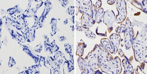
ab3461 labelling Adenosine Receptor A2a in the cytoplasm and membrane of Human placenta tissue (right) compared with a negative control (left) by Immunohistochemisty (formalin/PFA-fixed paraffin-embedded sections). To expose target proteins, antigen retrieval method was performed using 10mM sodium citrate (pH 6.0) microwaved for 8-15 min. Following antigen retrieval, tissues were blocked in 3% H2O2-methanol for 15 min at room temperature. Tissue sections were incubated with the primary antibody (1:100 in 3% BSA-PBS) overnight at 4°C. A HRP-conjugated anti-rabbit IgG was as the secondary antibody, followed by colorimetric detection using a DAB kit. Tissues were counterstained with hematoxylin and dehydrated with ethanol and xylene to prep for mounting.
-
 Immunocytochemistry - Anti-Adenosine Receptor A2a antibody (ab3461)ICC/IF image of ab3461 stained SKNSH cells. The cells were 100% methanol fixed (5 min) and then incubated in 1%BSA / 10% normal goat serum / 0.3M glycine in 0.1% PBS-Tween for 1h to permeabilise the cells and block non-specific protein-protein interactions. The cells were then incubated with the antibody (ab3461, 10µg/ml) overnight at +4°C. The secondary antibody (green) was ab96899 Dylight 488 goat anti-rabbit IgG (H+L) used at a 1/250 dilution for 1h. Alexa Fluor® 594 WGA was used to label plasma membranes (red) at a 1/200 dilution for 1h. DAPI was used to stain the cell nuclei (blue) at a concentration of 1.43µM.
Protocols
Datasheets and documents
-
SDS download
-
Datasheet download
References (32)
ab3461 has been referenced in 32 publications.
- Li D et al. Gut microbiota-derived inosine from dietary barley leaf supplementation attenuates colitis through PPAR? signaling activation. Microbiome 9:83 (2021). PubMed: 33820558
- Yang D et al. Methotrexate attenuates vascular inflammation through an adenosine-microRNA-dependent pathway. Elife 10:N/A (2021). PubMed: 33416495
- Jin H et al. Increased Extracellular Adenosine in Radiotherapy-Resistant Breast Cancer Cells Enhances Tumor Progression through A2AR-Akt-ß-Catenin Signaling. Cancers (Basel) 13:N/A (2021). PubMed: 33925516
- Dragić M et al. Downregulation of CD73/A2AR-Mediated Adenosine Signaling as a Potential Mechanism of Neuroprotective Effects of Theta-Burst Transcranial Magnetic Stimulation in Acute Experimental Autoimmune Encephalomyelitis. Brain Sci 11:N/A (2021). PubMed: 34205965
- Yi L et al. Sinomenine increases adenosine A2A receptor and inhibits NF-κB to inhibit arthritis in adjuvant-induced-arthritis rats and fibroblast-like synoviocytes through α7nAChR. J Leukoc Biol 110:1113-1120 (2021). PubMed: 34425026