Anti-CD3 zeta antibody (ab190728)
")
Key features and details
- Rabbit polyclonal to CD3 zeta
- Suitable for: WB, ICC/IF
- Reacts with: Human
- Isotype: IgG
Overview
-
Product name
Anti-CD3 zeta antibody
See all CD3 zeta primary antibodies -
Description
Rabbit polyclonal to CD3 zeta -
Host species
Rabbit -
Tested applications
Suitable for: WB, ICC/IFmore details -
Species reactivity
Reacts with: Human -
Immunogen
Recombinant full length protein corresponding to Human CD3 zeta aa 1 to the C-terminus.
Database link: P20963 -
Positive control
- Extracts of Jurkat cell line.
-
General notes
The Life Science industry has been in the grips of a reproducibility crisis for a number of years. Abcam is leading the way in addressing this with our range of recombinant monoclonal antibodies and knockout edited cell lines for gold-standard validation. Please check that this product meets your needs before purchasing.
If you have any questions, special requirements or concerns, please send us an inquiry and/or contact our Support team ahead of purchase. Recommended alternatives for this product can be found below, along with publications, customer reviews and Q&As
Properties
-
Form
Liquid -
Storage instructions
Shipped at 4°C. Store at +4°C short term (1-2 weeks). Upon delivery aliquot. Store at -20°C long term. Avoid freeze / thaw cycle. -
Storage buffer
Preservative: 0.1% Sodium azide
Constituents: 50% Glycerol, 49% PBS -
 Concentration information loading...
 -
Purity
Immunogen affinity purified -
Purification notes
ab190728 was affinity-purified from rabbit antiserum by affinity-chromatography using epitope-specific immunogen, and the purity is > 95% (by SDS-PAGE). -
Clonality
Polyclonal -
Isotype
IgG -
Research areas
Associated products
-
Compatible Secondaries
-
Isotype control
-
Positive Controls
-
Recombinant Protein
Applications
The Abpromise guarantee
Our Abpromise guarantee covers the use of ab190728 in the following tested applications.
The application notes include recommended starting dilutions; optimal dilutions/concentrations should be determined by the end user.
| Application | Abreviews | Notes |
|---|---|---|
| WB |
1/500 - 1/1000. Predicted molecular weight: 19 kDa.
|
|
| ICC/IF |
1/50 - 1/200.
|
| Notes |
|---|
|
WB
1/500 - 1/1000. Predicted molecular weight: 19 kDa. |
|
ICC/IF
1/50 - 1/200. |
Target
-
Function
Probable role in assembly and expression of the TCR complex as well as signal transduction upon antigen triggering. -
Involvement in disease
Defects in CD247 are the cause of immunodeficiency due to defect in CD3-zeta (CD3ZID) [MIM:610163]. An immunological deficiency characterized by T-cells impaired immune response to alloantigens, tetanus toxoid and mitogens. -
Sequence similarities
Belongs to the CD3Z/FCER1G family.
Contains 3 ITAM domains. -
Domain
The ITAM domains mediate interaction with SHB. -
Post-translational
modificationsPhosphorylated on Tyr residues after T-cell receptor triggering. -
Cellular localization
Membrane. - Information by UniProt
-
Database links
- Entrez Gene: 919 Human
- Omim: 186780 Human
- SwissProt: P20963 Human
- Unigene: 156445 Human
-
Alternative names
- 4930549J05Rik antibody
- A430104F18Rik antibody
- AW552088 antibody
see all
Images
-
Western blot - Anti-CD3 zeta antibody (ab190728)All lanes : Anti-CD3 zeta antibody (ab190728) at 1/500 dilution
Lane 1 : Wild-type Jurkat cell lysate
Lane 2 : CD247 knockout Jurkat cell lysate
Lane 3 : MOLT-4 cell lysate
Lane 4 : K562 cell lysate
Lysates/proteins at 20 µg per lane.
Performed under reducing conditions.
Predicted band size: 19 kDa
Observed band size: 16 kDa why is the actual band size different from the predicted?False colour image of Western blot: Anti-CD3 zeta antibody staining at 1/500 dilution, shown in green; Mouse anti-Alpha Tubulin [DM1A] (ab7291) loading control staining at 1/20000 dilution, shown in red. In Western blot, ab190728 was shown to bind specifically to CD3 zeta. A band was observed at 16 kDa in wild-type Jurkat cell lysates with no signal observed at this size in CD247 knockout cell line ab273856 (knockout cell lysate ab273810). To generate this image, wild-type and CD247 knockout Jurkat cell lysates were analysed. First, samples were run on an SDS-PAGE gel then transferred onto a nitrocellulose membrane. Membranes were blocked in 3% milk in TBS-0.1 % Tween® 20 (TBS-T) before incubation with primary antibodies overnight at 4°C. Blots were washed four times in TBS-T, incubated with secondary antibodies for 1 h at room temperature, washed again four times then imaged. Secondary antibodies used were Goat anti-Rabbit IgG H&L (IRDye® 800CW) preabsorbed (ab216773) and Goat anti-Mouse IgG H&L (IRDye® 680RD) preabsorbed (ab216776) at 1/20000 dilution.
-
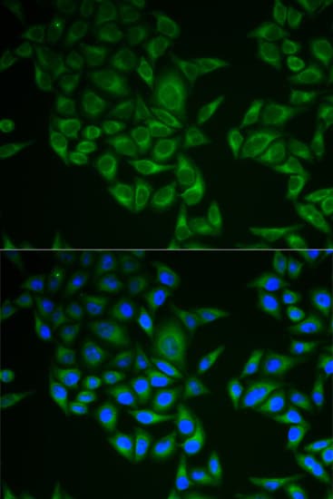 Immunocytochemistry/ Immunofluorescence - Anti-CD3 zeta antibody (ab190728)
Immunofluorescence analysis of A549 cells, using CD3 Zeta polyclonal antibody at 1:50.
-
 Western blot - Anti-CD3 zeta antibody (ab190728)Anti-CD3 zeta antibody (ab190728) + extracts of Jurkat cell line
Predicted band size: 19 kDa
Protocols
Datasheets and documents
-
SDS download
-
Datasheet download
References (2)
ab190728 has been referenced in 2 publications.
- Wang Y et al. NAD+ supplement potentiates tumor-killing function by rescuing defective TUB-mediated NAMPT transcription in tumor-infiltrated T cells. Cell Rep 36:109516 (2021). PubMed: 34380043
- Bustos-Morán E et al. Microtubule-associated protein-4 controls nanovesicle dynamics and T cell activation. J Cell Sci 130:1217-1223 (2017). PubMed: 28209780