Anti-Cofilin (phospho S3) antibody (ab12866)
 antibody (ab12866)")
Key features and details
- Rabbit polyclonal to Cofilin (phospho S3)
- Suitable for: WB, ICC/IF, Flow Cyt
- Reacts with: Mouse, Rat, Dog, Human, African green monkey
- Isotype: IgG
Overview
-
Product name
Anti-Cofilin (phospho S3) antibody
See all Cofilin-1 primary antibodies -
Description
Rabbit polyclonal to Cofilin (phospho S3) -
Host species
Rabbit -
Tested applications
Suitable for: WB, ICC/IF, Flow Cytmore details -
Species reactivity
Reacts with: Mouse, Rat, Dog, Human, African green monkey -
Immunogen
Synthetic peptide. This information is proprietary to Abcam and/or its suppliers.
-
General notes
The Life Science industry has been in the grips of a reproducibility crisis for a number of years. Abcam is leading the way in addressing this with our range of recombinant monoclonal antibodies and knockout edited cell lines for gold-standard validation. Please check that this product meets your needs before purchasing.
If you have any questions, special requirements or concerns, please send us an inquiry and/or contact our Support team ahead of purchase. Recommended alternatives for this product can be found below, along with publications, customer reviews and Q&As
Properties
-
Form
Liquid -
Storage instructions
Shipped at 4°C. Upon delivery aliquot and store at -20°C. Avoid freeze / thaw cycles. -
Storage buffer
pH: 7.3
Preservative: 0.05% Sodium azide
Constituents: PBS, 50% Glycerol, 0.1% BSA
PBS (without Mg2+ and Ca2+), BSA (IgG, protease free) -
 Concentration information loading...
 -
Purity
Immunogen affinity purified -
Clonality
Polyclonal -
Isotype
IgG -
Research areas
Associated products
-
Compatible Secondaries
-
Isotype control
-
Recombinant Protein
Applications
The Abpromise guarantee
Our Abpromise guarantee covers the use of ab12866 in the following tested applications.
The application notes include recommended starting dilutions; optimal dilutions/concentrations should be determined by the end user.
| Application | Abreviews | Notes |
|---|---|---|
| WB | (6) |
1/1000. Detects a band of approximately 20 kDa.
|
| ICC/IF | (3) |
1/250.
|
| Flow Cyt |
Use 3-5µg for 106 cells.
|
| Notes |
|---|
|
WB
1/1000. Detects a band of approximately 20 kDa. |
|
ICC/IF
1/250. |
|
Flow Cyt
Use 3-5µg for 106 cells. |
Target
-
Function
Controls reversibly actin polymerization and depolymerization in a pH-sensitive manner. It has the ability to bind G- and F-actin in a 1:1 ratio of cofilin to actin. It is the major component of intranuclear and cytoplasmic actin rods. -
Tissue specificity
Widely distributed in various tissues. -
Sequence similarities
Belongs to the actin-binding proteins ADF family.
Contains 1 ADF-H domain. -
Post-translational
modificationsPhosphorylated on Ser-3 in resting cells. -
Cellular localization
Nucleus matrix. Cytoplasm > cytoskeleton. Almost completely in nucleus in cells exposed to heat shock or 10% dimethyl sulfoxide. - Information by UniProt
-
Database links
- Entrez Gene: 476022 Dog
- Entrez Gene: 1072 Human
- Entrez Gene: 12631 Mouse
- Entrez Gene: 29271 Rat
- Omim: 601442 Human
- SwissProt: P23528 Human
- SwissProt: P18760 Mouse
- SwissProt: P45592 Rat
see all -
Alternative names
- 18 kDa phosphoprotein antibody
- CFL 1 antibody
- CFL antibody
see all
Images
-
Immunocytochemistry/ Immunofluorescence - Anti-Cofilin (phospho S3) antibody (ab12866)Immunofluorescence analysis of Phospho-Cofilin pSer3 was done on 70% confluent log phase PC-3 cells. The cells were fixed with 4% paraformaldehyde for 10 minutes, permeabilized with 0.1% Triton™ X-100 for 10 minutes, and blocked with 1% BSA for 1 hour at room temperature. The cells were labeled with ab12866 at 1:250 dilution in 0.1% BSA and incubated for 3 hours at room temperature and then labeled with Goat anti-Rabbit IgG (H+L) Superclonal™ Secondary Antibody, Alexa Fluor® 488 conjugate at a dilution of 1:2000 for 45 minutes at room temperature (Panel a: green). Nuclei (Panel b: blue) were stained with SlowFade® Gold Antifade Mountant with DAPI. F-actin (Panel c: red) was stained with Rhodamine Phalloidin (1:300). Panel d is a merged image showing cytoplasmic and nuclear localization. Panel e is a no primary antibody control. The images were captured at 60X magnification.
-
 Western blot - Anti-Cofilin (phospho S3) antibody (ab12866)Peptide Competition and Phosphatase Treatment
Lysates prepared from MDCK cells treated with staurosporine (1) or left untreated (2-6) were resolved by SDS-PAGE on a 10% polyacrylamide gel and transferred to PVDF. Membranes were either left untreated (1-5) or treated with lambda phosphatase (6), blocked with a 5% BSA-TBST buffer for one hour at room temperature, and incubated with the ab12866 antibody for two hours at room temperature in a 3% BSA-TBST buffer, following prior incubation with: no peptide (1, 2, 6), the non phosphopeptide corresponding to the immunogen (3), a generic phosphoserine-containing peptide (4) or, the phosphopeptide immunogen (5). After washing, membranes were incubated with goat F(ab)2 anti-rabbit IgG HRP conjugate and bands were detected using the Pierce SuperSignal method. The data show that only the peptide corresponding to cofilin [pS3] blocks the antibody signal. The data also show that phosphatase stripping eliminates the signal, verifying that the anti -
 Immunocytochemistry/ Immunofluorescence - Anti-Cofilin (phospho S3) antibody (ab12866)ICC/IF image of ab12866 stained MCF7 cells. The cells were 4% PFA fixed (10 min) and then incubated in 1%BSA / 10% normal goat serum / 0.3M glycine in 0.1% PBS-Tween for 1h to permeabilise the cells and block non-specific protein-protein interactions. The cells were then incubated with the antibody (ab12866, 1µg/ml) overnight at +4°C. The secondary antibody (green) was Alexa Fluor® 488 goat anti-rabbit IgG (H+L) used at a 1/1000 dilution for 1h. Alexa Fluor® 594 WGA was used to label plasma membranes (red) at a 1/200 dilution for 1h. DAPI was used to stain the cell nuclei (blue) at a concentration of 1.43µM.
 -
 Immunocytochemistry/ Immunofluorescence - Anti-Cofilin (phospho S3) antibody (ab12866)This image is courtesy of an anonymous abreview.
Immunocytochemistry/ Immunofluorescence analysis of human neuroblastoma cells labeling Cofilin (phospho S3) with ab12866 at 1/500 dilution. Cells were fixed in paraformaldehyde, permeabilized for 15 minutes in 0.01% Triton X-100, blocked using 5% serum for 30 minutes at 20°C, then incubated with ab12866 at a 1/500 dilution for 2 hours at 20°C. The secondary used was a Dylight 488 conjugated donkey anti-rabbit IgG (H+L) used at a 1/500 dilution.
-
 Western blot - Anti-Cofilin (phospho S3) antibody (ab12866)All lanes : Anti-Cofilin (phospho S3) antibody (ab12866) at 1 µg/ml
Lane 1 : HeLa whole cell extract
Lane 2 : HeLa treated for overnight with 150 uM of H2O2 whole cell extract
Lane 3 : HeLa treated for overnight with 3 uM of Staurosporine whole cell extract
Lane 4 : COS-7 whole cell extract
Lane 5 : NIH/3T3 (Mouse embryo fibroblast cell line) whole cell extract
Lane 6 : MCF7 whole cell extract
Lane 7 : Rat Skeletal Muscle whole cell extract
Lane 8 : A-431 whole cell extract
Lane 9 : A-431 treated for overnight with 150 uM of H2O2 whole cell extract
Lane 10 : Jurkat whole cell extract
Lane 11 : Jurkat treated for overnight with 3 uM of Staurosporine whole cell extract
Lysates/proteins at 20 µg per lane.
Secondary
All lanes : Goat anti-rabbit IgG (H+L), HRP conjugate at 1/2500 dilution
Observed band size: 18 kDa why is the actual band size different from the predicted? -
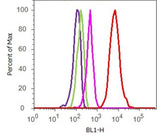 Flow Cytometry - Anti-Cofilin (phospho S3) antibody (ab12866)
Flow Cytometry analysis of U-87 MG cells labeling Cofilin (phospho S3) with ab12866. Cells were fixed with 70% ethanol for 10 minutes, permeabilized with 0.25% Triton™ X-100 for 20 minutes, and blocked with 5% BSA for 30 minutes at room temperature. Cells were labeled with Anti-Cofilin (phospho S3) antibody (ab12866, red) or with rabbit isotype control (pink) at 3-5 ug/million cells in 2.5% BSA. After incubation at room temperature for 2 hours, the cells were labeled with Alexa Fluor® 488 Goat Anti-Rabbit Secondary Antibody at a dilution of 1/400 for 30 minutes at room temperature. The representative 10,000 cells were acquired and analyzed for each sample. The purple histogram represents unstained control cells and the green histogram represents no-primary-antibody control.
Protocols
Datasheets and documents
-
SDS download
-
Datasheet download
References (69)
ab12866 has been referenced in 69 publications.
- Fu R et al. Neuroprotective Effects of Tetrahydroxystilbene Glucoside against Rotenone-Induced Toxicity in PC12 Cells. Biol Pharm Bull 45:143-149 (2022). PubMed: 34707025
- Feng L et al. Chaperonin-Containing TCP1 Subunit 5 Protects Against the Effect of Mer Receptor Tyrosine Kinase Knockdown in Retinal Pigment Epithelial Cells by Interacting With Filamentous Actin and Activating the LIM-Kinase 1/Cofilin Pathway. Front Med (Lausanne) 9:861371 (2022). PubMed: 35492354
- Keiser AA et al. Systemic HDAC3 inhibition ameliorates impairments in synaptic plasticity caused by simulated galactic cosmic radiation exposure in male mice. Neurobiol Learn Mem 178:107367 (2021). PubMed: 33359392
- Xu GJ et al. Environmental enrichment combined with fasudil treatment inhibits neuronal death in the hippocampal CA1 region and ameliorates memory deficits. Neural Regen Res 16:1460-1466 (2021). PubMed: 33433459
- Zhu YT et al. Environmental enrichment combined with fasudil promotes motor function recovery and axonal regeneration after stroke. Neural Regen Res 16:2512-2520 (2021). PubMed: 33907042