Anti-SOCS3 antibody (ab16030)
")
Key features and details
- Rabbit polyclonal to SOCS3
- Suitable for: WB, ICC/IF
- Reacts with: Human
- Isotype: IgG
Overview
-
Product name
Anti-SOCS3 antibody
See all SOCS3 primary antibodies -
Description
Rabbit polyclonal to SOCS3 -
Host species
Rabbit -
Tested applications
Suitable for: WB, ICC/IFmore details -
Species reactivity
Reacts with: Human
Predicted to work with: Mouse, Rat, Chicken, Cow, Dog
-
Immunogen
Synthetic peptide corresponding to Human SOCS3 aa 200 to the C-terminus (C terminal) conjugated to keyhole limpet haemocyanin.
(Peptide available asab16199) -
General notes
The Life Science industry has been in the grips of a reproducibility crisis for a number of years. Abcam is leading the way in addressing this with our range of recombinant monoclonal antibodies and knockout edited cell lines for gold-standard validation. Please check that this product meets your needs before purchasing.
If you have any questions, special requirements or concerns, please send us an inquiry and/or contact our Support team ahead of purchase. Recommended alternatives for this product can be found below, along with publications, customer reviews and Q&As
Properties
-
Form
Liquid -
Storage instructions
Shipped at 4°C. Store at +4°C short term (1-2 weeks). Upon delivery aliquot. Store at -20°C or -80°C. Avoid freeze / thaw cycle. -
Storage buffer
pH: 7.40
Preservative: 0.02% Sodium azide
Constituent: PBS
Batches of this product that have a concentration < 1mg/ml may have BSA added as a stabilising agent. If you would like information about the formulation of a specific lot, please contact our scientific support team who will be happy to help. -
 Concentration information loading...
 -
Purity
Immunogen affinity purified -
Clonality
Polyclonal -
Isotype
IgG -
Research areas
Associated products
-
Compatible Secondaries
-
Isotype control
-
Recombinant Protein
Applications
The Abpromise guarantee
Our Abpromise guarantee covers the use of ab16030 in the following tested applications.
The application notes include recommended starting dilutions; optimal dilutions/concentrations should be determined by the end user.
| Application | Abreviews | Notes |
|---|---|---|
| WB | (9) |
Use at an assay dependent concentration. Predicted molecular weight: 24 kDa.
|
| ICC/IF | (2) |
Use a concentration of 1 µg/ml.
|
| Notes |
|---|
|
WB
Use at an assay dependent concentration. Predicted molecular weight: 24 kDa. |
|
ICC/IF
Use a concentration of 1 µg/ml. |
Target
-
Function
SOCS family proteins form part of a classical negative feedback system that regulates cytokine signal transduction. SOCS3 is involved in negative regulation of cytokines that signal through the JAK/STAT pathway. Inhibits cytokine signal transduction by binding to tyrosine kinase receptors including gp130, LIF, erythropoietin, insulin, IL12, GCSF and leptin receptors. Binding to JAK2 inhibits its kinase activity. Suppresses fetal liver erythropoiesis. Regulates onset and maintenance of allergic responses mediated by T-helper type 2 cells. Regulates IL-6 signaling in vivo (By similarity). Probable substrate recognition component of a SCF-like ECS (Elongin BC-CUL2/5-SOCS-box protein) E3 ubiquitin-protein ligase complex which mediates the ubiquitination and subsequent proteasomal degradation of target proteins. Seems to recognize IL6ST. -
Tissue specificity
Widely expressed with high expression in heart, placenta, skeletal muscle, peripheral blood leukocytes, fetal and adult lung, and fetal liver and kidney. Lower levels in thymus. -
Pathway
Protein modification; protein ubiquitination. -
Involvement in disease
Note=There is some evidence that SOCS3 may be a susceptibility gene for atopic dermatitis linked to 17q25. SOCS3 messenger RNA is significantly more highly expressed in skin from patients with atopic dermatitis than in skin from healthy controls. Furthermore, a genetic association between atopic dermatitis and a haplotype in the SOCS3 gene has been found in two independent groups of patients. -
Sequence similarities
Contains 1 SH2 domain.
Contains 1 SOCS box domain. -
Domain
The ESS and SH2 domains are required for JAK phosphotyrosine binding. Further interaction with the KIR domain is necessary for signal and kinase inhibition.
The SOCS box domain mediates the interaction with the Elongin BC complex, an adapter module in different E3 ubiquitin ligase complexes. -
Post-translational
modificationsPhosphorylated on tyrosine residues after stimulation by the cytokines, IL-2, EPO or IGF1. - Information by UniProt
-
Database links
- Entrez Gene: 395299 Chicken
- Entrez Gene: 282081 Cow
- Entrez Gene: 442949 Dog
- Entrez Gene: 9021 Human
- Entrez Gene: 12702 Mouse
- Entrez Gene: 89829 Rat
- Omim: 604176 Human
- SwissProt: Q90X67 Chicken
see all -
Alternative names
- ATOD4 antibody
- CIS 3 antibody
- CIS-3 antibody
see all
Images
-
Western blot - Anti-SOCS3 antibody (ab16030)The 30kDa band observed is comparable to the molecular weight seen with other commercially available antibodies to SOCS3.
-
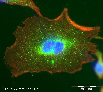 Immunocytochemistry/ Immunofluorescence - Anti-SOCS3 antibody (ab16030)
ICC/IF image of ab16030 stained human HeLa cells. The cells were PFA fixed (10 min) and incubated with the antibody (ab16030, 1µg/ml) for 1h at room temperature. The secondary antibody (green) was Alexa Fluor® 488 goat anti-rabbit IgG (H+L) used at a 1/1000 dilution for 1h. Image-iTTM FX Signal Enhancer was used as the primary blocking agent, 5% BSA (TBS-T) was used for all other blocking steps. DAPI was used to stain the cell nuclei (blue). Alexa Fluor® 594 WGA was used to label plasma membranes (red).
-
 Western blot - Anti-SOCS3 antibody (ab16030)This image is courtesy of an anonymous AbreviewAll lanes : Anti-SOCS3 antibody (ab16030) at 1 µg/ml
Lane 1 : Untreated Human peripheral blood mononuclear cell lysate
Lane 2 : Human peripheral blood mononuclear cell lysate (treated with aCD3/28)
Lane 3 : Human peripheral blood mononuclear cell lysate (treated with IFNγ)
Lane 4 : SOCS3 protein
Lysates/proteins at 15 µg per lane.
Secondary
All lanes : HRP-conjugated goat anti-rabbit polyclonal IgG
at 1/2000 dilution
Developed using the ECL technique.
Performed under reducing conditions.
Predicted band size: 24 kDa
Observed band size: 25 kDa why is the actual band size different from the predicted?
Exposure time: 15 minutes
Protocols
Datasheets and documents
-
SDS download
-
Datasheet download
References (124)
ab16030 has been referenced in 124 publications.
- Qian C et al. Opening KATP channels induces inflammatory tolerance and prevents chronic pain. Brain Behav Immun 107:76-86 (2023). PubMed: 36198341
- Hirani D et al. Macrophage-derived IL-6 trans-signalling as a novel target in the pathogenesis of bronchopulmonary dysplasia. Eur Respir J 59:N/A (2022). PubMed: 34446466
- Li J et al. Ginsenoside Rg1 Reduced Microglial Activation and Mitochondrial Dysfunction to Alleviate Depression-Like Behaviour Via the GAS5/EZH2/SOCS3/NRF2 Axis. Mol Neurobiol 59:2855-2873 (2022). PubMed: 35230663
- Zhou Z et al. IL-17A Mediates Demyelination by Activating A1 Astrocytes via SOCS3 During Angiostrongylus cantonensis Infection. Front Immunol 13:845011 (2022). PubMed: 35296090
- Hollingsworth TJ et al. Chronic Proinflammatory Signaling Accelerates the Rate of Degeneration in a Spontaneous Polygenic Model of Inherited Retinal Dystrophy. Front Pharmacol 13:839424 (2022). PubMed: 35387333