Anti-STAT5a (phospho S780) antibody (ab30649)
 antibody (ab30649)")
Key features and details
- Rabbit polyclonal to STAT5a (phospho S780)
- Suitable for: ICC/IF, WB, IHC-P
- Reacts with: Human
- Isotype: IgG
Overview
-
Product name
Anti-STAT5a (phospho S780) antibody
See all STAT5a primary antibodies -
Description
Rabbit polyclonal to STAT5a (phospho S780) -
Host species
Rabbit -
Tested applications
Suitable for: ICC/IF, WB, IHC-Pmore details -
Species reactivity
Reacts with: Human
Predicted to work with: Mouse, Rat
-
Immunogen
Synthetic peptide corresponding to Human STAT5a (phospho S780).
Database link: P42229 -
Positive control
- IHC: human breast carcinoma. WB: Jurkat treated with 125 ng/mL PMA for 30 minutes lysate. 293T treated with 125 ng/mL PMA for 30 minutes lysate. ICC/IF: HeLa cells.
-
General notes
The Life Science industry has been in the grips of a reproducibility crisis for a number of years. Abcam is leading the way in addressing this with our range of recombinant monoclonal antibodies and knockout edited cell lines for gold-standard validation. Please check that this product meets your needs before purchasing.
If you have any questions, special requirements or concerns, please send us an inquiry and/or contact our Support team ahead of purchase. Recommended alternatives for this product can be found below, along with publications, customer reviews and Q&As
Properties
-
Form
Liquid -
Storage instructions
Shipped at 4°C. Upon delivery aliquot and store at -20°C. Avoid freeze / thaw cycles. -
Storage buffer
pH: 7.40
Preservative: 0.02% Sodium azide
Constituents: PBS, 50% Glycerol (glycerin, glycerine), 0.87% Sodium chloride
Without Mg2+ and Ca2+ -
 Concentration information loading...
 -
Purity
Immunogen affinity purified -
Purification notes
ab30649 was affinity-purified from rabbit antiserum by affinity-chromatography using epitope-specific phosphopeptide. The antibody against non-phosphopeptide was removed by chromatography using non-phosphopeptide corresponding to the phosphorylation site -
Clonality
Polyclonal -
Isotype
IgG -
Research areas
Associated products
-
Compatible Secondaries
-
Isotype control
-
Recombinant Protein
Applications
The Abpromise guarantee
Our Abpromise guarantee covers the use of ab30649 in the following tested applications.
The application notes include recommended starting dilutions; optimal dilutions/concentrations should be determined by the end user.
| Application | Abreviews | Notes |
|---|---|---|
| ICC/IF |
Use a concentration of 1 µg/ml.
|
|
| WB |
1/500 - 1/1000. Detects a band of approximately 91 kDa (predicted molecular weight: 91 kDa).
|
|
| IHC-P |
1/50 - 1/100.
|
| Notes |
|---|
|
ICC/IF
Use a concentration of 1 µg/ml. |
|
WB
1/500 - 1/1000. Detects a band of approximately 91 kDa (predicted molecular weight: 91 kDa). |
|
IHC-P
1/50 - 1/100. |
Target
-
Function
Carries out a dual function: signal transduction and activation of transcription. Binds to the GAS element and activates PRL-induced transcription. -
Sequence similarities
Belongs to the transcription factor STAT family.
Contains 1 SH2 domain. -
Post-translational
modificationsTyrosine phosphorylated in response to IL-2, IL-3, IL-7, IL-15, GM-CSF, growth hormone, prolactin, erythropoietin and thrombopoietin. Tyrosine phosphorylation is required for DNA-binding activity and dimerization. Serine phosphorylation is also required for maximal transcriptional activity. -
Cellular localization
Cytoplasm. Nucleus. Translocated into the nucleus in response to phosphorylation. - Information by UniProt
-
Database links
- Entrez Gene: 6776 Human
- Entrez Gene: 20850 Mouse
- Entrez Gene: 24918 Rat
- Omim: 601511 Human
- SwissProt: P42229 Human
- SwissProt: P42230 Mouse
- SwissProt: Q62771 Rat
- Unigene: 437058 Human
see all -
Alternative names
- Mammary gland factor antibody
- MGF antibody
- Signal transducer and activator of transcription 5A antibody
see all
Images
-
Western blot - Anti-STAT5a (phospho S780) antibody (ab30649)All lanes : Anti-STAT5a (phospho S780) antibody (ab30649) at 1/1000 dilution
Lane 1 : Jurkat untreated lysate
Lane 2 : Jurkat treated with 125 ng/mL PMA for 30 minutes lysate
Lane 3 : 293T untreated lysate
Lane 4 : 293T treated with 125 ng/mL PMA for 30 minutes lysate
Lysates/proteins at 20 µg per lane.
Secondary
All lanes : Goat Anti-Rabbit IgG (H+L) HRP at 1/10000 dilution
Predicted band size: 91 kDaLoading control: beta Actin.
-
 Immunohistochemistry (Formalin/PFA-fixed paraffin-embedded sections) - Anti-STAT5a (phospho S780) antibody (ab30649)
Immunohistochemistry analysis of paraffin-embedded human breast carcinoma, using ab30649. The picture on the right is blocked with the phospho peptide.
-
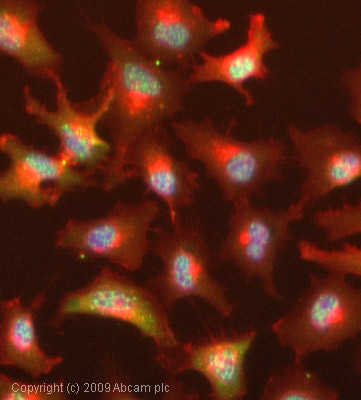 Immunocytochemistry/ Immunofluorescence - Anti-STAT5a (phospho S780) antibody (ab30649)ICC/IF image of ab30649 stained HeLa cells. The cells were 100% methanol fixed (5 min) and then incubated in 1%BSA / 10% normal goat serum / 0.3M glycine in 0.1% PBS-Tween for 1h to permeabilise the cells and block non-specific protein-protein interactions. The cells were then incubated with the antibody (ab30649, 1µg/ml) overnight at +4°C. The secondary antibody (green) was Alexa Fluor® 488 goat anti-rabbit IgG (H+L) used at a 1/1000 dilution for 1h. Alexa Fluor® 594 WGA was used to label plasma membranes (red) at a 1/200 dilution for 1h. DAPI was used to stain the cell nuclei (blue) at a concentration of 1.43µM.
 -
 Western blot - Anti-STAT5a (phospho S780) antibody (ab30649)All lanes : Anti-STAT5a (phospho S780) antibody (ab30649)
Lane 1 : HeLa cells extract
Lane 2 : HeLa cells extract with synthesized non-phosphopeptide
Lane 3 : HeLa cells extract with synthesized phosphopeptide
Predicted band size: 91 kDa
Observed band size: 91 kDa
Protocols
Datasheets and documents
-
SDS download
-
Datasheet download
References (1)
ab30649 has been referenced in 1 publication.
- Li RE et al. Quantitative Phosphoproteomic Analysis Reveals Dendritic Cell- Specific STAT Signaling After a2-3-Linked Sialic Acid Ligand Binding. Front Immunol 12:673454 (2021). PubMed: 33968084