Anti-USP10 antibody (ab70895)
")
Key features and details
- Rabbit polyclonal to USP10
- Suitable for: IHC-P, WB, IP, ICC/IF
- Reacts with: Mouse, Human
- Isotype: IgG
Overview
-
Product name
Anti-USP10 antibody
See all USP10 primary antibodies -
Description
Rabbit polyclonal to USP10 -
Host species
Rabbit -
Tested applications
Suitable for: IHC-P, WB, IP, ICC/IFmore details -
Species reactivity
Reacts with: Mouse, Human -
Immunogen
Synthetic peptide within Human USP10 aa 50-150. The exact immunogen sequence used to generate this antibody is proprietary information. If additional detail on the immunogen is needed to determine the suitability of the antibody for your needs, please contact our Scientific Support team to discuss your requirements.
Database link: Q14694 -
Positive control
- WB: HeLa, TCMK-1 and HEK-293T whole cell lysate. IHC-P: Human breast carcinoma tissue. Human normal tonsil tissue. IP: HeLa whole cell lysate. ICC/IF: HeLa cells.
-
General notes
The Life Science industry has been in the grips of a reproducibility crisis for a number of years. Abcam is leading the way in addressing this with our range of recombinant monoclonal antibodies and knockout edited cell lines for gold-standard validation. Please check that this product meets your needs before purchasing.
If you have any questions, special requirements or concerns, please send us an inquiry and/or contact our Support team ahead of purchase. Recommended alternatives for this product can be found below, along with publications, customer reviews and Q&As
Properties
-
Form
Liquid -
Storage instructions
Shipped at 4°C. Upon delivery aliquot and store at -20°C. Avoid freeze / thaw cycles. -
Storage buffer
pH: 6.8
Preservative: 0.09% Sodium azide
Constituents: 0.1% BSA, Tris buffered saline -
 Concentration information loading...
 -
Purity
Immunogen affinity purified -
Clonality
Polyclonal -
Isotype
IgG -
Research areas
Associated products
-
Compatible Secondaries
-
Isotype control
-
Positive Controls
-
Recombinant Protein
Applications
The Abpromise guarantee
Our Abpromise guarantee covers the use of ab70895 in the following tested applications.
The application notes include recommended starting dilutions; optimal dilutions/concentrations should be determined by the end user.
| Application | Abreviews | Notes |
|---|---|---|
| IHC-P |
Use a concentration of 5 µg/ml. Perform heat mediated antigen retrieval with citrate buffer pH 6 before commencing with IHC staining protocol.
|
|
| WB |
1/2000 - 1/10000. Detects a band of approximately 105 kDa (predicted molecular weight: 87 kDa).
|
|
| IP |
Use at 2-5 µg/mg of lysate.
|
|
| ICC/IF |
Use a concentration of 1 µg/ml.
|
| Notes |
|---|
|
IHC-P
Use a concentration of 5 µg/ml. Perform heat mediated antigen retrieval with citrate buffer pH 6 before commencing with IHC staining protocol. |
|
WB
1/2000 - 1/10000. Detects a band of approximately 105 kDa (predicted molecular weight: 87 kDa). |
|
IP
Use at 2-5 µg/mg of lysate. |
|
ICC/IF
Use a concentration of 1 µg/ml. |
Target
-
Function
Hydrolase that can remove conjugated ubiquitin from target proteins such as p53/TP53, SNX3 and CFTR. Acts as an essential regulator of p53/TP53 stability: in unstressed cells, specifically deubiquitinates p53/TP53 in the cytoplasm, leading to counteract MDM2 action and stabilize p53/TP53. Following DNA damage, translocates to the nucleus and deubiquitinates p53/TP53, leading to regulate the p53/TP53-dependent DNA damage response. Does not deubiquitinate MDM2. Deubiquitinates CFTR in early endosomes, enhancing its endocytic recycling. -
Tissue specificity
Widely expressed. -
Sequence similarities
Belongs to the peptidase C19 family. USP10 subfamily. -
Post-translational
modificationsPhosphorylated by ATM following DNA damage, leading to stablization and translocation it to the nucleus. -
Cellular localization
Cytoplasm. Nucleus. Early endosome. Cytoplasmic in normal conditions. After DNA damage, translocates to the nucleus following phosphorylation by ATM. - Information by UniProt
-
Database links
- Entrez Gene: 9100 Human
- Entrez Gene: 22224 Mouse
- Omim: 609818 Human
- SwissProt: Q14694 Human
- SwissProt: P52479 Mouse
- Unigene: 136778 Human
- Unigene: 256910 Mouse
- Unigene: 421337 Mouse
-
Alternative names
- Deubiquitinating enzyme 10 antibody
- KIAA0190 antibody
- MGC124997 antibody
see all
Images
-
Western blot - Anti-USP10 antibody (ab70895)All lanes : Anti-USP10 antibody (ab70895) at 0.1 µg/ml
Lane 1 : HeLa (Human epithelial cell line from cervix adenocarcinoma) whole cell lysate
Lane 2 : HEK-293T (Human epithelial cell line from embryonic kidney transformed with large T antigen) whole cell lysate
Lysates/proteins at 50 µg per lane.
Predicted band size: 87 kDa
Exposure time: 30 secondsLysates prepared using NETN lysis buffer.
-
 Immunohistochemistry (Formalin/PFA-fixed paraffin-embedded sections) - Anti-USP10 antibody (ab70895)
Immunohistochemistry (Formalin/PFA-fixed paraffin-embedded sections) analysis of human breast carcinoma tissue labelling USP10 with ab70895 at 1/200 (1 µg/ml). Detection: DAB.
-
 Immunocytochemistry/ Immunofluorescence - Anti-USP10 antibody (ab70895)
ICC image of ab70895 stained HeLa cells. The cells were 4% formaldehyde fixed (10 min) and then incubated in 1%BSA / 10% normal goat serum / 0.3M glycine in 0.1% PBS-Tween for 1h to permeabilise the cells and block non-specific protein-protein interactions. The cells were then incubated with the antibody (ab70895, 1µg/ml) overnight at +4°C. The secondary antibody (green) was Alexa Fluor® 488 goat anti-rabbit IgG (H+L) used at a 1/1000 dilution for 1h. Alexa Fluor® 594 WGA was used to label plasma membranes (red) at a 1/200 dilution for 1h. DAPI was used to stain the cell nuclei (blue) at a concentration of 1.43µM.
-
 Immunoprecipitation - Anti-USP10 antibody (ab70895)
Detection of human USP10 by western blot of Immunoprecipitates.
Samples: Lane 1 is whole cell lysate (1.0 mg per IP reaction; 20% of IP loaded) from HeLa cells prepared using NETN lysis buffer and Lane 2 is control IgG.
Antibodies: ab70895 used for IP at 3 µg per reaction. For blotting immunoprecipitated USP10, ab70895 was used at 1 µg/ml. -
 Western blot - Anti-USP10 antibody (ab70895)Anti-USP10 antibody (ab70895) at 0.1 µg/ml + TCMK-1 (Mouse kidney epithelial cell line) whole cell lysate at 50 µg
Predicted band size: 87 kDa
Exposure time: 3 minutesLysates prepared using NETN lysis buffer.
-
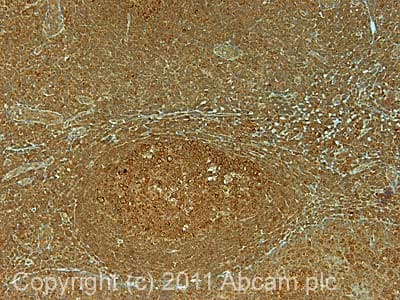 Immunohistochemistry (Formalin/PFA-fixed paraffin-embedded sections) - Anti-USP10 antibody (ab70895)IHC image of ab70895 staining in human normal tonsil formalin fixed paraffin embedded tissue section, performed on a Leica BondTM system using the standard protocol F. The section was pre-treated using heat mediated antigen retrieval with sodium citrate buffer (pH6, epitope retrieval solution 1) for 20 mins. The section was then incubated with ab70895, 5µg/ml, for 15 mins at room temperature and detected using an HRP conjugated compact polymer system. DAB was used as the chromogen. The section was then counterstained with haematoxylin and mounted with DPX.
For other IHC staining systems (automated and non-automated) customers should optimize variable parameters such as antigen retrieval conditions, primary antibody concentration and antibody incubation times.
Protocols
Datasheets and documents
-
SDS download
-
Datasheet download
References (3)
ab70895 has been referenced in 3 publications.
- Lu L et al. FSTL1-USP10-Notch1 Signaling Axis Protects Against Cardiac Dysfunction Through Inhibition of Myocardial Fibrosis in Diabetic Mice. Front Cell Dev Biol 9:757068 (2021). PubMed: 34957094
- Cirillo L et al. UBAP2L Forms Distinct Cores that Act in Nucleating Stress Granules Upstream of G3BP1. Curr Biol 30:698-707.e6 (2020). PubMed: 31956030
- Katoh H et al. Japanese encephalitis virus core protein inhibits stress granule formation through an interaction with Caprin-1 and facilitates viral propagation. J Virol 87:489-502 (2013). PubMed: 23097442