Anti-TBR2 / Eomes antibody (ab23345)
 - Anti-TBR2 / Eomes antibody (ab23345)")
Key features and details
- Rabbit polyclonal to TBR2 / Eomes
- Suitable for: IHC-Fr, WB
- Reacts with: Mouse, Human
- Isotype: IgG
Get better batch-to-batch reproducibility with a recombinant antibody


- Research with confidence – consistent and reproducible results with every batch
- Long-term and scalable supply – powered by recombinant technology for fast production
- Success from the first experiment – confirmed specificity through extensive validation
- Ethical standards compliant – production is animal-free
Overview
-
Product name
Anti-TBR2 / Eomes antibody
See all TBR2 / Eomes primary antibodies -
Description
Rabbit polyclonal to TBR2 / Eomes -
Host species
Rabbit -
Specificity
From Jan 2024, QC testing of replenishment batches of this polyclonal changed. All tested and expected application and reactive species combinations are still covered by our Abcam product promise. However, we no longer test all applications. For more information on a specific batch, please contact our Scientific Support who will be happy to help.
-
Tested applications
Suitable for: IHC-Fr, WBmore details -
Species reactivity
Reacts with: Mouse, Human
Predicted to work with: Rat, Cow, Common marmoset
-
Immunogen
Synthetic peptide corresponding to Mouse TBR2/ Eomes aa 650 to the C-terminus (C terminal) conjugated to keyhole limpet haemocyanin.
(Peptide available asab25698) -
Positive control
- IHC-Fr: C57/BL6 and Mash1 mouse forebrain coronal sections. Mouse brain E14 frozen tissue section. Mouse developing cerebral cortex tissue sections. Mouse embryonic brain tissue section. WB: E14 Mouse Embryo Brain Tissue Lysate. EL4 cells. Human PTA-6967 Whole Cell Lysate. Human ES Cells. Human Mesendoderm (Day 2) Whole Cell Lysate.
-
General notes
Tbr2 expression is observed in neuron progenitor compartments in development (the subventricular zone and ventricular zone) and expression rises and falls with cortical plate neurogenesis. Transition from radial glia to intermediate progenitor cell is associated with upregulation of Tbr2.
The Life Science industry has been in the grips of a reproducibility crisis for a number of years. Abcam is leading the way in addressing this with our range of recombinant monoclonal antibodies and knockout edited cell lines for gold-standard validation. Please check that this product meets your needs before purchasing.
If you have any questions, special requirements or concerns, please send us an inquiry and/or contact our Support team ahead of purchase. Recommended alternatives for this product can be found below, along with publications, customer reviews and Q&As
Properties
-
Form
Liquid -
Storage instructions
Shipped at 4°C. Store at +4°C short term (1-2 weeks). Upon delivery aliquot. Store at -20°C or -80°C. Avoid freeze / thaw cycle. -
Storage buffer
pH: 7.40
Preservative: 0.02% Sodium azide
Constituent: PBS
Batches of this product that have a concentration < 1mg/ml may have BSA added as a stabilising agent. If you would like information about the formulation of a specific lot, please contact our scientific support team who will be happy to help. -
 Concentration information loading...
 -
Purity
Immunogen affinity purified -
Primary antibody notes
Tbr2 expression is observed in neuron progenitor compartments in development (the subventricular zone and ventricular zone) and expression rises and falls with cortical plate neurogenesis. Transition from radial glia to intermediate progenitor cell is associated with upregulation of Tbr2. -
Clonality
Polyclonal -
Isotype
IgG -
Research areas
Associated products
-
ChIP Related Products
-
Compatible Secondaries
-
Isotype control
-
Recombinant Protein
-
Related Products
Applications
The Abpromise guarantee
Our Abpromise guarantee covers the use of ab23345 in the following tested applications.
The application notes include recommended starting dilutions; optimal dilutions/concentrations should be determined by the end user.
| Application | Abreviews | Notes |
|---|---|---|
| IHC-Fr | (10) |
1/100 - 1/500.
Abcam recommends the following antigen retrieval method: Heated citrate solution (10mM citrate pH 6.0 + 0.05% Tween-20). We recommend using Goat Anti-Rabbit IgG H&L (Alexa Fluor® 488) (ab150077) secondary antibody. |
| WB | (5) |
Use a concentration of 0.4 - 2.5 µg/ml. Detects a band of approximately 85 kDa (predicted molecular weight: 72 kDa).
Abcam recommends using milk as the blocking agent. In our hands, when tested in western blot, this product typically gives a weaker signal in mouse tissue lysates compared to human cell lines. However, this product gives clean, specific staining in IHC-Fr on mouse E14.5 cortex. Abcam welcomes customer feedback and would appreciate any comments regarding this product and the data presented. |
| Notes |
|---|
|
IHC-Fr
1/100 - 1/500. Abcam recommends the following antigen retrieval method: Heated citrate solution (10mM citrate pH 6.0 + 0.05% Tween-20). We recommend using Goat Anti-Rabbit IgG H&L (Alexa Fluor® 488) (ab150077) secondary antibody. |
|
WB
Use a concentration of 0.4 - 2.5 µg/ml. Detects a band of approximately 85 kDa (predicted molecular weight: 72 kDa). Abcam recommends using milk as the blocking agent. In our hands, when tested in western blot, this product typically gives a weaker signal in mouse tissue lysates compared to human cell lines. However, this product gives clean, specific staining in IHC-Fr on mouse E14.5 cortex. Abcam welcomes customer feedback and would appreciate any comments regarding this product and the data presented. |
Target
-
Function
Functions as a transcriptional activator playing a crucial role during development. Functions in trophoblast differentiation and later in gastrulation, regulating both mesoderm delamination and endoderm specification. Plays a role in brain development being required for the specification and the proliferation of the intermediate progenitor cells and their progeny in the cerebral cortex. Also involved in the differentiation of CD8+ T-cells during immune response regulating the expression of lytic effector genes. -
Tissue specificity
Expressed in CD8+ T-cells. -
Involvement in disease
Note=A translocation t(3;10)(p24;q23) located 215 kb 3' to the EOMES gene but leading to loss of its expression was identified in a large consanguineous family. Homozygous silencing produces microcephaly associated with corpus callosum agenesis, bilateral polymicrogyria, ventricular dilatation and a small cerebellum. -
Sequence similarities
Contains 1 T-box DNA-binding domain. -
Developmental stage
Detected at 7 weeks of development in the forebrain floorplate of the CNS. Expressed within the mantle layer and migrating neuroblasts of the telencephalon at 12.5 weeks of development. -
Cellular localization
Nucleus. - Information by UniProt
-
Database links
- Entrez Gene: 8320 Human
- Entrez Gene: 13813 Mouse
- Entrez Gene: 233242 Rat
- Entrez Gene: 316052 Rat
- Omim: 604615 Human
- SwissProt: O95936 Human
- SwissProt: O54839 Mouse
- Unigene: 591663 Human
see all -
Alternative names
- eomes antibody
- EOMES_HUMAN antibody
- Eomesodermin antibody
see all
Images
-
Immunohistochemistry (Frozen sections) - Anti-TBR2 / Eomes antibody (ab23345)Image from Azim K et al., PLoS One. 2012;7(11):e49087. Fig 5.; doi: 10.1371/journal.pone.0049087. Reproduced under the Creative Commons license http://creativecommons.org/licenses/by/4.0/
Dlx2 and Tbr2 identify non overlapping progenitor lineages in the adult mouse SVZ (subventricular zone).
(A–B) Adult C57/BL6 mouse forebrain coronal sections at rostro-caudal point 1.2 relative to the bregma were immunostained for Dcx (red), Dlx2 or Tbr2 (green) and Ki67 (blue). Both progenitor populations show characteristics of migrating neuroblasts, as indicated by their Dcx expression (C–D). Adult Mash1 mouse forebrain coronal sections at rostro-caudal point 1.2 relative to the bregma were immunostained for Tbr2 (red) and Dlx2 (blue). Both Tbr2 and Dlx2 exhibited EGFP expression, but showed no colocalisation. Right side captions show cropped individual channels and the merges. Full panel insets are zoomed and cropped DAB stained photomicrographs of rostral periventricular sections for Tbr2 in the dorsal SVZ (A), Dlx2 in the dorso-lateral SVZ (B) and Mash1 in the ventro-lateral SVZ (C). Yellow arrows and arrowheads show respectively positive stained cell and low level TF staining. Dotted lines mark approximate boundaries of ventricular space. Flattened confocal z-stacks are of 14–15 µm thickness, including captions. Scale bars: 15 µm in full panels, 20 µm in captions and 25 µm in insets.
-
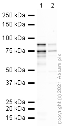 Western blot - Anti-TBR2 / Eomes antibody (ab23345)All lanes : Anti-TBR2 / Eomes antibody (ab23345) at 1 µg/ml
Lane 1 : Human PTA-6967 Whole Cell Lysate at 20 µg
Lane 2 : E14 Mouse Embryo Brain Tissue Lysate at 40 µg
Secondary
All lanes : Goat polyclonal to Rabbit IgG - H&L - Pre-Adsorbed (HRP) at 1/50000 dilution
Predicted band size: 72 kDa
Observed band size: 85 kDa why is the actual band size different from the predicted?
Additional bands at: 75 kDa. We are unsure as to the identity of these extra bands.
Exposure time: 12 minutesGel type: MOPS
Blocking buffer: 1% milk
-
 Western blot - Anti-TBR2 / Eomes antibody (ab23345)ab23345 at 1 mg/ml + Human ES Cells treated with Retinoic Acid (48h) at 20 µg
Secondary
Goat Anti-Rabbit IgG H&L (HRP) preadsorbed at 1/50000 dilution
Developed using the ECL technique.
Performed under reducing conditions.
Predicted band size: 72 kDa
Observed band size: 85 kDa why is the actual band size different from the predicted?
Additional bands at: 50 kDa, 65 kDa, 75 kDa. We are unsure as to the identity of these extra bands.
Exposure time: 20 minutesThis blot was produced using a 4-12% Bis-tris gel under the MOPS buffer system. The gel was run at 200V for 50 minutes before being transferred onto a Nitrocellulose membrane at 30V for 70 minutes. The membrane was then blocked for an hour using 3% Milk before being incubated with ab23345 overnight at 4°C. Antibody binding was detected using an anti-rabbit antibody conjugated to HRP, and visualised using ECL development solution ab133406.
Abcam recommends using milk as the blocking agent.
-
 Western blot - Anti-TBR2 / Eomes antibody (ab23345)Anti-TBR2 / Eomes antibody (ab23345) at 1 µg/ml + Human ES Cells treated with Retinoic Acid (24h) at 20 µg
Secondary
Goat Anti-Rabbit IgG H&L (HRP) preadsorbed at 1/50000 dilution
Developed using the ECL technique.
Performed under reducing conditions.
Predicted band size: 72 kDa
Observed band size: 85 kDa why is the actual band size different from the predicted?
Additional bands at: 75 kDa. We are unsure as to the identity of these extra bands.
Exposure time: 20 minutesThis blot was produced using a 4-12% Bis-tris gel under the MOPS buffer system. The gel was run at 200V for 50 minutes before being transferred onto a Nitrocellulose membrane at 30V for 70 minutes. The membrane was then blocked for an hour using 3% Milk before being incubated with ab23345 overnight at 4°C. Antibody binding was detected using an anti-rabbit antibody conjugated to HRP, and visualised using ECL development solution ab133406.
Abcam recommends using milk as the blocking agent.
-
 Immunohistochemistry (Frozen sections) - Anti-TBR2 / Eomes antibody (ab23345)
IHC image of TRB2 staining in a mouse brain E14 frozen tissue section. The section was pre-treated using pressure cooker heat mediated antigen retrieval with sodium citrate buffer (pH6). Non-specific protein-protein interactions were then blocked in TBS containing 0.2% (v/v) Triton X-100 for 1h at room temperature. The section was then incubated overnight at +4°C in TBS containing 0.05% (v/v) Triton X-100 and 1% (w/v) BSA with ab23345 at 1/100 dilution. Goat Anti-Rabbit IgG H&L (Alexa Fluor® 488) (ab150077) secondary antibody was used to detect the primary antibody. The section was mounted with DPX.
For other IHC staining systems (automated and non-automated) customers should optimize variable parameters such as antigen retrieval conditions, primary antibody concentration and antibody incubation times.
-
 Western blot - Anti-TBR2 / Eomes antibody (ab23345)All lanes : Anti-TBR2 / Eomes antibody (ab23345) at 1 µg/ml (BLOCKED WITH 3% MILK)
Lane 1 : Human Mesendoderm (Day 2) Whole Cell Lysate
Lane 2 : E14 Mouse Embryo Brain Tissue Lysate
Lysates/proteins at 10 µg per lane.
Secondary
All lanes : Goat Anti-Rabbit IgG H&L (HRP) preadsorbed (ab97080) at 1/5000 dilution
Performed under reducing conditions.
Predicted band size: 72 kDa
Observed band size: 85 kDa why is the actual band size different from the predicted?
Additional bands at: 75 kDa. We are unsure as to the identity of these extra bands.
Exposure time: 4 minutes -
 Immunohistochemistry (Frozen sections) - Anti-TBR2 / Eomes antibody (ab23345)This image is courtesy of an Abreview submitted by Dr Guillermo Estivill-Torrus
ab23345 staining mouse developing cerebral cortex tissue sections by IHC-Fr. Sections were PFA fixed and permeabilized in TX-100 prior to blocking with 2.5% serum for 1 hour at RT. The primary antibody was diluted 1/500 and incubated with the sample for 18 hours. A biotinylated pig anti-rabbit IgG antibody, diluted 1/500, was used as the secondary.
-
 Immunohistochemistry (Frozen sections) - Anti-TBR2 / Eomes antibody (ab23345)This image is a courtesy of Anonymous Abreview
ab23345 staining TBR2 / Eomes in mouse embryonic brain tissue section by Immunohistochemistry (Frozen sections). Tissue samples were fixed with formaldehyde and blocking with 1% BSA and normal Goat serum for 30 minutes at RT. The sample was incubated with primary antibody (1/1000 in TBS + BSA 1%) for 10 hours at 40C. An Alexa Fluor® 555-conjugated Goat polyclonal to rabbit IgG was used as secondary antibody at 1/800 dilution.
-
 Western blot - Anti-TBR2 / Eomes antibody (ab23345)This image was submitted as part of a review published on 9th May 2006Lanes 1-3 : Anti-TBR2 / Eomes antibody (ab23345) at 1/2000 dilution
Lanes 4-6 : V5 antibody
Lane 1 : EL4 cells + empty vector
Lane 2 : EL4 cells + vector expressing V5 tagged Eomesodermin
Lanes 3 & 6 : EL4 cells + V5 tagged vector
Lane 4 : EL4 cells + empty vector
Lane 5 : EL4 cells + vector expressing V5 tagged Eomesodermin
Secondary
All lanes : Goat anti Rabbit at 1/2500 dilution
Predicted band size: 72 kDa
Observed band size: 72 kDaab23345 detects a clear band of ~ 72 kDa in lysates from EL4 cells expressing V5 tagged Eomesodermin (lane 2). Lanes 1 and 3 contain lysates from EL4 cells expressing empty vector or V5 tag alone. Lanes 4-6 show the same lysates blotted with anti-V5 tag antibody. GAPDH was used as a loading control.
-
 Western blot - Anti-TBR2 / Eomes antibody (ab23345)All lanes : Anti-TBR2 / Eomes antibody (ab23345) at 1 µg/ml
Lane 1 : Human Mesendoderm (Day 2) Whole Cell Lysate
Lane 2 : Human Mesendoderm (Day 2) Whole Cell Lysate with Mouse TBR2 / Eomes peptide (ab25698) at 1 µg/ml
Lysates/proteins at 20 µg per lane.
Secondary
All lanes : Goat polyclonal to Rabbit IgG - H&L - Pre-Adsorbed (HRP) at 1/3000 dilution
Performed under reducing conditions.
Predicted band size: 72 kDa
Observed band size: 85 kDa why is the actual band size different from the predicted?
Additional bands at: 74 kDa. We are unsure as to the identity of these extra bands. -
 Western blot - Anti-TBR2 / Eomes antibody (ab23345)This image is a courtesy of Anonymous AbreviewAnti-TBR2 / Eomes antibody (ab23345) at 1/1000 dilution + Lysate prepared from mouse embryonic brain tissue at 20 µg
Secondary
HRP-conjugated goat polyclonal to rabbit IgG
Developed using the ECL technique.
Performed under reducing conditions.
Predicted band size: 72 kDa
Observed band size: 72 kDa
Exposure time: 5 minutes
Protocols
Datasheets and documents
-
SDS download
-
Datasheet download
References (474)
ab23345 has been referenced in 474 publications.
- Junaković A et al. Laminar dynamics of deep projection neurons and mode of subplate formation are hallmarks of histogenetic subdivisions of the human cingulate cortex before onset of arealization. Brain Struct Funct 228:613-633 (2023). PubMed: 36592215
- Lange J et al. Dystrophin deficiency affects human astrocyte properties and response to damage. Glia 70:466-490 (2022). PubMed: 34773297
- Notaras M et al. Schizophrenia is defined by cell-specific neuropathology and multiple neurodevelopmental mechanisms in patient-derived cerebral organoids. Mol Psychiatry 27:1416-1434 (2022). PubMed: 34789849
- Yu L et al. The transcription factor Eomes promotes expression of inhibitory receptors on hepatic CD8+ T cells during HBV persistence. FEBS J 289:3241-3261 (2022). PubMed: 34986510
- Chew L et al. Generating Cerebral Organoids from Human Pluripotent Stem Cells. Methods Mol Biol 2389:177-199 (2022). PubMed: 34558011