Anti-Histone H3 (di methyl K4) antibody - ChIP Grade (ab7766)
 antibody - ChIP Grade (ab7766)")
Key features and details
- Rabbit polyclonal to Histone H3 (di methyl K4) - ChIP Grade
- Suitable for: ChIP, WB, PepArr, ICC/IF
- Reacts with: Mouse, Rat, Cow, Human
- Isotype: IgG
Get better batch-to-batch reproducibility with a recombinant antibody


- Research with confidence – consistent and reproducible results with every batch
- Long-term and scalable supply – powered by recombinant technology for fast production
- Success from the first experiment – confirmed specificity through extensive validation
- Ethical standards compliant – production is animal-free
Overview
-
Product name
Anti-Histone H3 (di methyl K4) antibody - ChIP Grade
See all Histone H3 primary antibodies -
Description
Rabbit polyclonal to Histone H3 (di methyl K4) - ChIP Grade -
Host species
Rabbit -
Specificity
From Jan 2024, QC testing of replenishment batches of this polyclonal changed. All tested and expected application and reactive species combinations are still covered by our Abcam product promise. However, we no longer test all applications. For more information on a specific batch, please contact our Scientific Support who will be happy to help. You may also be interested in our alternative recombinant antibody, ab176878.
-
Tested applications
Suitable for: ChIP, WB, PepArr, ICC/IFmore details -
Species reactivity
Reacts with: Mouse, Rat, Cow, Human
Predicted to work with: Pig, Saccharomyces cerevisiae, Tetrahymena, Drosophila melanogaster, Schizosaccharomyces pombe, Mammals, Plasmodium falciparum, Common marmoset, Candida albicans
-
Immunogen
Synthetic peptide. This information is proprietary to Abcam and/or its suppliers.
(Peptide available asab7768) -
Positive control
- ChIP: Chromatin prepared from HeLa cells. WB: HeLa, NIH/3T3 and PC12 nuclear lysate (triton enriched). Calf Thymus Histone Preparation Nuclear Lysate. ICC/IF: HeLa cells.
-
General notes
For detection of Histone H3 specifically methylated at position Lys 4. This antibody was used in a screen by Dover et al, (2002) to isolate yeast mutants that are unable to methylate Lysine 4. In immunofluorescence, this antibody detects foci in the nucleus that are non-colocalising with condensed chromatin. The perinuclear and perinucleolar heterochromatin are not stained with this antibody.
Learn about ChIP assay kits, other ChIP antibodies, protocols and more in the ChIP assay guide.
The Life Science industry has been in the grips of a reproducibility crisis for a number of years. Abcam is leading the way in addressing this with our range of recombinant monoclonal antibodies and knockout edited cell lines for gold-standard validation. Please check that this product meets your needs before purchasing.
If you have any questions, special requirements or concerns, please send us an inquiry and/or contact our Support team ahead of purchase. Recommended alternatives for this product can be found below, along with publications, customer reviews and Q&As
Properties
-
Form
Liquid -
Storage instructions
Shipped at 4°C. Store at +4°C short term (1-2 weeks). Upon delivery aliquot. Store at -20°C or -80°C. Avoid freeze / thaw cycle. -
Storage buffer
pH: 7.40
Preservative: 0.02% Sodium azide
Constituent: PBS
Batches of this product that have a concentration < 1mg/ml may have BSA added as a stabilising agent. If you would like information about the formulation of a specific lot, please contact our scientific support team who will be happy to help. -
 Concentration information loading...
 -
Purity
Immunogen affinity purified -
Primary antibody notes
For detection of Histone H3 specifically methylated at position Lys 4. This antibody was used in a screen by Dover et al, (2002) to isolate yeast mutants that are unable to methylate Lysine 4. In immunofluorescence, this antibody detects foci in the nucleus that are non-colocalising with condensed chromatin. The perinuclear and perinucleolar heterochromatin are not stained with this antibody. -
Clonality
Polyclonal -
Isotype
IgG -
Research areas
Associated products
-
ChIP Related Products
-
Compatible Secondaries
-
Isotype control
-
Multiplex Immunoassays
-
Recombinant Protein
-
Related Products
- Histone H3 (K4) Methyltransferase Activity Quantification Assay Kit (ab113452)
- Histone Demethylase (H3K4) Activity Quantification Assay Kit (ab113455)
- Histone H3 (di-methyl K4) Quantification Kit (Colorimetric) (ab115052)
- Prestained Protein Ladder - Broad molecular weight (10 - 245 kDa) (ab116028)
- Tranylcypromine hydrochloride (2-PCPA), Irreversible monoamine oxidase (MAO) inhibitor (ab120606)
Applications
The Abpromise guarantee
Our Abpromise guarantee covers the use of ab7766 in the following tested applications.
The application notes include recommended starting dilutions; optimal dilutions/concentrations should be determined by the end user.
| Application | Abreviews | Notes |
|---|---|---|
| ChIP | (3) |
Use 2 µg for 25 µg of chromatin.
Use ALDOA ChIP primer pair ab269260 as positive control. |
| WB | (11) |
Use a concentration of 1 µg/ml. Detects a band of approximately 17 kDa (predicted molecular weight: 15 kDa).
|
| PepArr |
Use a concentration of 0.2 - 2 µg/ml.
|
|
| ICC/IF | (6) |
Use a concentration of 5 µg/ml.
|
| Notes |
|---|
|
ChIP
Use 2 µg for 25 µg of chromatin. Use ALDOA ChIP primer pair ab269260 as positive control. |
|
WB
Use a concentration of 1 µg/ml. Detects a band of approximately 17 kDa (predicted molecular weight: 15 kDa). |
|
PepArr
Use a concentration of 0.2 - 2 µg/ml. |
|
ICC/IF
Use a concentration of 5 µg/ml. |
Target
-
Function
Core component of nucleosome. Nucleosomes wrap and compact DNA into chromatin, limiting DNA accessibility to the cellular machineries which require DNA as a template. Histones thereby play a central role in transcription regulation, DNA repair, DNA replication and chromosomal stability. DNA accessibility is regulated via a complex set of post-translational modifications of histones, also called histone code, and nucleosome remodeling. -
Sequence similarities
Belongs to the histone H3 family. -
Developmental stage
Expressed during S phase, then expression strongly decreases as cell division slows down during the process of differentiation. -
Post-translational
modificationsAcetylation is generally linked to gene activation. Acetylation on Lys-10 (H3K9ac) impairs methylation at Arg-9 (H3R8me2s). Acetylation on Lys-19 (H3K18ac) and Lys-24 (H3K24ac) favors methylation at Arg-18 (H3R17me).
Citrullination at Arg-9 (H3R8ci) and/or Arg-18 (H3R17ci) by PADI4 impairs methylation and represses transcription.
Asymmetric dimethylation at Arg-18 (H3R17me2a) by CARM1 is linked to gene activation. Symmetric dimethylation at Arg-9 (H3R8me2s) by PRMT5 is linked to gene repression. Asymmetric dimethylation at Arg-3 (H3R2me2a) by PRMT6 is linked to gene repression and is mutually exclusive with H3 Lys-5 methylation (H3K4me2 and H3K4me3). H3R2me2a is present at the 3' of genes regardless of their transcription state and is enriched on inactive promoters, while it is absent on active promoters.
Methylation at Lys-5 (H3K4me), Lys-37 (H3K36me) and Lys-80 (H3K79me) are linked to gene activation. Methylation at Lys-5 (H3K4me) facilitates subsequent acetylation of H3 and H4. Methylation at Lys-80 (H3K79me) is associated with DNA double-strand break (DSB) responses and is a specific target for TP53BP1. Methylation at Lys-10 (H3K9me) and Lys-28 (H3K27me) are linked to gene repression. Methylation at Lys-10 (H3K9me) is a specific target for HP1 proteins (CBX1, CBX3 and CBX5) and prevents subsequent phosphorylation at Ser-11 (H3S10ph) and acetylation of H3 and H4. Methylation at Lys-5 (H3K4me) and Lys-80 (H3K79me) require preliminary monoubiquitination of H2B at 'Lys-120'. Methylation at Lys-10 (H3K9me) and Lys-28 (H3K27me) are enriched in inactive X chromosome chromatin.
Phosphorylated at Thr-4 (H3T3ph) by GSG2/haspin during prophase and dephosphorylated during anaphase. Phosphorylation at Ser-11 (H3S10ph) by AURKB is crucial for chromosome condensation and cell-cycle progression during mitosis and meiosis. In addition phosphorylation at Ser-11 (H3S10ph) by RPS6KA4 and RPS6KA5 is important during interphase because it enables the transcription of genes following external stimulation, like mitogens, stress, growth factors or UV irradiation and result in the activation of genes, such as c-fos and c-jun. Phosphorylation at Ser-11 (H3S10ph), which is linked to gene activation, prevents methylation at Lys-10 (H3K9me) but facilitates acetylation of H3 and H4. Phosphorylation at Ser-11 (H3S10ph) by AURKB mediates the dissociation of HP1 proteins (CBX1, CBX3 and CBX5) from heterochromatin. Phosphorylation at Ser-11 (H3S10ph) is also an essential regulatory mechanism for neoplastic cell transformation. Phosphorylated at Ser-29 (H3S28ph) by MLTK isoform 1, RPS6KA5 or AURKB during mitosis or upon ultraviolet B irradiation. Phosphorylation at Thr-7 (H3T6ph) by PRKCBB is a specific tag for epigenetic transcriptional activation that prevents demethylation of Lys-5 (H3K4me) by LSD1/KDM1A. At centromeres, specifically phosphorylated at Thr-12 (H3T11ph) from prophase to early anaphase, by DAPK3 and PKN1. Phosphorylation at Thr-12 (H3T11ph) by PKN1 is a specific tag for epigenetic transcriptional activation that promotes demethylation of Lys-10 (H3K9me) by KDM4C/JMJD2C. Phosphorylation at Tyr-42 (H3Y41ph) by JAK2 promotes exclusion of CBX5 (HP1 alpha) from chromatin.
Monoubiquitinated by RAG1 in lymphoid cells, monoubiquitination is required for V(D)J recombination (By similarity). Ubiquitinated by the CUL4-DDB-RBX1 complex in response to ultraviolet irradiation. This may weaken the interaction between histones and DNA and facilitate DNA accessibility to repair proteins. -
Cellular localization
Nucleus. Chromosome. - Information by UniProt
-
Database links
- Entrez Gene: 8350 Human
- Entrez Gene: 8351 Human
- Entrez Gene: 8352 Human
- Entrez Gene: 8353 Human
- Entrez Gene: 8354 Human
- Entrez Gene: 8355 Human
- Entrez Gene: 8356 Human
- Entrez Gene: 8357 Human
see all -
Alternative names
- H3 histone family member E pseudogene antibody
- H3 histone family, member A antibody
- H3/A antibody
see all
Images
-
ChIP - Anti-Histone H3 (di methyl K4) antibody - ChIP Grade (ab7766)
Chromatin was prepared from HeLa cells according to the Abcam X-ChIP protocol. Cells were fixed with formaldehyde for 10 minutes. The ChIP was performed with 25µg of chromatin, 2µg of ab7766 (blue), and 20µl of Protein A/G sepharose beads. No antibody was added to the beads control (yellow). The immunoprecipitated DNA was quantified by real time PCR (Taqman approach for active and inactive loci, Sybr green approach for heterochromatic loci). Primers and probes are located in the first kb of the transcribed region.
-
 Peptide Array - Anti-Histone H3 (di methyl K4) antibody - ChIP Grade (ab7766)
All batches of ab7766 are tested in Peptide Array against peptides to different Histone H3 modifications. Six dilutions of each peptide are printed on to the Peptide Array in triplicate and results are averaged before being plotted on to a graph. Results show strong binding to Histone H3 - di methyl K4 peptide (ab7768), indicating that this antibody specifically recognises the Histone H3 - di methyl K4 modification.
- ab1340 - Histone H3 - mono methyl K4
- ab1342 - Histone H3 - tri methyl K4
- ab1771 - Histone H3 - mono methyl K9
- ab1772 - Histone H3 - di methyl K9
- ab1773 - Histone H3 - tri methyl K9
- ab1780 - Histone H3 - mono methyl K27
- ab1781 - Histone H3 - di methyl K27
- ab1782 - Histone H3 - tri methyl K27
- ab7228 - Histone H3 - unmodified
- ab7768 - Histone H3 - di methyl K4
-
 Immunocytochemistry/ Immunofluorescence - Anti-Histone H3 (di methyl K4) antibody - ChIP Grade (ab7766)
ab7766 staining Histone H3 (di methyl K4) in HeLa cells. The cells were fixed with 4% paraformaldehyde (10 min), permeabilized with 0.1% PBS-Triton X-100 for 5 minutes and then blocked with 1% BSA/10% normal goat serum/0.3M glycine in 0.1% PBS-Tween for 1h. The cells were then incubated overnight at 4°C with ab7766 at 5 µg/ml and ab7291, Mouse monoclonal [DM1A] to alpha Tubulin - Loading Control. Cells were then incubated with ab150081, Goat polyclonal Secondary Antibody to Rabbit IgG - H&L (Alexa Fluor® 488), pre-adsorbed at 1/1000 dilution (shown in green) and ab150120, Goat polyclonal Secondary Antibody to Mouse IgG - H&L (Alexa Fluor® 594), pre-adsorbed at 1/1000 dilution (shown in pseudocolour red). Nuclear DNA was labelled with DAPI (shown in blue).
Also suitable in cells fixed with 100% methanol (5 min).
Image was acquired with a high-content analyser (Operetta CLS, Perkin Elmer) and a maximum intensity projection of confocal sections is shown.
-
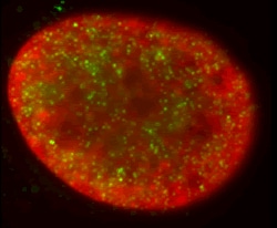 Immunocytochemistry/ Immunofluorescence - Anti-Histone H3 (di methyl K4) antibody - ChIP Grade (ab7766)This image is courtesy of Kirk McManus in the lab of Michael Hendzel, Univeristy of AlbertaDimethylated lysine 4 (green) is found in several hundred small nuclear foci that do not colocalize with condensed regions of chromatin (DAPI stained, red). The perinuclear and perinucleolar heterochromatin do not stain with this antibody.
 -
 Western blot - Anti-Histone H3 (di methyl K4) antibody - ChIP Grade (ab7766)Anti-Histone H3 (di methyl K4) antibody - ChIP Grade (ab7766) at 1 µg/ml + Calf Thymus Histone Preparation Nuclear Lysate (ab121) at 0.5 µg
Secondary
Goat Anti-Rabbit IgG H&L (HRP) preadsorbed (ab97080) at 1/5000 dilution
Developed using the ECL technique.
Performed under reducing conditions.
Predicted band size: 15 kDa
Observed band size: 17 kDa why is the actual band size different from the predicted?
Additional bands at: 14 kDa. We are unsure as to the identity of these extra bands.
Exposure time: 4 minutes -
 Immunocytochemistry/ Immunofluorescence - Anti-Histone H3 (di methyl K4) antibody - ChIP Grade (ab7766)This image is courtesy of an Abreview submitted by Dr Alexander Rapp
ab7766 at 1/200 staining human U2OS (osteosarcoma) cells by ICC/IF. The cells were paraformaldehyde fixed and then stained with the antibody for 1 hour. A Cy2 ® conjugated donkey anti-rabbit antibody was used as the secondary (green). The image shows uniformal staining of the whole nucleus, with several specles found. The insert shows H3-di methyl K4 of tw2 cells only. DAPI nuclear staining is shown in blue.
-
 Western blot - Anti-Histone H3 (di methyl K4) antibody - ChIP Grade (ab7766)All lanes : Anti-Histone H3 (di methyl K4) antibody - ChIP Grade (ab7766) at 1 µg/ml
Lane 1 : HeLa nuclear lysate (triton enriched)
Lane 2 : NIH 3T3 nuclear lysate (triton enriched)
Lane 3 : PC12 nuclear lysate (triton enriched
Lysates/proteins at 20 µg per lane.
Secondary
All lanes : Goat polyclonal to Rabbit IgG - H&L - Pre-Adsorbed (HRP) at 1/50000 dilution
Predicted band size: 15 kDa
Observed band size: 17 kDa why is the actual band size different from the predicted?Blocking buffer: 2% BSA
Gel type: MES
Exposure Time: 1 minute
-
 Western blot - Anti-Histone H3 (di methyl K4) antibody - ChIP Grade (ab7766)
MCF7 cells were incubated at 37°C for 24h with vehicle control (0 µM) and different concentrations of tranylcypromine hydrochloride (ab120606). Increased expression of Histone 3 K4 di-methyl (ab7766) in MCF7 cells correlates with an increase in tranylcypromine hydrochloride concentration, as described in literature.
Nuclear extracts were prepared with RIPA buffer (containing protease inhibitors and sodium orthovanadate), 10 µg of each were loaded on the gel and the WB was run under reducing conditions. After transfer the membrane was blocked for an hour using 5% BSA before being incubated with ab7766 at 1 µg /ml and ab1791 at 1 µg /ml overnight at 4°C. Antibody binding was detected using an anti-rabbit HRP secondary antibody (ab97051) at 1/10000 dilution and visualized using ECL development solution.
Protocols
Datasheets and documents
-
SDS download
-
Datasheet download
References (304)
ab7766 has been referenced in 304 publications.
- Hu R et al. A NR2E1-interacting peptide of LSD1 inhibits the proliferation of brain tumour initiating cells. Cell Prolif 56:e13350 (2023). PubMed: 36321378
- Barsoum M et al. Loss of the Ash2l subunit of histone H3K4 methyltransferase complexes reduces chromatin accessibility at promoters. Sci Rep 12:21506 (2022). PubMed: 36513698
- Kerschbamer E et al. CHD8 suppression impacts on histone H3 lysine 36 trimethylation and alters RNA alternative splicing. Nucleic Acids Res 50:12809-12828 (2022). PubMed: 36537238
- Fenclová T et al. Nursing Exposure to Bisphenols as a Cause of Male Idiopathic Infertility. Front Physiol 13:725442 (2022). PubMed: 35283775
- Matsuo M et al. Identification and characterization of a novel enhancer in the HTLV-1 proviral genome. Nat Commun 13:2405 (2022). PubMed: 35504920