Anti-Cytokeratin 8 antibody (ab59400)
")
Key features and details
- Rabbit polyclonal to Cytokeratin 8
- Suitable for: ICC/IF, WB, IHC-P
- Reacts with: Human
- Isotype: IgG
Overview
-
Product name
Anti-Cytokeratin 8 antibody
See all Cytokeratin 8 primary antibodies -
Description
Rabbit polyclonal to Cytokeratin 8 -
Host species
Rabbit -
Tested applications
Suitable for: ICC/IF, WB, IHC-Pmore details -
Species reactivity
Reacts with: Human -
Immunogen
Synthetic non-phosphopeptide derived from human Cytokeratin 8 around the phosphorylation site of serine 431 (L-T-SP-P-G).
-
General notes
The Life Science industry has been in the grips of a reproducibility crisis for a number of years. Abcam is leading the way in addressing this with our range of recombinant monoclonal antibodies and knockout edited cell lines for gold-standard validation. Please check that this product meets your needs before purchasing.
If you have any questions, special requirements or concerns, please send us an inquiry and/or contact our Support team ahead of purchase. Recommended alternatives for this product can be found below, along with publications, customer reviews and Q&As
Properties
-
Form
Liquid -
Storage instructions
Shipped at 4°C. Store at -20°C. Stable for 12 months at -20°C. -
Storage buffer
pH: 7.40
Preservative: 0.02% Sodium azide
Constituents: PBS, 50% Glycerol, 0.87% Sodium chloride -
 Concentration information loading...
 -
Purity
Immunogen affinity purified -
Clonality
Polyclonal -
Isotype
IgG -
Research areas
Associated products
-
Compatible Secondaries
-
Isotype control
-
Recombinant Protein
Applications
The Abpromise guarantee
Our Abpromise guarantee covers the use of ab59400 in the following tested applications.
The application notes include recommended starting dilutions; optimal dilutions/concentrations should be determined by the end user.
| Application | Abreviews | Notes |
|---|---|---|
| ICC/IF |
Use a concentration of 1 µg/ml.
|
|
| WB | (1) |
1/500 - 1/1000. Detects a band of approximately 54 kDa (predicted molecular weight: 54 kDa).
|
| IHC-P | (5) |
Use at an assay dependent concentration.
|
| Notes |
|---|
|
ICC/IF
Use a concentration of 1 µg/ml. |
|
WB
1/500 - 1/1000. Detects a band of approximately 54 kDa (predicted molecular weight: 54 kDa). |
|
IHC-P
Use at an assay dependent concentration. |
Target
-
Function
Together with KRT19, helps to link the contractile apparatus to dystrophin at the costameres of striated muscle. -
Tissue specificity
Observed in muscle fibers accumulating in the costameres of myoplasm at the sarcolemma membrane in structures that contain dystrophin and spectrin. Expressed in gingival mucosa and hard palate of the oral cavity. -
Involvement in disease
Cirrhosis -
Sequence similarities
Belongs to the intermediate filament family. -
Post-translational
modificationsPhosphorylation on serine residues is enhanced during EGF stimulation and mitosis. Ser-74 phosphorylation plays an important role in keratin filament reorganization.
O-glycosylated. O-GlcNAcylation at multiple sites increases solubility, and decreases stability by inducing proteasomal degradation.
O-glycosylated (O-GlcNAcylated), in a cell cycle-dependent manner. -
Cellular localization
Cytoplasm. Nucleus, nucleoplasm. Nucleus matrix. - Information by UniProt
-
Database links
- Entrez Gene: 3856 Human
- Omim: 148060 Human
- SwissProt: P05787 Human
- Unigene: 533782 Human
- Unigene: 708445 Human
-
Alternative names
- CARD2 antibody
- CK 8 antibody
- CK-8 antibody
see all
Images
-
Immunocytochemistry/ Immunofluorescence - Anti-Cytokeratin 8 antibody (ab59400)ICC/IF image of ab59400 stained MCF7 cells. The cells were 100% methanol fixed (5 min) and then incubated in 1%BSA / 10% normal goat serum / 0.3M glycine in 0.1% PBS-Tween for 1h to permeabilise the cells and block non-specific protein-protein interactions. The cells were then incubated with the antibody (ab59400, 1µg/ml) overnight at +4°C. The secondary antibody (green) was Alexa Fluor® 488 goat anti-rabbit IgG (H+L) used at a 1/1000 dilution for 1h. Alexa Fluor® 594 WGA was used to label plasma membranes (red) at a 1/200 dilution for 1h. DAPI was used to stain the cell nuclei (blue) at a concentration of 1.43µM.
-
 Immunohistochemistry (Formalin/PFA-fixed paraffin-embedded sections) - Anti-Cytokeratin 8 antibody (ab59400)This image is courtesy of an Abreview provided by Megha Rajaram.ab59400 staining Cytokeratin 8 in Human breast cancer cells xenografted into nude mice by Immunohistochemistry (Formalin/ PFA-fixed paraffin-embedded tissue sections). The sections were fixed in formalin and subjected to heat-mediated antigen retrieval in citrate buffer (0.1M Sodium Citrate) prior to blocking with 5% serum for 1 hour at 4°C. The primary antibody was diluted 1/200 in 5% goat serum in PBS and incubated with the sample for 12 hours at 4°C. A Biotin-conjugated Goat anti-Rabbit polyclonal was used as the secondary antibody, diluted 1/1000.
 -
 Immunohistochemistry (Formalin/PFA-fixed paraffin-embedded sections) - Anti-Cytokeratin 8 antibody (ab59400)Immunohistochemical analysis of paraffin embedded human colon carcinoma tissue using ab59400 at 1/50 dilution, in the presence (right) and absence (left) of immunising peptide. Then, a polymer secondary antibody was used for detection.
 -
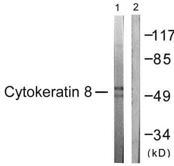 Western blot - Anti-Cytokeratin 8 antibody (ab59400)All lanes : Anti-Cytokeratin 8 antibody (ab59400) at 1/500 dilution
Lane 1 : EGF treated (200ng/ml, 30mins) 293 cell extracts
Lane 2 : EGF treated (200ng/ml, 30mins) 293 cell extracts with immunising peptide
Predicted band size: 54 kDa
Observed band size: 54 kDa -
 Immunocytochemistry/ Immunofluorescence - Anti-Cytokeratin 8 antibody (ab59400)
Immunocytochemistry/ Immunofluorescence analysis of HeLa cells, labeling Cytokeratin 8 with ab59400. The picture on the right is blocked with the synthesized peptide.
-
 Immunohistochemistry (Formalin/PFA-fixed paraffin-embedded sections) - Anti-Cytokeratin 8 antibody (ab59400)This image is courtesy of an abreview submitted by Carl Hobbs, King's College London, United Kingdom
Immunohistochemistry (Formalin/PFA-fixed paraffin-embedded sections) analysis of human kidney tissue sections labeling Cytokeratin 8 with ab59400 at 1/100 dilution. Tissue sections were fixed with formaldehyde; heat mediated antigen retrieval was performed using a citric acid. 2% BSA was used to block, followed by incubation with ab59400 in TBS/BSA/azide for 2 hours at 21°C. a polyclonal goat anti-rabbit IgG bitin conjugated secondary antibody was used at 1/300 dilution.
Protocols
Datasheets and documents
-
SDS download
-
Datasheet download
References (48)
ab59400 has been referenced in 48 publications.
- Koster S et al. Modelling Chlamydia and HPV co-infection in patient-derived ectocervix organoids reveals distinct cellular reprogramming. Nat Commun 13:1030 (2022). PubMed: 35210413
- Liu R et al. Dormant Nfatc1 reporter-marked basal stem/progenitor cells contribute to mammary lobuloalveoli formation. iScience 25:103982 (2022). PubMed: 35310332
- Gurumurthy RK et al. Patient-derived and mouse endo-ectocervical organoid generation, genetic manipulation and applications to model infection. Nat Protoc 17:1658-1690 (2022). PubMed: 35546639
- Wu T et al. Targeting HIC1/TGF-β axis-shaped prostate cancer microenvironment restrains its progression. Cell Death Dis 13:624 (2022). PubMed: 35853880
- Padalhin A et al. Recovery of sweet taste preference in adult rats following bilateral chorda tympani nerve transection. PeerJ 10:e14455 (2022). PubMed: 36452076