DRAQ5™ (ab108410)
")
Overview
-
Product name
DRAQ5™ -
Tested applications
Suitable for: FM, Flow Cyt, ICC/IFmore details -
General notes
DRAQ5™ is a cell permeable far-red fluorescent DNA dye that can be used in fixed or non-fixed/ live cells in combination with common labels such as GFP or FITC.
As with any cell-permeant DNA intercalating probe, DRAQ5 may inhibit cell division in long-term assays and should be tested for any effect.
DRAQ5 staining can be used in flow cytometry, live cell imaging and cell-based assays and the dye is highly compatible with standard protocols across many instrumentation platforms.
The chemical name of DRAQ5 is 1, 5-bis{[2-(di-methylamino)ethyl]amino}-4, 8-dihydroxyanthracene-9, 10-dione.
The advantages of DRAQ5 staining include
- convenient ready-to-use aqueous solution
- rapid uptake into living cells, providing a high level of nuclear discrimination
- no photobleaching effect
- can be used in most cell types, eukaryotic and prokaryotic: mammalian, bacterial, parasitic, plant, etc.
- no compensation needed with common FITC/GFP + PE combinations in flow cytometry
- no RNase treatment required
- no fluorescence enhancement upon DNA binding
- compatible with optics of benchtop flow, laser scanning cytometers and non-UV laser scanning and lamp-based confocal microscopesSPECTRAL PROPERTIES:
Excitation
- 647 nm line optimal (Exmax 646 nm)
- 488, 514, 568 and 633 nm lines, sub-optimal
- Two-photon excitation (1047 nm) and excitation dark (700-850 nm)
Emission (instrument dependent):
- 665 nm to infra-red max 681 nm / 697 nm intercalated with dsDNA)
- minimal overlap with vis range e.g. GFP and FITC
- Em. filters may include 695L, 715LP or 780 LP
Concentration: 5 mM
Properties
-
Form
Liquid -
Storage instructions
Store at +4°C. Do Not Freeze. Store In the Dark. -
 Concentration information loading...
 -
Research areas
Applications
The Abpromise guarantee
Our Abpromise guarantee covers the use of ab108410 in the following tested applications.
The application notes include recommended starting dilutions; optimal dilutions/concentrations should be determined by the end user.
| Application | Abreviews | Notes |
|---|---|---|
| FM |
Use at an assay dependent concentration.
|
|
| Flow Cyt |
1/250 - 1/1000.
For Gating of Nucleated cells = 5µM; For Cell Cycle Analysis = 20µM |
|
| ICC/IF |
1/1000. For Cell-based assays, Immunofluorescence microscopy and In-Cell WB = 5µM
It is highly recommended that the concentration and labelling conditions are carefully determined by each investigator for optimal performance in the assay of interest. For more specific information about the applications, please refer to the Protocol Booklet section. |
| Notes |
|---|
|
FM
Use at an assay dependent concentration. |
|
Flow Cyt
1/250 - 1/1000. For Gating of Nucleated cells = 5µM; For Cell Cycle Analysis = 20µM |
|
ICC/IF
1/1000. For Cell-based assays, Immunofluorescence microscopy and In-Cell WB = 5µM It is highly recommended that the concentration and labelling conditions are carefully determined by each investigator for optimal performance in the assay of interest. For more specific information about the applications, please refer to the Protocol Booklet section. |
Images
-
Immunocytochemistry/ Immunofluorescence - DRAQ5™ (ab108410)
HeLa cells were stained with Lamin B1 antibody - Nuclear Envelope Marker (ab16048) and alpha Tubulin antibody [DM1A] - Loading Control (ab7291). The cells were 100% methanol fixed (5 min) and then incubated in 1% BSA in 0.1% PBS-Tween for 1h to permeabilize the cells and block non-specific protein-protein interactions. The cells were then incubated with the primary antibodies (ab16048 & ab7291) at 1µg/ml overnight at 4C. The secondary antibodies were Goat polyclonal Secondary Antibody to Rabbit IgG - H&L (DyLight® 488), pre-adsorbed (ab96899) (green) and Goat polyclonal Secondary Antibody to Mouse IgG - H&L (DyLight® 594), pre-adsorbed (ab96881) (red) used at 1/250 dilution for 1h at room temperature. 5µM DRAQ5 was added to the secondary antibody mixture to label nuclear DNA (pseudocolor purple).
-
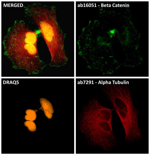 Immunocytochemistry/ Immunofluorescence - DRAQ5™ (ab108410)
HeLa cells were stained with beta Catenin antibody (ab16051) and alpha Tubulin antibody [DM1A] - Loading Control (ab7291). The cells were 100% methanol fixed (5 min) and then incubated in 1% BSA in 0.1% PBS-Tween for 1h to permeabilize the cells and block non-specific protein-protein interactions. The cells were then incubated with the primary antibodies (ab16051 & ab7921) at 1µg/ml overnight at 4C. The secondary antibodies were Goat polyclonal Secondary Antibody to Rabbit IgG - H&L (DyLight® 488), pre-adsorbed (ab96899) (green) and Goat polyclonal Secondary Antibody to Rabbit IgG - H&L (DyLight® 594), pre-adsorbed (ab96899) (red) used at 1/250 dilution for 1h at room temperature. 5µM DRAQ5 was added to the secondary antibody mixture to label nuclear DNA (pseudocolor orange).
-
 Immunocytochemistry/ Immunofluorescence - DRAQ5™ (ab108410)DRAQ5™-stained nuclei in a adult Drosophila brain.
Datasheets and documents
-
SDS download
-
Datasheet download
References (53)
ab108410 has been referenced in 53 publications.
- Yap EL et al. Bidirectional perisomatic inhibitory plasticity of a Fos neuronal network. Nature 590:115-121 (2021). PubMed: 33299180
- Zhang YW et al. Human Plasma In-Cell Western Assays-An In vitro Predictor for In vivo Pharmacology in Oncology Drug Discovery. Curr Protoc 1:e51 (2021). PubMed: 33587334
- SenGupta S et al. Triple-Negative Breast Cancer Cells Recruit Neutrophils by Secreting TGF-ß and CXCR2 Ligands. Front Immunol 12:659996 (2021). PubMed: 33912188
- Senatore E et al. The TBC1D31/praja2 complex controls primary ciliogenesis through PKA-directed OFD1 ubiquitylation. EMBO J 40:e106503 (2021). PubMed: 33934390
- Martínez AL et al. Development of a novel in vitro assay to screen for neuroprotective drugs against iatrogenic neurite shortening. PLoS One 16:e0248139 (2021). PubMed: 33690613