Anti-GRP78 BiP antibody (ab32618)
")
Key features and details
- Rabbit polyclonal to GRP78 BiP
- Suitable for: WB, IHC-P, ICC/IF
- Reacts with: Mouse, Human, Chinese hamster
- Isotype: IgG
Get better batch-to-batch reproducibility with a recombinant antibody


- Research with confidence – consistent and reproducible results with every batch
- Long-term and scalable supply – powered by recombinant technology for fast production
- Success from the first experiment – confirmed specificity through extensive validation
- Ethical standards compliant – production is animal-free
Overview
-
Product name
Anti-GRP78 BiP antibody
See all GRP78 BiP primary antibodies -
Description
Rabbit polyclonal to GRP78 BiP -
Host species
Rabbit -
Tested applications
Suitable for: WB, IHC-P, ICC/IFmore details -
Species reactivity
Reacts with: Mouse, Human, Chinese hamster
Predicted to work with: Rat, Chicken, Hamster, Xenopus laevis
-
Immunogen
Synthetic peptide within Human GRP78 BiP aa 1-100. The exact sequence is proprietary.
Database link: P11021 -
Positive control
- HeLa cells, breast carcinoma.
-
General notes
This product is FOR RESEARCH USE ONLY. For commercial use, please contact partnerships@abcam.com.
The Life Science industry has been in the grips of a reproducibility crisis for a number of years. Abcam is leading the way in addressing this with our range of recombinant monoclonal antibodies and knockout edited cell lines for gold-standard validation. Please check that this product meets your needs before purchasing.
If you have any questions, special requirements or concerns, please send us an inquiry and/or contact our Support team ahead of purchase. Recommended alternatives for this product can be found below, along with publications, customer reviews and Q&As
Properties
-
Form
Liquid -
Storage instructions
Shipped at 4°C. Upon delivery aliquot and store at -20°C. Avoid freeze / thaw cycles. -
Storage buffer
pH: 7.60
Preservative: 0.1% Sodium azide
Constituents: PBS, 1% BSA -
 Concentration information loading...
 -
Purity
Immunogen affinity purified -
Clonality
Polyclonal -
Isotype
IgG -
Research areas
Associated products
-
Compatible Secondaries
-
Isotype control
-
Positive Controls
-
Recombinant Protein
Applications
The Abpromise guarantee
Our Abpromise guarantee covers the use of ab32618 in the following tested applications.
The application notes include recommended starting dilutions; optimal dilutions/concentrations should be determined by the end user.
| Application | Abreviews | Notes |
|---|---|---|
| WB |
Use at an assay dependent concentration. Detects a band of approximately 75 kDa (predicted molecular weight: 78 kDa).
|
|
| IHC-P |
1/100. Perform heat mediated antigen retrieval before commencing with IHC staining protocol.
|
|
| ICC/IF |
1/100.
|
| Notes |
|---|
|
WB
Use at an assay dependent concentration. Detects a band of approximately 75 kDa (predicted molecular weight: 78 kDa). |
|
IHC-P
1/100. Perform heat mediated antigen retrieval before commencing with IHC staining protocol. |
|
ICC/IF
1/100. |
Target
-
Function
Probably plays a role in facilitating the assembly of multimeric protein complexes inside the endoplasmic reticulum. Involved in the correct folding of proteins and degradation of misfolded proteins via its interaction with DNAJC10, probably to facilitate the release of DNAJC10 from its substrate. -
Involvement in disease
Autoantigen in rheumatoid arthritis. -
Sequence similarities
Belongs to the heat shock protein 70 family. -
Cellular localization
Endoplasmic reticulum lumen. Melanosome. Cytoplasm. Identified by mass spectrometry in melanosome fractions from stage I to stage IV. - Information by UniProt
-
Database links
- Entrez Gene: 396487 Chicken
- Entrez Gene: 100689305 Chinese hamster
- Entrez Gene: 3309 Human
- Entrez Gene: 14828 Mouse
- Entrez Gene: 25617 Rat
- Entrez Gene: 397850 Xenopus laevis
- Omim: 138120 Human
- SwissProt: Q90593 Chicken
see all -
Alternative names
- 78 kDa glucose regulated protein antibody
- 78 kDa glucose-regulated protein antibody
- AL022860 antibody
see all
Images
-
Western blot - Anti-GRP78 BiP antibody (ab32618)All lanes : Anti-GRP78 BiP antibody (ab32618) at 1 µg/ml
Lane 1 : Liver (Mouse) Tissue Lysate
Lane 2 : CHO-K1 cell lysate Whole Cell Lysate
Lane 3 : HeLa (Human epithelial carcinoma cell line) Whole Cell Lysate
Lysates/proteins at 10 µg per lane.
Secondary
All lanes : Goat Anti-Rabbit IgG H&L (HRP) preadsorbed (ab97080) at 1/5000 dilution
Developed using the ECL technique.
Performed under reducing conditions.
Predicted band size: 78 kDa
Observed band size: 75 kDa why is the actual band size different from the predicted?
Exposure time: 30 seconds
The band observed at 75 kDa could potentially be a cleaved form of GRP78 BiP due to the presence of a 18 amino acid signal peptide. -
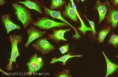 Immunocytochemistry/ Immunofluorescence - Anti-GRP78 BiP antibody (ab32618)ICC/IF image of ab32618 stained HeLa cells. The cells were 100% methanol fixed (5 min) and then incubated in 1%BSA / 10% normal goat serum / 0.3M glycine in 0.1% PBS-Tween for 1h to permeabilise the cells and block non-specific protein-protein interactions. The cells were then incubated with the antibody (ab32618, 1µg/ml) overnight at +4°C. The secondary antibody (green) was Alexa Fluor® 488 goat anti-rabbit IgG (H+L) used at a 1/1000 dilution for 1h. Alexa Fluor® 594 WGA was used to label plasma membranes (red) at a 1/200 dilution for 1h. DAPI was used to stain the cell nuclei (blue) at a concentration of 1.43µM.
 -
 Immunohistochemistry (Formalin/PFA-fixed paraffin-embedded sections) - Anti-GRP78 BiP antibody (ab32618)This image shows human breast carcinoma stained with ab32618 diluted 1/100.
 -
 Immunohistochemistry (Formalin/PFA-fixed paraffin-embedded sections) - Anti-GRP78 BiP antibody (ab32618)Ab32618 staining Human normal liver parenchyma. Staining is localised to endoplasmic reticulum compartment.
Left panel: with primary antibody at 2 ug/ml. Right panel: isotype control.
Sections were stained using an automated system DAKO Autostainer Plus , at room temperature: sections were rehydrated and antigen retrieved with the Dako 3 in 1 AR buffers citrate pH6.0 in a DAKO PT Link. Slides were peroxidase blocked in 3% H2O2 in methanol for 10 mins. They were then blocked with Dako Protein block for 10 minutes (containing casein 0.25% in PBS) then incubated with primary antibody for 20 min and detected with Dako envision flex amplification kit for 30 minutes. Colorimetric detection was completed with Diaminobenzidine for 5 minutes. Slides were counterstained with Haematoxylin and coverslipped under DePeX. Please note that for manual staining we recommend to optimize the primary antibody concentration and incubation time (overnight incubation), and amplification may be requi
Protocols
Datasheets and documents
-
SDS download
-
Datasheet download
References (50)
ab32618 has been referenced in 50 publications.
- Wu Z et al. sGRP78 enhances selective autophagy of monomeric TLR4 to regulate myeloid cell death. Cell Death Dis 13:587 (2022). PubMed: 35798718
- Zhou L et al. Transcription factor EB-mediated autophagy promotes dermal fibroblast differentiation and collagen production by regulating endoplasmic reticulum stress and autophagy-dependent secretion. Int J Mol Med 47:547-560 (2021). PubMed: 33416091
- Khongwichit S et al. A functional interaction between GRP78 and Zika virus E protein. Sci Rep 11:393 (2021). PubMed: 33432092
- Kavalakatt S et al. Urocortin 3 overexpression reduces ER stress and heat shock response in 3T3-L1 adipocytes. Sci Rep 11:15666 (2021). PubMed: 34341463
- Wu L et al. Apigenin Ameliorates Insulin Resistance and Lipid Accumulation by Endoplasmic Reticulum Stress and SREBP-1c/SREBP-2 Pathway in Palmitate-Induced HepG2 Cells and High-Fat Diet-Fed Mice. J Pharmacol Exp Ther 377:146-156 (2021). PubMed: 33509902