Anti-HSF1 antibody (ab2923)
")
Key features and details
- Rabbit polyclonal to HSF1
- Suitable for: IP, WB, ICC/IF
- Reacts with: Mouse, Human, Caenorhabditis elegans
- Isotype: IgG
Get better batch-to-batch reproducibility with a recombinant antibody


- Research with confidence – consistent and reproducible results with every batch
- Long-term and scalable supply – powered by recombinant technology for fast production
- Success from the first experiment – confirmed specificity through extensive validation
- Ethical standards compliant – production is animal-free
Overview
-
Product name
Anti-HSF1 antibody
See all HSF1 primary antibodies -
Description
Rabbit polyclonal to HSF1 -
Host species
Rabbit -
Tested applications
Suitable for: IP, WB, ICC/IFmore details -
Species reactivity
Reacts with: Mouse, Human, Caenorhabditis elegans -
Immunogen
Recombinant full length protein corresponding to Human HSF1. Recombinant human HSF1 expressed in E. coli.
-
Positive control
- WB: HEK-293T, HeLa, K562, A431, and HepG2 whole cell lysate. C. elegans whole worm tissue. ICC/IF: HeLa and NIH3T3 cells. IP: HeLa whole cell lysate.
-
General notes
The Life Science industry has been in the grips of a reproducibility crisis for a number of years. Abcam is leading the way in addressing this with our range of recombinant monoclonal antibodies and knockout edited cell lines for gold-standard validation. Please check that this product meets your needs before purchasing.
If you have any questions, special requirements or concerns, please send us an inquiry and/or contact our Support team ahead of purchase. Recommended alternatives for this product can be found below, along with publications, customer reviews and Q&As
Properties
-
Form
Liquid -
Storage instructions
Shipped at 4°C. Store at +4°C short term (1-2 weeks). Upon delivery aliquot. Store at -20°C. Avoid freeze / thaw cycle. -
Storage buffer
Preservative: 0.05% Sodium azide
Constituent: 99% PBS -
 Concentration information loading...
 -
Purity
Whole antiserum -
Primary antibody notes
All organisms respond to elevated temperatures and a variety of environmental stresses by rapid synthesis of heat shock RNAs and proteins. The regulation of heat shock gene transcription is mediated by the transcriptional activator, heat shock factor (HSF), which binds to heat shock response elements (HSEs). These HSEs are found as three repeats of a 5-nucleotide {nGAAn} module, arranged in alternating orientation and present upstream of all heat shock genes. The HSEs are highly conserved among species yet HSF purified from yeast, Drosophila and human have different molecular weights and the proteins do not show significant immunological cross reaction. Two HSFs have been identified in human cells, HSF 1 and HSF 2, which bind to the same HSEs and have 38% sequence identity. These factors are activated by distinct stimuli, HSF 1 is responsive to classical stress signals such as heat, heavy metals and oxidative reagents, whereas HSF 2 is activated during hemin-mediated differentiation of human erythroleukemia cells. HSF 1 exists constitutively in the cytoplasm and the nucleus of unstressed cells as a monomer which lacks DNA binding activity. Through an unknown signal generated during stress, HSF 1 becomes activated to a nuclear localized, trimeric state which binds to DNA. The phosphorylation of HSF 1 is necessary for maximal transcription of heat shock genes. -
Clonality
Polyclonal -
Isotype
IgG -
Research areas
Associated products
-
Compatible Secondaries
-
Isotype control
-
Positive Controls
-
Recombinant Protein
Applications
The Abpromise guarantee
Our Abpromise guarantee covers the use of ab2923 in the following tested applications.
The application notes include recommended starting dilutions; optimal dilutions/concentrations should be determined by the end user.
| Application | Abreviews | Notes |
|---|---|---|
| IP |
Use at an assay dependent concentration.
2 μL |
|
| WB | (1) |
1/1000 - 1/10000. Detects a band of approximately 83 kDa (predicted molecular weight: 57 kDa).
|
| ICC/IF |
1/50.
|
| Notes |
|---|
|
IP
Use at an assay dependent concentration. 2 μL |
|
WB
1/1000 - 1/10000. Detects a band of approximately 83 kDa (predicted molecular weight: 57 kDa). |
|
ICC/IF
1/50. |
Target
-
Function
DNA-binding protein that specifically binds heat shock promoter elements (HSE) and activates transcription. In higher eukaryotes, HSF is unable to bind to the HSE unless the cells are heat shocked. -
Sequence similarities
Belongs to the HSF family. -
Domain
the 9aaTAD motif is a transactivation domain present in a large number of yeast and animal transcription factors. -
Post-translational
modificationsPhosphorylated on multiple serine residues, a subset of which are involved in stress-related regulation of transcription activation. Constitutive phosphorylation represses transcriptional activity at normal temperatures. Levels increase on specific residues heat-shock and enhance HSF1 transactivation activity. Phosphorylation on Ser-307 derepresses activation on heat-stress and in combination with Ser-303 phosphorylation appears to be involved in recovery after heat-stress. Phosphorylated on Ser-230 by CAMK2, in vitro. Cadmium also enhances phosphorylation at this site. Phosphorylation on Ser-303 is a prerequisite for HSF1 sumoylation. Phosphorylation on Ser-121 inhibits transactivation and promotes HSP90 binding. Phosphorylation on Thr-142 also mediates transcriptional activity induced by heat. Phosphorylation on Ser-326 plays an important role in heat activation of HSF1 transcriptional activity.
Sumoylated with SUMO1 and SUMO2 on heat-shock. Heat-inducible sumoylation occurs after 15 min of heat-shock, after which levels decrease and at 4 hours, levels return to control levels. Sumoylation has no effect on HSE binding nor on transcriptional activity. Phosphorylation on Ser-303 is a prerequisite for sumoylation. -
Cellular localization
Cytoplasm. Nucleus. Cytoplasmic during normal growth. On activation, translocates to nuclear stress granules. Colocalizes with SUMO1 in nuclear stress granules. - Information by UniProt
-
Database links
- Entrez Gene: 3297 Human
- Entrez Gene: 15499 Mouse
- Omim: 140580 Human
- SwissProt: Q00613 Human
- SwissProt: P38532 Mouse
- Unigene: 530227 Human
- Unigene: 347444 Mouse
-
Alternative names
- Heat shock factor 1 antibody
- Heat shock factor protein 1 antibody
- Heat shock transcription factor 1 antibody
see all
Images
-
Western blot - Anti-HSF1 antibody (ab2923)All lanes : Anti-HSF1 antibody (ab2923) at 1/1000 dilution
Lane 1 : HEK-293T (Human epithelial cell line from embryonic kidney transformed with large T antigen) whole cell lysate
Lane 2 : HeLa (Human epithelial cell line from cervix adenocarcinoma) whole cell lysate
Lane 3 : K562 (Human chronic myelogenous leukemia cell line from bone marrow ) whole cell lysate
Lane 4 : A431 (Human epidermoid carcinoma cell line) whole cell lysate
Lane 5 : HepG2 (Human liver hepatocellular carcinoma cell line) whole cell lysate
Lane 6 : COS-7 (African green monkey kidney fibroblast-like cell line) whole cell lysate
Lane 7 : NIH/3T3 (Mouse embryo fibroblast cell line) whole cell lysate
Lane 8 : NRK (Rat kidney normal tissue) whole cell lysate
Lysates/proteins at 50 µg per lane.
Secondary
All lanes : HRP-conjugated goat anti-rabbit IgG secondary antibody at 1/20000 dilution
Predicted band size: 57 kDaWestern blot analysis of Heat Shock Factor 1 (HSF1) was performed by loading samples onto a 4-20% Tris-HCl polyacrylamide gel. Proteins were transferred to a PVDF membrane and blocked with 5% BSA/TBST for at least 1 hour. The membrane was incubated with ab2923 overnight at 4°C on a rocking platform, washed in TBS-0.1% Tween 20, and probed with a secondary antibody for at least one hour. Chemiluminescent detection was performed.
-
 Immunocytochemistry/ Immunofluorescence - Anti-HSF1 antibody (ab2923)
Immunocytochemistry/Immunofluorescence analysis of HSF1 (green) in HeLa (Human epithelial cell line from cervix adenocarcinoma) and NIH/3T3 (Mouse embryo fibroblast cell line) cells. Formalin fixed cells were permeabilized with 0.1% Triton X-100 in TBS for 10 minutes at room temperature and blocked with 1% BSA for 15 minutes at room temperature. Cells were probed with ab2923 (1:50) for at least 1 hour at room temperature, washed with PBS, and incubated with a DyLight 488-conjugated goat-anti rabbit IgG secondary antibody (1:400) for 30 minutes at room temperature. Nuclei (blue) were stained with Hoechst 33342 dye. Images were taken at 20X magnification.
-
 Immunoprecipitation - Anti-HSF1 antibody (ab2923)
Immunoprecipitation of HSF1 was performed on HeLa (Human epithelial cell line from cervix adenocarcinoma) cells. Antigen:antibody complexes were formed by incubating 500µg whole cell lysate with 2µg of ab2923 overnight on a rocking platform at 4°C. Immune complexes were captured on 50µl Protein A/G Plus Agarose, washed extensively, and eluted with buffer. Samples were resolved on a 4-20% Tris-HCl polyacrylamide gel, transferred to a PVDF membrane, and blocked with 5% BSA/TBST for at least 1 hour. The membrane was incubated with ab2923 (1:1000) overnight rotating at 4°C, washed in TBST, and probed with detection reagent (1:1000) for at least one hour. Chemiluminescent detection was performed.
-
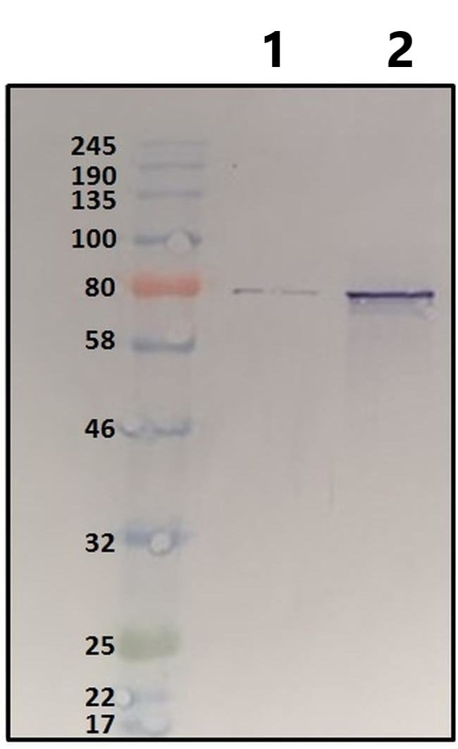 Western blot - Anti-HSF1 antibody (ab2923)All lanes : Anti-HSF1 antibody (ab2923) at 1/5000 dilution
Lane 1 : HSF1 RNAi treated C. elegans whole worm tissue
Lane 2 : Untreated C. elegans whole worm tissue
Lysates/proteins at 10 µg per lane.
Secondary
All lanes : anti-rabbit IgG HRP-linked at 1/5000 dilution
Predicted band size: 57 kDaWestern blot analysis of HSF1 was performed by loading each sample onto an SDS-PAGE gel. Proteins were transferred to PVDF membrane and followed by blocking in TBST+5% non-fat milk. HSF1 was detected using the primary anitbody (ab2923) in TBST+2% non-fat milk overnight at 4°C on a rocking platform, followed by secondary antibody for 30 min. Detection was performed using and 1-Step™ TMB-Blotting Substrate Solution.
Protocols
Datasheets and documents
-
Datasheet download
References (13)
ab2923 has been referenced in 13 publications.
- Chen L et al. Transcription factor YY1 inhibits the expression of THY1 to promote interstitial pulmonary fibrosis by activating the HSF1/miR-214 axis. Aging (Albany NY) 12:8339-8351 (2020). PubMed: 32396525
- Li T et al. HSF1 Attenuates LPS-Induced Acute Lung Injury in Mice by Suppressing Macrophage Infiltration. Oxid Med Cell Longev 2020:1936580 (2020). PubMed: 33381262
- Zhang L et al. Proteomics analysis of proteins interacting with heat shock factor 1 in squamous cell carcinoma of the cervix. Oncol Lett 18:2568-2575 (2019). PubMed: 31402952
- Grossi V et al. The longevity SNP rs2802292 uncovered: HSF1 activates stress-dependent expression of FOXO3 through an intronic enhancer. Nucleic Acids Res 46:5587-5600 (2018). PubMed: 29733381
- Neueder A et al. Novel Isoforms of Heat Shock Transcription Factor 1, HSF1?a and HSF1?ß, Regulate Chaperone Protein Gene Transcription. J Biol Chem 289:19894-906 (2014). PubMed: 24855652