Human APP (Amyloid Precursor Protein) knockout HEK-293T cell line (ab255362)
")
Overview
-
Product name
Human APP (Amyloid Precursor Protein) knockout HEK-293T cell line
See all Amyloid Precursor Protein lysates -
Parental Cell Line
HEK293T -
Organism
Human -
Mutation description
Knockout achieved by using CRISPR/Cas9, Homozygous: 1 bp insertion in exon 5 -
Passage number
<20 -
Knockout validation
Immunocytochemistry (ICC), Sanger Sequencing, Western Blot (WB) -
Tested applications
Suitable for: WB, ICCmore details -
Biosafety level
2 -
General notes
Recommended control: Human wild-type HEK293T cell line (ab255449). Please note a wild-type cell line is not automatically included with a knockout cell line order, if required please add recommended wild-type cell line at no additional cost using the code WILDTYPE-TMTK1.
Cryopreservation cell medium: Cell Freezing Medium-DMSO Serum free media, contains 8.7% DMSO in MEM supplemented with methyl cellulose.
Culture medium: DMEM (High Glucose) + 10% FBS
Initial handling guidelines: Upon arrival, the vial should be stored in liquid nitrogen vapor phase and not at -80°C. Storage at -80°C may result in loss of viability.
1. Thaw the vial in 37°C water bath for approximately 1-2 minutes.
2. Transfer the cell suspension (0.8 mL) to a 15 mL/50 mL conical sterile polypropylene centrifuge tube containing 8.4 mL pre-warmed culture medium, wash vial with an additional 0.8 mL culture medium (total volume 10 mL) to collect remaining cells, and centrifuge at 201 x g (rcf) for 5 minutes at room temperature. 10 mL represents minimum recommended dilution. 20 mL represents maximum recommended dilution.
3. Resuspend the cell pellet in 5 mL pre-warmed culture medium and count using a haemocytometer or alternative cell counting method. Based on cell count, seed cells in an appropriate cell culture flask at a density of 2x10⁴ cells/cm2. Seeding density is given as a guide only and should be scaled to align with individual lab schedules.
4. Incubate the culture at 37°C incubator with 5% CO2. Cultures should be monitored daily.Subculture guidelines:
- All seeding densities should be based on cell counts gained by established methods.
- A guide seeding density of 2x10⁴ cells/cm2 is recommended.
- A partial media change 24 hours prior to subculture may be helpful to encourage growth, if required.
- Cells should be passaged when they have achieved 80-90% confluence.
This product is subject to limited use licenses from The Broad Institute, ERS Genomics Limited and Sigma-Aldrich Co. LLC, and is developed with patented technology. For full details of the licenses and patents please refer to our limited use license and patent pages.
We will provide viable cells that proliferate on revival.
Properties
-
Number of cells
1 x 10⁶ cells/vial, 1 mL -
Adherent /Suspension
Adherent -
Tissue
Kidney -
Cell type
epithelial -
STR Analysis
Amelogenin X D5S818: 8, 9 D13S317: 12, 14 D7S820: 11 D16S539: 9, 13 vWA: 16, 19 TH01: 7, 9.3 TPOX: 11 CSF1PO: 11, 12 -
Mycoplasma free
Yes -
Storage instructions
Shipped on Dry Ice. Store in liquid nitrogen. -
Storage buffer
Constituents: 8.7% Dimethylsulfoxide, 2% Cellulose, methyl ether -
Research areas
Target
-
Function
Functions as a cell surface receptor and performs physiological functions on the surface of neurons relevant to neurite growth, neuronal adhesion and axonogenesis. Involved in cell mobility and transcription regulation through protein-protein interactions. Can promote transcription activation through binding to APBB1-KAT5 and inhibits Notch signaling through interaction with Numb. Couples to apoptosis-inducing pathways such as those mediated by G(O) and JIP. Inhibits G(o) alpha ATPase activity (By similarity). Acts as a kinesin I membrane receptor, mediating the axonal transport of beta-secretase and presenilin 1. Involved in copper homeostasis/oxidative stress through copper ion reduction. In vitro, copper-metallated APP induces neuronal death directly or is potentiated through Cu(2+)-mediated low-density lipoprotein oxidation. Can regulate neurite outgrowth through binding to components of the extracellular matrix such as heparin and collagen I and IV. The splice isoforms that contain the BPTI domain possess protease inhibitor activity. Induces a AGER-dependent pathway that involves activation of p38 MAPK, resulting in internalization of amyloid-beta peptide and leading to mitochondrial dysfunction in cultured cortical neurons. Provides Cu(2+) ions for GPC1 which are required for release of nitric oxide (NO) and subsequent degradation of the heparan sulfate chains on GPC1.
Beta-amyloid peptides are lipophilic metal chelators with metal-reducing activity. Bind transient metals such as copper, zinc and iron. In vitro, can reduce Cu(2+) and Fe(3+) to Cu(+) and Fe(2+), respectively. Beta-amyloid 42 is a more effective reductant than beta-amyloid 40. Beta-amyloid peptides bind to lipoproteins and apolipoproteins E and J in the CSF and to HDL particles in plasma, inhibiting metal-catalyzed oxidation of lipoproteins. Beta-APP42 may activate mononuclear phagocytes in the brain and elicit inflammatory responses. Promotes both tau aggregation and TPK II-mediated phosphorylation. Interaction with overexpressed HADH2 leads to oxidative stress and neurotoxicity. Also binds GPC1 in lipid rafts.
Appicans elicit adhesion of neural cells to the extracellular matrix and may regulate neurite outgrowth in the brain.
The gamma-CTF peptides as well as the caspase-cleaved peptides, including C31, are potent enhancers of neuronal apoptosis.
N-APP binds TNFRSF21 triggering caspase activation and degeneration of both neuronal cell bodies (via caspase-3) and axons (via caspase-6). -
Tissue specificity
Expressed in all fetal tissues examined with highest levels in brain, kidney, heart and spleen. Weak expression in liver. In adult brain, highest expression found in the frontal lobe of the cortex and in the anterior perisylvian cortex-opercular gyri. Moderate expression in the cerebellar cortex, the posterior perisylvian cortex-opercular gyri and the temporal associated cortex. Weak expression found in the striate, extra-striate and motor cortices. Expressed in cerebrospinal fluid, and plasma. Isoform APP695 is the predominant form in neuronal tissue, isoform APP751 and isoform APP770 are widely expressed in non-neuronal cells. Isoform APP751 is the most abundant form in T-lymphocytes. Appican is expressed in astrocytes. -
Involvement in disease
Alzheimer disease 1
Cerebral amyloid angiopathy, APP-related -
Sequence similarities
Belongs to the APP family.
Contains 1 BPTI/Kunitz inhibitor domain. -
Domain
The basolateral sorting signal (BaSS) is required for sorting of membrane proteins to the basolateral surface of epithelial cells.
The NPXY sequence motif found in many tyrosine-phosphorylated proteins is required for the specific binding of the PID domain. However, additional amino acids either N- or C-terminal to the NPXY motif are often required for complete interaction. The PID domain-containing proteins which bind APP require the YENPTY motif for full interaction. These interactions are independent of phosphorylation on the terminal tyrosine residue. The NPXY site is also involved in clathrin-mediated endocytosis. -
Post-translational
modificationsProteolytically processed under normal cellular conditions. Cleavage either by alpha-secretase, beta-secretase or theta-secretase leads to generation and extracellular release of soluble APP peptides, S-APP-alpha and S-APP-beta, and the retention of corresponding membrane-anchored C-terminal fragments, C80, C83 and C99. Subsequent processing of C80 and C83 by gamma-secretase yields P3 peptides. This is the major secretory pathway and is non-amyloidogenic. Alternatively, presenilin/nicastrin-mediated gamma-secretase processing of C99 releases the amyloid beta proteins, amyloid-beta 40 (Abeta40) and amyloid-beta 42 (Abeta42), major components of amyloid plaques, and the cytotoxic C-terminal fragments, gamma-CTF(50), gamma-CTF(57) and gamma-CTF(59). Many other minor beta-amyloid peptides, beta-amyloid 1-X peptides, are found in cerebral spinal fluid (CSF) including the beta-amyloid X-15 peptides, produced from the cleavage by alpha-secretase and all terminating at Gln-686.
Proteolytically cleaved by caspases during neuronal apoptosis. Cleavage at Asp-739 by either caspase-6, -8 or -9 results in the production of the neurotoxic C31 peptide and the increased production of beta-amyloid peptides.
N- and O-glycosylated. O-glycosylation on Ser and Thr residues with core 1 or possibly core 8 glycans. Partial tyrosine glycosylation (Tyr-681) is found on some minor, short beta-amyloid peptides (beta-amyloid 1-15, 1-16, 1-17, 1-18, 1-19 and 1-20) but not found on beta-amyloid 38, beta-amyloid 40 nor on beta-amyloid 42. Modification on a tyrosine is unusual and is more prevelant in AD patients. Glycans had Neu5AcHex(Neu5Ac)HexNAc-O-Tyr, Neu5AcNeu5AcHex(Neu5Ac)HexNAc-O-Tyr and O-AcNeu5AcNeu5AcHex(Neu5Ac)HexNAc-O-Tyr structures, where O-Ac is O-acetylation of Neu5Ac. Neu5AcNeu5Ac is most likely Neu5Ac 2,8Neu5Ac linked. O-glycosylations in the vicinity of the cleavage sites may influence the proteolytic processing. Appicans are L-APP isoforms with O-linked chondroitin sulfate.
Phosphorylation in the C-terminal on tyrosine, threonine and serine residues is neuron-specific. Phosphorylation can affect APP processing, neuronal differentiation and interaction with other proteins. Phosphorylated on Thr-743 in neuronal cells by Cdc5 kinase and Mapk10, in dividing cells by Cdc2 kinase in a cell-cycle dependent manner with maximal levels at the G2/M phase and, in vitro, by GSK-3-beta. The Thr-743 phosphorylated form causes a conformational change which reduces binding of Fe65 family members. Phosphorylation on Tyr-757 is required for SHC binding. Phosphorylated in the extracellular domain by casein kinases on both soluble and membrane-bound APP. This phosphorylation is inhibited by heparin.
Extracellular binding and reduction of copper, results in a corresponding oxidation of Cys-144 and Cys-158, and the formation of a disulfide bond. In vitro, the APP-Cu(+) complex in the presence of hydrogen peroxide results in an increased production of beta-amyloid-containing peptides.
Trophic-factor deprivation triggers the cleavage of surface APP by beta-secretase to release sAPP-beta which is further cleaved to release an N-terminal fragment of APP (N-APP).
Beta-amyloid peptides are degraded by IDE. -
Cellular localization
Membrane. Membrane, clathrin-coated pit. Cell surface protein that rapidly becomes internalized via clathrin-coated pits. During maturation, the immature APP (N-glycosylated in the endoplasmic reticulum) moves to the Golgi complex where complete maturation occurs (O-glycosylated and sulfated). After alpha-secretase cleavage, soluble APP is released into the extracellular space and the C-terminal is internalized to endosomes and lysosomes. Some APP accumulates in secretory transport vesicles leaving the late Golgi compartment and returns to the cell surface. Gamma-CTF(59) peptide is located to both the cytoplasm and nuclei of neurons. It can be translocated to the nucleus through association with APBB1 (Fe65). Beta-APP42 associates with FRPL1 at the cell surface and the complex is then rapidly internalized. APP sorts to the basolateral surface in epithelial cells. During neuronal differentiation, the Thr-743 phosphorylated form is located mainly in growth cones, moderately in neurites and sparingly in the cell body. Casein kinase phosphorylation can occur either at the cell surface or within a post-Golgi compartment. Associates with GPC1 in perinuclear compartments. Colocalizes with SORL1 in a vesicular pattern in cytoplasm and perinuclear regions. - Information by UniProt
Associated products
-
KO cell lysates
-
Related Products
Applications
The Abpromise guarantee
Our Abpromise guarantee covers the use of ab255362 in the following tested applications.
The application notes include recommended starting dilutions; optimal dilutions/concentrations should be determined by the end user.
| Application | Abreviews | Notes |
|---|---|---|
| WB |
Use at an assay dependent concentration. Predicted molecular weight: 86 kDa.
|
|
| ICC |
Use at an assay dependent concentration.
|
| Notes |
|---|
|
WB
Use at an assay dependent concentration. Predicted molecular weight: 86 kDa. |
|
ICC
Use at an assay dependent concentration. |
Images
-
Western blot - Human APP knockout HEK293T cell line (ab255362)Lanes 1-2 : Anti-Amyloid Precursor Protein antibody [DE2B4] (ab12266) at 1/500 dilution
Lanes 3-4 : Anti-Amyloid Precursor Protein antibody [DE2B4] (ab12266)
Lane 1 : Wild-type HeLa cell lysate
Lane 2 : SH-SY5Y cell lysate
Lane 3 : Wild-type HEK-293T cell lysate
Lane 4 : APP knockout HEK-293T cell lysate
Lysates/proteins at 20 µg per lane.
Secondary
All lanes : Goat anti-Mouse IgG H&L (IRDye® 800CW) preadsorbed (ab216772) at 1/20000 dilution
Performed under reducing conditions.
Predicted band size: 86 kDa
Observed band size: 37 kDa why is the actual band size different from the predicted?Lanes 1 - 4: Green signal. Green - ab12266 observed at 110 kDa.
Lanes 5 - 8: Red signal. Red - loading control, ab181602 observed at 37 kDa.
ab12266 was shown to react with Amyloid Precursor Protein in wild-type HEK-293T cells. Loss of signal was observed when knockout cell line ab255362 (knockout cell lysate ab263777) was used. Wild-type and Amyloid Precursor Protein knockout samples were subjected to SDS-PAGE. ab12266 and Anti-GAPDH antibody [EPR16891] - Loading Control (ab181602) were incubated overnight at 4°C at 1 in 500 dilution and 1 in 20000 dilution respectively. Blots were developed with Goat anti-Mouse IgG H&L (IRDye® 800CW) preadsorbed (ab216772) and Goat Anti-Rabbit IgG H&L (IRDye® 680RD) preadsorbed (ab216777) secondary antibodies at 1 in 20000 dilution for 1 hour at room temperature before imaging.
-
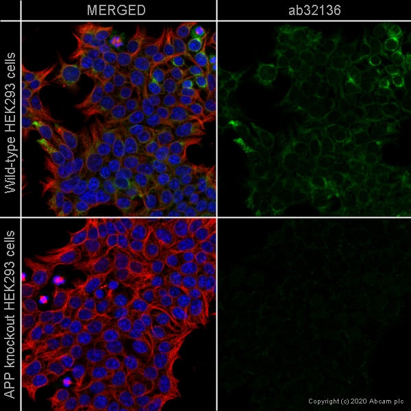 Immunocytochemistry/ Immunofluorescence - Human APP (Amyloid Precursor Protein) knockout HEK-293T cell line (ab255362)ab32136 staining Amyloid Precursor Protein in wild-type HEK293 cells (top panel) and APP knockout HEK293 cells (ab255362) (bottom panel). The cells were fixed with 100% methanol (5 min) then permeabilized with 0.1% Triton X-100 for 5 minutes and then blocked with 1% BSA/10% normal goat serum/0.3M glycine in 0.1% PBS-Tween for 1h. The cells were then incubated with ab32136 at 1/500 dilution and ab7291 (Mouse monoclonal to alpha Tubulin) at 1/1000 dilution overnight at 4°C followed by a further incubation at room temperature for 1h with a goat secondary antibody to rabbit IgG (Alexa Fluor® 488) (ab150081) at 2 μg/ml (shown in green) and a goat secondary antibody to mouse IgG (Alexa Fluor® 594) (ab150120) at 2 μg/ml (shown in red). Nuclear DNA was labelled in blue with DAPI.
Image was taken with a confocal microscope (Leica-Microsystems TCS SP8). -
 Sanger Sequencing - Human APP knockout HEK293T cell line (ab255362)Homozygous: 1 bp insertion in exon 5
Protocols
Datasheets and documents
-
SDS download
-
Datasheet download
References (0)
ab255362 has not yet been referenced specifically in any publications.