Anti-APC antibody (ab265597)
 - Anti-APC antibody (ab265597)")
Key features and details
- Rabbit polyclonal to APC
- Suitable for: IHC-P, IP
- Reacts with: Human
- Isotype: IgG
Overview
-
Product name
Anti-APC antibody
See all APC primary antibodies -
Description
Rabbit polyclonal to APC -
Host species
Rabbit -
Tested applications
Suitable for: IHC-P, IPmore details -
Species reactivity
Reacts with: Human -
Immunogen
Synthetic peptide within Human APC aa 2793-2843. The exact sequence is proprietary. NP_000029.2
Database link: P25054 -
Positive control
- IP: HeLa whole cell lysate. IHC-P: Human colon carcinoma tissue.
-
General notes
Anti-APC antibody (ab265597) is not recommended for WB of crude lysate. Use at 1 μg/ml to blot enriched sources (e.g. Immunoprecipitates) of APC.
The Life Science industry has been in the grips of a reproducibility crisis for a number of years. Abcam is leading the way in addressing this with our range of recombinant monoclonal antibodies and knockout edited cell lines for gold-standard validation. Please check that this product meets your needs before purchasing.
If you have any questions, special requirements or concerns, please send us an inquiry and/or contact our Support team ahead of purchase. Recommended alternatives for this product can be found below, along with publications, customer reviews and Q&As
Properties
-
Form
Liquid -
Storage instructions
Shipped at 4°C. Store at +4°C short term (1-2 weeks). Upon delivery aliquot. Store at -20°C long term. Avoid freeze / thaw cycle. -
Storage buffer
pH: 7
Preservative: 0.09% Sodium azide
Constituent: Tris citrate/phosphate
pH 7 to 8 -
 Concentration information loading...
 -
Purity
Immunogen affinity purified -
Clonality
Polyclonal -
Isotype
IgG -
Research areas
Associated products
-
Compatible Secondaries
-
Positive Controls
Applications
The Abpromise guarantee
Our Abpromise guarantee covers the use of ab265597 in the following tested applications.
The application notes include recommended starting dilutions; optimal dilutions/concentrations should be determined by the end user.
| Application | Abreviews | Notes |
|---|---|---|
| IHC-P |
1/500 - 1/2000. Perform heat mediated antigen retrieval with citrate buffer pH 6 before commencing with IHC staining protocol.
|
|
| IP |
Use at 2-5 µg/mg of lysate.
|
| Notes |
|---|
|
IHC-P
1/500 - 1/2000. Perform heat mediated antigen retrieval with citrate buffer pH 6 before commencing with IHC staining protocol. |
|
IP
Use at 2-5 µg/mg of lysate. |
Target
-
Function
Tumor suppressor. Promotes rapid degradation of CTNNB1 and participates in Wnt signaling as a negative regulator. APC activity is correlated with its phosphorylation state. Activates the GEF activity of SPATA13 and ARHGEF4. Plays a role in hepatocyte growth factor (HGF)-induced cell migration. Required for MMP9 up-regulation via the JNK signaling pathway in colorectal tumor cells. Acts as a mediator of ERBB2-dependent stabilization of microtubules at the cell cortex. It is required for the localization of MACF1 to the cell membrane and this localization of MACF1 is critical for its function in microtubule stabilization. -
Tissue specificity
Expressed in a variety of tissues. -
Involvement in disease
Defects in APC are a cause of familial adenomatous polyposis (FAP) [MIM:175100]; which includes also Gardner syndrome (GS). FAP and GS contribute to tumor development in patients with uninherited forms of colorectal cancer. FAP is characterized by adenomatous polyps of the colon and rectum, but also of upper gastrointestinal tract (ampullary, duodenal and gastric adenomas). This is a viciously premalignant disease with one or more polyps progressing through dysplasia to malignancy in untreated gene carriers with a median age at diagnosis of 40 years.
Defects in APC are a cause of hereditary desmoid disease (HDD) [MIM:135290]; also known as familial infiltrative fibromatosis (FIF). HDD is an autosomal dominant trait with 100% penetrance and possible variable expression among affected relatives. HDD patients show multifocal fibromatosis of the paraspinal muscles, breast, occiput, arms, lower ribs, abdominal wall, and mesentery. Desmoid tumors appears also as a complication of familial adenomatous polyposis.
Defects in APC are a cause of medulloblastoma (MDB) [MIM:155255]. MDB is a malignant, invasive embryonal tumor of the cerebellum with a preferential manifestation in children. Although the majority of medulloblastomas occur sporadically, some manifest within familial cancer syndromes such as Turcot syndrome and basal cell nevus syndrome (Gorlin syndrome).
Defects in APC are a cause of mismatch repair cancer syndrome (MMRCS) [MIM:276300]; also known as Turcot syndrome or brain tumor-polyposis syndrome 1 (BTPS1). MMRCS is an autosomal dominant disorder characterized by malignant tumors of the brain associated with multiple colorectal adenomas. Skin features include sebaceous cysts, hyperpigmented and cafe au lait spots.
Defects in APC are a cause of gastric cancer (GASC) [MIM:613659]; also called gastric cancer intestinal or stomach cancer. Gastric cancer is a malignant disease which starts in the stomach, can spread to the esophagus or the small intestine, and can extend through the stomach wall to nearby lymph nodes and organs. It also can metastasize to other parts of the body. The term gastric cancer or gastric carcinoma refers to adenocarcinoma of the stomach that accounts for most of all gastric malignant tumors. Two main histologic types are recognized, diffuse type and intestinal type carcinomas. Diffuse tumors are poorly differentiated infiltrating lesions, resulting in thickening of the stomach. In contrast, intestinal tumors are usually exophytic, often ulcerating, and associated with intestinal metaplasia of the stomach, most often observed in sporadic disease.
Defects in APC are a cause of hepatocellular carcinoma (HCC) [MIM:114550]. This defect includes also the disease entity termed hepatoblastoma. -
Sequence similarities
Belongs to the adenomatous polyposis coli (APC) family.
Contains 7 ARM repeats. -
Domain
The microtubule tip localization signal (MtLS) motif; mediates interaction with MAPRE1 and targeting to the growing microtubule plus ends. -
Post-translational
modificationsPhosphorylated by GSK3B.
Ubiquitinated, leading to its degradation by the proteasome. Ubiquitination is facilitated by Axin. Deubiquitinated by ZRANB1/TRABID. -
Cellular localization
Cell junction > adherens junction. Cytoplasm > cytoskeleton. Cell projection > lamellipodium. Cell projection > ruffle membrane. Cytoplasm. Cell membrane. Associated with the microtubule network at the growing distal tip of microtubules. Accumulates in the lamellipodium and ruffle membrane in response to hepatocyte growth factor (HGF) treatment. The MEMO1-RHOA-DIAPH1 signaling pathway controls localization of the phosophorylated form to the cell membrane. - Information by UniProt
-
Database links
- Entrez Gene: 324 Human
- Omim: 611731 Human
- SwissProt: P25054 Human
- Unigene: 158932 Human
-
Alternative names
- Adenomatous Polyposis Coli antibody
- Adenomatous polyposis coli protein antibody
- Apc antibody
see all
Images
-
Immunohistochemistry (Formalin/PFA-fixed paraffin-embedded sections) - Anti-APC antibody (ab265597)
Immunohistochemical analysis of formalin-fixed, paraffin-embedded human colon carcinoma tissue labeling APC with ab265597 at 1/1000 dilution. Detection: DAB.
-
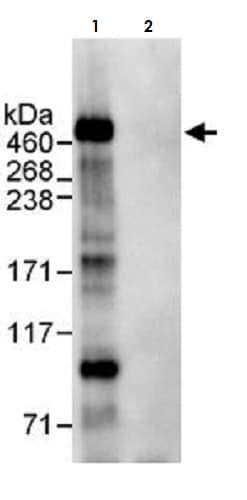 Immunoprecipitation - Anti-APC antibody (ab265597)
APC was immunoprecipitated from HeLa (Human epithelial cell line from cervix adenocarcinoma) whole cell lysate (1.0 mg per IP reaction; 20% of IP loaded) with ab265597 at 3 µg/mg lysate. Western blot was performed from the immunoprecipitates using ab265597 at 1 µg/ml .
Lane 1: ab265597 IP in HeLa whole cell lysate.
Lane 2: Control IgG IP in HeLa whole cell lysate.Detection: Chemiluminescence with an exposure time of 30 seconds.
Protocols
To our knowledge, customised protocols are not required for this product. Please try the standard protocols listed below and let us know how you get on.
Datasheets and documents
-
SDS download
-
Datasheet download
References (0)
ab265597 has not yet been referenced specifically in any publications.