Human GNA13 knockout HeLa cell line
- Advanced Validation
- What is this?
Be the first to review this product! Submit a review
|
(0 Publication)
- WB
Lab
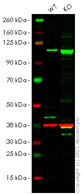
Western blot - Human GNA13 knockout HeLa cell line (AB264846)
Lanes 1 - 2 : Merged signal (red and green). Green - ab128900 observed at 45 kDa. Red - loading control ab8245 (Mouse anti-GAPDH antibody [6C5]) observed at 37 kDa.
ab128900 was shown to react with GNA13 in wild-type HeLa cells in Western blot with loss of signal observed in GNA13 knockout cell line ab264846 (GNA13 knockout cell lysate ab257451). Wild-type HeLa and GNA13 knockout cell lysates were subjected to SDS-PAGE. Membranes were blocked in 3 % milk in TBS-T (0.1 % Tween®) before incubation with ab128900 and ab8245 (Mouse anti-GAPDH antibody [6C5]) overnight at 4 °C at a 1 in 1000 dilution and a 1 in 20000 dilution respectively. Blots were incubated with Goat anti-Rabbit IgG H&L (IRDye® 800CW) preabsorbed (ab216773) and Goat anti-Mouse IgG H&L (IRDye® 680RD) preabsorbed (ab216776) secondary antibodies at 1 in 20000 dilution for 1 h at room temperature before imaging.
All lanes:
Western blot - Anti-GNA13 antibody [EPR5436] (<a href='/en-us/products/primary-antibodies/gna13-antibody-epr5436-ab128900'>ab128900</a>) at 1/1000 dilution
Lane 1:
Wild-type HeLa cell lysate at 20 µg
Lane 2:
GNA13 knockout HeLa cell lysate at 20 µg
Lane 2:
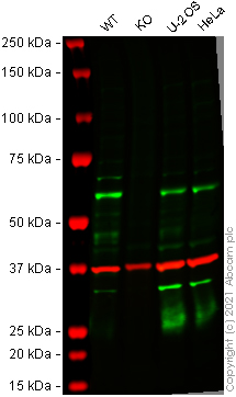
Western blot - Human GNA13 knockout HeLa cell line (ab264846)
Predicted band size: 44 kDa
Observed band size: 45 kDa
false

- Sanger seq
Unknown
Sanger Sequencing - Human GNA13 knockout HeLa cell line (AB264846)
Allele-2 : Insertion of the selection cassette in exon 2.

- Sanger seq
Unknown
Sanger Sequencing - Human GNA13 knockout HeLa cell line (AB264846)
Allele-1 : Insertion of the selection cassette in exon 2.
Complementary Products

Primary Antibodies
AB283946
Anti-SARS-CoV-2 Spike Glycoprotein S1 (RBD) antibody [EPR24852-174]
20ul selling size|RabMAb|Recombinant

- 0
- 1
Cell Lines & Lysates
AB264921
Human FOLR1 (Folate Binding Protein/FBP) knockout HeLa cell line
Advanced Validation

- 0
- 0
Reactivity data
Product details
We will provide viable cells that proliferate on revival.
This product is subject to limited use licenses from The Broad Institute, ERS Genomics Limited and Sigma-Aldrich Co. LLC, and is developed with patented technology. For full details of the licenses and patents please refer to our limited use license and patent pages.
What's included?
Properties and storage information
Gene name
Gene editing type
Gene editing method
Knockout validation
Quality control - STR analysis
Shipped at conditions
Appropriate short-term storage conditions
Appropriate long-term storage conditions
Supplementary information
This supplementary information is collated from multiple sources and compiled automatically.
Biological function summary
The role of G protein subunit GNA13 involves participating in critical cellular functions such as cytoskeletal reorganization cell migration and proliferation. It is an integral part of the G12/G13 subunits' complex which plays a role in controlling cell dynamics and morphology. Through these activities GNA13 contributes to the regulation of processes like embryonic development and tissue repair. Additionally it influences cell behavior through the RhoA pathway affecting actin cytoskeleton and cell shape changes.
Pathways
GNA13 interacts with a variety of important signaling systems. One primary pathway where GNA13 is active is the Rho signaling pathway where it activates RhoA leading to changes in the actin cytoskeleton. This pathway is necessary for cellular processes like adhesion and motility. Additionally GNA13 participates in the mitogen-activated protein kinase (MAPK) pathway influencing cellular proliferation. Through these pathways GNA13 is related to proteins such as RhoGEFs which act as guanine nucleotide exchange factors and MAPKs essential in transmitting mitogenic signals.
Cell culture
Biosafety level
EU: 2 US: 2
Adherent/suspension
Adherent
Gender
Female
Initial handling guidelines
Upon arrival, the vial should be stored in liquid nitrogen vapor phase and not at -80°C. Storage at -80°C may result in loss of viability. <p>1. Thaw the vial in 37°C water bath for approximately 1-2 minutes.<br>2. Transfer the cell suspension (0.8 mL) to a 15 mL/50 mL conical sterile polypropylene centrifuge tube containing 8.4 mL pre-warmed culture medium, wash vial with an additional 0.8 mL culture medium (total volume 10 mL) to collect remaining cells, and centrifuge at 201 x g (rcf) for 5 minutes at room temperature. 10 mL represents minimum recommended dilution. 20 mL represents maximum recommended dilution.<br>3. Resuspend the cell pellet in 5 mL pre-warmed culture medium and count using a haemocytometer or alternative cell counting method seed all remaining cells into a T25.<br>4. Incubate the culture at 37°C incubator with 5% CO<sub>2</sub>. Check the culture one day after revival and continue to check until 80% confluent. Media change can be given if needed.<br>5. Once confluent passage into an appropriate flask at a density of 2x10<sup>4</sup> cells/cm<sup>2</sup>. Seeding density is given as a guide only and should be scaled to align with individual lab schedules. Cultures should be monitored daily.</p>
Subculture guidelines
- All seeding densities should be based on cell counts gained by established methods.
- A guide seeding density of 2x10⁴ cells/cm2 is recommended.
- Cells should be passaged when they have achieved 80-90% confluence.
Culture medium
DMEM (High Glucose) + 10% FBS
Cryopreservation medium
Cell Freezing Medium-DMSO Serum free media, contains 8.7% DMSO in MEM supplemented with methyl cellulose.
Product promise
Please note: All products are 'FOR RESEARCH USE ONLY. NOT FOR USE IN DIAGNOSTIC OR THERAPEUTIC PROCEDURES'.
For licensing inquiries, please contact partnerships@abcam.com