Human RAB10 knockout A549 cell line
- Advanced Validation
- What is this?
Be the first to review this product! Submit a review
|
(0 Publication)
- ICC/IF
Collaborator
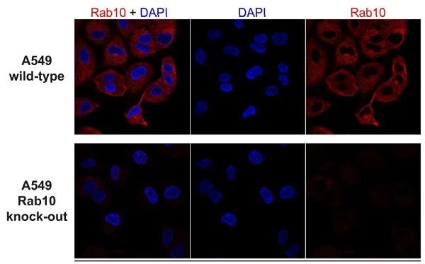
Immunocytochemistry/ Immunofluorescence - Human RAB10 knockout A549 cell line (AB261868)
Immunofluorescent analysis of 4% paraformaldehyde-fixed, 0.1% saponin permeabilized A549 wild-type and knockout cells (ab261868) labeling RAB10 (red) with ab237703 at 0.5 μg/ml, followed by anti-Rabbit secondary at 1/1000 dilution. The nuclear counter stain is DAPI (blue).
Image courtesy of Dr. Dario Alessi from University of Dundee
- ICC/IF
Collaborator
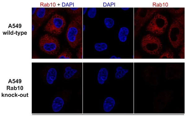
Immunocytochemistry/ Immunofluorescence - Human RAB10 knockout A549 cell line (AB261868)
Immunofluorescent analysis of 4% paraformaldehyde-fixed, 0.1% saponin permeabilized A549 wild-type and knockout cells (ab261868) labeling RAB10 (red) with ab237703 at 0.5 μg/ml, followed by anti-Rabbit secondary at 1/1000 dilution. The nuclear counter stain is DAPI (blue).
Image courtesy of Dr. Dario Alessi from University of Dundee
- ICC/IF
Lab
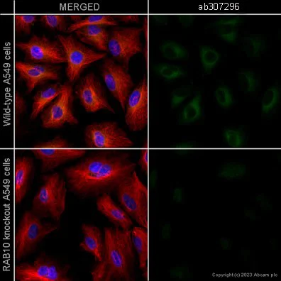
Immunocytochemistry/ Immunofluorescence - Human RAB10 knockout A549 cell line (AB261868)
Immunofluorescent analysis of 4% Paraformaldehyde fixed, 0.1% Saponin/PBS permeabilized wild-type A549 cells and RAB10 KO A549 (RAB10 knockout human lung carcinoma epithelial cell) (ab261868) labelling RAB10 with ab307296 at 1/25000 (0.04 ug/ml) dilution, followed by ab150117 Goat Anti-Mouse IgG H&L (Alexa Fluor® 488) preadsorbed antibody at 1/1000 (2ug/ml) dilution (Green). ab202272 Recombinant Alexa Fluor® 594 Tubulin [EP1332Y] was used to counterstain tubulin at 1/200 (2.5ug/ml) dilution (Red). The Nuclear counterstain was DAPI (Blue). Confocal image showing cytoplasmic staining in wild-type A549 cell line, and no staining in RAB10 KO A549 cell line. Images were acquired on the Perkin Elmer® Operetta HCA and a maximum intensity projection of 7 confocal planes is shown for the representative images.
- ICC/IF
Lab
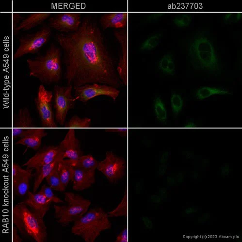
Immunocytochemistry/ Immunofluorescence - Human RAB10 knockout A549 cell line (AB261868)
Immunofluorescent analysis of 4% paraformaldehyde-fixed, 0.1% saponin permeabilized RAB10 KO A549 (RAB10 knockout human lung carcinoma epithelial cell) (ab261868) labelling RAB10 (red) with ab237703 at 0.04 μg/ml, followed by Goat Anti-Rabbit IgG H&L (Alexa Fluor® 488) preadsorbed (ab150081) at 1/1000 dilution. The nuclear counter stain is DAPI (blue). Confocal image showing cytoplasmic staining in wild-type A549 cell line, and no staining in RAB10 KO A549 cell line. Images were acquired on the Perkin Elmer® Operetta HCA and a maximum intensity projection of 7 confocal planes is shown for the representative images.
- WB
Lab
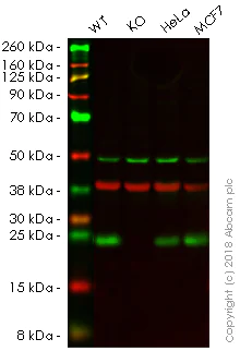
Western blot - Human RAB10 knockout A549 cell line (AB261868)
Lanes 1 - 4 : Merged signal (red and green). Green - ab237703 observed at 25 kDa. Red - loading control ab8245 observed at 37 kDa.
ab237703 was shown to recognize Rab10 in wild-type A549 cells as signal was lost at the expected MW in Rab10 knockout cells. Additional cross-reactive bands were observed in the wild-type and knockout cells. Wild-type and Rab10 knockout samples were subjected to SDS-PAGE. ab237703 and ab8245 (Mouse anti GAPDH loading control) were incubated overnight at 4°C at 1/1000 dilution and 1/20000 dilution respectively. Blots were developed with Goat anti-Rabbit IgG H&L (IRDye® 800CW) preabsorbed ab216773 and Goat anti-Mouse IgG H&L (IRDye® 680RD) preabsorbed ab216776 secondary antibodies at 1/20000 dilution for 1 hour at room temperature before imaging.
All lanes:
Western blot - Anti-RAB10 antibody [MJF-R23] (<a href='/en-us/products/primary-antibodies/rab10-antibody-mjf-r23-ab237703'>ab237703</a>) at 1/1000 dilution
Lane 1:
Wild-type A549 (Human lung carcinoma cell line) whole cell lysate at 20 µg
Lane 2:
RAB10 knockout A549 (Human lung carcinoma cell line) whole cell lysate at 20 µg
Lane 2:
Western blot - Human RAB10 knockout A549 cell line (ab261868)
Lane 3:
HeLa (Human epithelial cell line from cervix adenocarcinoma) whole cell lysate at 20 µg
Lane 4:
MCF7 (Human breast adenocarcinoma cell line) whole cell lysate at 20 µg
Predicted band size: 22 kDa
Observed band size: 25 kDa
false
- WB
Lab
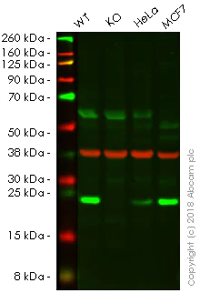
Western blot - Human RAB10 knockout A549 cell line (AB261868)
Lanes 1 - 4 : Merged signal (red and green). Green - ab181367 observed at 25 kDa. Red - loading control ab8245 observed at 37 kDa.
ab181367 was shown to specifically react with Rab10 in wild-type A549 cells as signal was lost in Rab10 knockout cell line ab261868 (knockout cell lysate ab261677). Wild-type and Rab10 knockout samples were subjected to SDS-PAGE. ab181367 and ab8245 (Mouse anti GAPDH loading control) were incubated overnight at 4°C at 1/1000 dilution and 1/20000 dilution respectively. Blots were developed with Goat anti-Rabbit IgG H&L (IRDye® 800CW) preabsorbed ab216773 and Goat anti-Mouse IgG H&L (IRDye® 680RD) preabsorbed ab216776 secondary antibodies at 1/20000 dilution for 1 hour at room temperature before imaging.
All lanes:
Western blot - Anti-RAB10 antibody [EPR13242] (<a href='/en-us/products/primary-antibodies/rab10-antibody-epr13242-ab181367'>ab181367</a>) at 1/1000 dilution
Lane 1:
Wild-type A549 (Human lung carcinoma cell line) whole cell lysate at 20 µg
Lane 2:
Western blot - Human RAB10 knockout A549 cell lysate (<a href='/en-us/products/cell-lysates/human-rab10-knockout-a549-cell-lysate-ab261677'>ab261677</a>) at 20 µg
Lane 2:
Western blot - Human RAB10 knockout A549 cell line (ab261868)
Lane 3:
HeLa (human epithelial cell line from cervix adenocarcinoma) whole cell lysate at 20 µg
Lane 4:
MCF7 (Human breast adenocarcinoma cell line) whole cell lysate at 20 µg
Predicted band size: 22 kDa
Observed band size: 25 kDa
false

- NGS
Lab
Next Generation Sequencing - Human RAB10 knockout A549 cell line (AB261868)
X = 1 bp insertion
Complementary Products
Primary Antibodies
AB237703
20ul selling size|KO Validated|RabMAb|Recombinant

- 0
- 0


Reactivity data
Product details
We will provide viable cells that proliferate on revival.
This product is subject to limited use licenses from The Broad Institute, ERS Genomics Limited and Sigma-Aldrich Co. LLC, and is developed with patented technology. For full details of the licenses and patents please refer to our limited use license and patent pages.
What's included?
Properties and storage information
Gene name
Gene editing type
Gene editing method
Knockout validation
Quality control - STR analysis
Shipped at conditions
Appropriate short-term storage conditions
Appropriate long-term storage conditions
Supplementary information
This supplementary information is collated from multiple sources and compiled automatically.
Biological function summary
RAB10 participates in vesicular trafficking and transport processes. It is part of the endosomal and exocytic complexes contributing to the sorting and delivery of cellular cargo. RAB10's activity affects the delivery of glucose transporter type 4 (GLUT4) to the plasma membrane impacting glucose uptake and metabolism. It also plays a critical role in the polarized transport of cargo in neuronal cells influencing neuronal signaling and function.
Pathways
RAB10 integrates into the insulin signaling and polarized epithelial cell transport pathways. Within these RAB10 cooperates with proteins like GLUT4 and the TBC1D4 a RAB GAP protein essential for GLUT4 vesicle translocation. These interactions ensure efficient response and coordination of cellular trafficking required for metabolic regulation and cellular growth processes.
Cell culture
Biosafety level
EU: 1 US: 1
Adherent/suspension
Adherent
Gender
Male
Initial handling guidelines
Upon arrival, the vial should be stored in liquid nitrogen vapor phase and not at -80°C. Storage at -80°C may result in loss of viability. <p>1. Thaw the vial in 37°C water bath for approximately 1-2 minutes.<br>2. Transfer the cell suspension (0.8 mL) to a 15 mL/50 mL conical sterile polypropylene centrifuge tube containing 8.4 mL pre-warmed culture medium, wash vial with an additional 0.8 mL culture medium (total volume 10 mL) to collect remaining cells, and centrifuge at 201 x g (rcf) for 5 minutes at room temperature. 10 mL represents minimum recommended dilution. 20 mL represents maximum recommended dilution.<br>3. Resuspend the cell pellet in 5 mL pre-warmed culture medium and count using a haemocytometer or alternative cell counting method seed all remaining cells into a T25.<br>4. Incubate the culture at 37°C incubator with 5% CO<sub>2</sub>. Check the culture one day after revival and continue to check until 80% confluent. Media change can be given if needed.<br>5. Once confluent passage into an appropriate flask at a density of 2x10<sup>4</sup> cells/cm<sup>2</sup>. Seeding density is given as a guide only and should be scaled to align with individual lab schedules. Cultures should be monitored daily.</p>
Subculture guidelines
- All seeding densities should be based on cell counts gained by established methods.
- A guide seeding density of 2x10⁴ cells/cm2 is recommended.
- Cells should be passaged when they have achieved 80-90% confluence.
- Do not allow the cell density to exceed 7x10⁴ cells/cm2.
Culture medium
F-12K + 10% FBS
Cryopreservation medium
Cell Freezing Medium-DMSO Serum free media, contains 8.7% DMSO in MEM supplemented with methyl cellulose.
Product promise
Please note: All products are 'FOR RESEARCH USE ONLY. NOT FOR USE IN DIAGNOSTIC OR THERAPEUTIC PROCEDURES'.
For licensing inquiries, please contact partnerships@abcam.com