Human BAX knockout HeLa cell lysate
Be the first to review this product! Submit a review
|
(0 Publication)

- WB
Lab
Western blot - Human BAX knockout HeLa cell lysate (AB263841)
Lane 1 : Wild-type HeLa cell lysate (20µg)
Lane 2 : BAX knockout HeLa cell lysate (20µg)
Lanes 1- 2 : Merged signal (red and green). Green - ab182734 observed at 21 kDa. Red - loading control ab8245 observed at 37 kDa.
ab182734 Recombinant Anti-Bax antibody [EPR18284 was shown to specifically react with BAX in wild-type HeLa cells in western blot. Loss of signal was observed when knockout cell line ab255363 (knockout cell lysate ab263841) was used. Wild-type and BAX knockout samples were subjected to SDS-PAGE. Membrane was blocked for 1 hour at room temperature in 0.1% TBST with 3% non-fat dried milk. ab182734 and Anti-GAPDH antibody [6C5] - Loading Control (ab8245) were incubated overnight at 4°C at 1 in 1000 dilution and 1 in 20000 dilution respectively. Blots were developed with Goat anti-Rabbit IgG H&L (IRDye® 800CW) preadsorbed (ab216773) and Goat anti-Mouse IgG H&L (IRDye® 680RD) preadsorbed (ab216776) secondary antibodies at 1 in 20000 dilution for 1 hour at room temperature before imaging.
All lanes:
Western blot - Anti-Bax antibody [EPR18284] (<a href='/en-us/products/primary-antibodies/bax-antibody-epr18284-ab182734'>ab182734</a>) at 1/1000 dilution
Lane 1:
Wild-type HeLa cell lysate at 20 µg
Lane 2:
BAX knockout HeLa cell lysate at 20 µg
Lane 2:
Western blot - Human BAX knockout HeLa cell line (<a href='/en-us/products/cell-lines/human-bax-knockout-hela-cell-line-ab255363'>ab255363</a>)
Predicted band size: 21 kDa
Observed band size: 21 kDa
false

- Sanger seq
Unknown
Sanger Sequencing - Human BAX knockout HeLa cell lysate (AB263841)
Allele-2 : Insertion of the selection cassette in exon 2

- Sanger seq
Unknown
Sanger Sequencing - Human BAX knockout HeLa cell lysate (AB263841)
Allele-1 : 1 bp deletion in exon 2
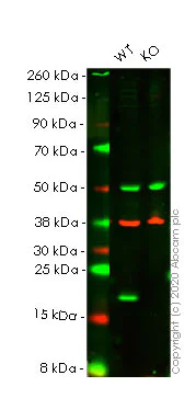
- WB
Lab
Western blot - Human BAX knockout HeLa cell lysate (AB263841)
Lane 1 : Wild-type HeLa cell lysate (20µg)
Lane 2 : BAX knockout HeLa cell lysate (20µg)
Lanes 1- 2 : Merged signal (red and green). Green - ab32503. Red - loading control ab8245 observed at 37 kDa.
ab32503 Recombinant Anti-Bax antibody [E63] was shown to specifically react with BAX in wild-type HeLa cells in western blot. Loss of signal was observed when knockout cell line ab255363 (knockout cell lysate ab263841) was used. Wild-type and BAX knockout samples were subjected to SDS-PAGE. Membrane was blocked for 1 hour at room temperature in 0.1% TBST with 3% non-fat dried milk. ab32503 and Anti-GAPDH antibody [6C5] - Loading Control (ab8245) were incubated overnight at 4°C at 1 in 1000 dilution and 1 in 20000 dilution respectively. Blots were developed with Goat anti-Rabbit IgG H&L (IRDye® 800CW) preadsorbed (ab216773) and Goat anti-Mouse IgG H&L (IRDye® 680RD) preadsorbed (ab216776) secondary antibodies at 1 in 20000 dilution for 1 hour at room temperature before imaging.
All lanes:
Western blot - Anti-Bax antibody [E63] (<a href='/en-us/products/primary-antibodies/bax-antibody-e63-ab32503'>ab32503</a>) at 1/1000 dilution
Lane 1:
Wild-type HeLa cell lysate at 20 µg
Lane 2:
Western blot - Human BAX knockout HeLa cell lysate (ab263841) at 20 µg
Lane 2:
Western blot - Human BAX knockout HeLa cell line (<a href='/en-us/products/cell-lines/human-bax-knockout-hela-cell-line-ab255363'>ab255363</a>)
Predicted band size: 21 kDa
false
Complementary Products

Primary Antibodies
AB210655
Alexa Fluor® 594 Anti-Integrin alpha 2 antibody [EPR17338] - C-terminal
RabMAb|Recombinant|KO Validated

- 0
- 0


Reactivity data
Product details
Knockout cell lysate achieved by CRISPR/Cas9.
REACH authorisation
Abcam has not and does not intend to apply for the REACH Authorisation of customers' uses of products that contain European Authorisation list (Annex XIV) substances.
It is the responsibility of our customers to check the necessity of application of REACH Authorisation, and any other relevant authorisations, for their intended uses.
Lysate preparation: Our lysates are made using RIPA buffer to which we add a protease inhibitor cocktail and phosphatase inhibitor cocktail (ratio: 300:100:10). This means that the protein of interest is denatured. If you require a native form of the protein please use the live cell version. Please refer to our lysis protocol for further details on how our lysates are prepared.
User storage instructions: Lyophilizate may be stored at 4°C. After reconstitution, store at -20°C for short-term storage or -80°C for long-term storage.
This product is subject to limited use licenses from The Broad Institute, ERS Genomics Limited and Sigma-Aldrich Co. LLC, and is developed with patented technology. For full details of the licenses and patents please refer to our limited use license and patent pages.
What's included?
Properties and storage information
Gene name
Gene editing type
Gene editing method
Knockout validation
Quality control - STR analysis
Shipped at conditions
Appropriate short-term storage conditions
Appropriate long-term storage conditions
Supplementary information
This supplementary information is collated from multiple sources and compiled automatically.
Biological function summary
The Bax protein forms homodimers or heterodimers with other Bcl-2 family proteins to facilitate apoptosis. This protein is part of a larger complex involving several members of the Bcl-2 family which finely tunes the balance between cell survival and programmed cell death. Through a change in its conformation Bax translocates to the outer mitochondrial membrane where it promotes the release of cytochrome c and other apoptogenic factors into the cytosol.
Pathways
Bax plays a significant role in the intrinsic pathway of apoptosis also known as the mitochondrial pathway. This pathway involves the release of cytochrome c which further activates downstream proteins such as caspase-9 and caspase-3. Bax interacts closely with proteins like Bak another pro-apoptotic member of the Bcl-2 family providing a cooperative function in mitochondrial membrane permeabilization. Together Bax and Bak act as gatekeepers of the mitochondrial pathway determining the cell's fate in response to apoptotic stimuli.
Cell culture
Biosafety level
EU: 2 US: 2
Adherent/suspension
Adherent
Gender
Female
Product promise
Please note: All products are 'FOR RESEARCH USE ONLY. NOT FOR USE IN DIAGNOSTIC OR THERAPEUTIC PROCEDURES'.
For licensing inquiries, please contact partnerships@abcam.com