Anti-68kDa Neurofilament/NF-L antibody [Nfl21] - BSA and Azide free
- BOND RX™ Validated
- Recombinant
- What is this?
Be the first to review this product! Submit a review
|
(0 Publication)
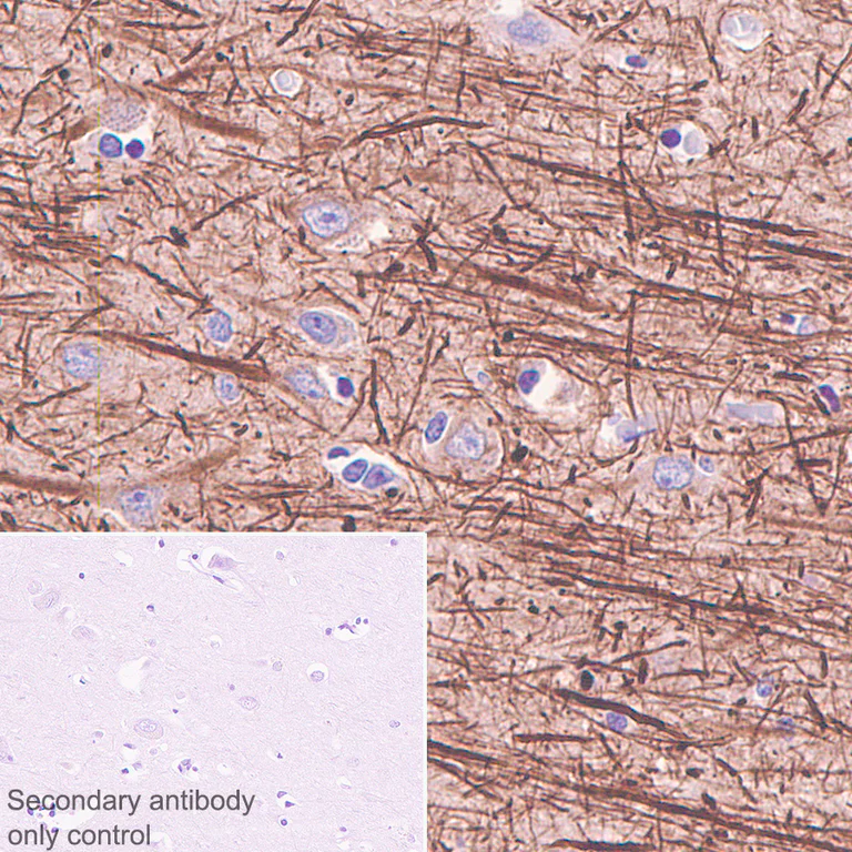
- IHC-P
Unknown
Immunohistochemistry (Formalin/PFA-fixed paraffin-embedded sections) - Anti-68kDa Neurofilament/NF-L antibody [Nfl21] - BSA and Azide free (AB273479)
This data was developed using ab273441 the same antibody clone in a different buffer formulation.
Immunohistochemical analysis of paraffin-embedded human cerebrum tissue labeling Neurofilament light with ab273441 at 1/500 (2.116 μg/ml) dilution followed by a ready to use LeicaDS9800 (Bond® Polymer Refine Detection). Positive staining on human cerebrum is observed. The section was incubated with ab273441 for 30 mins at room temperature and blocked mouse IgG with specific antibody ab125913 for 8min. The immunostaining was performed on a Leica Biosystems BOND® RX instrument. Counterstained with Hematoxylin.
Secondary antibody only control : Secondary antibody is a ready to use LeicaDS9800 (Bond® Polymer Refine Detection).
Heat mediated antigen retrieval with Tris-EDTA buffer (pH 9.0, epitope retrieval solution2) for 20 mins.
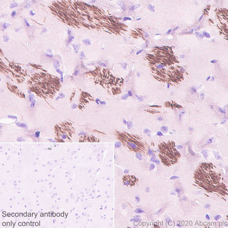
- IHC-P
Unknown
Immunohistochemistry (Formalin/PFA-fixed paraffin-embedded sections) - Anti-68kDa Neurofilament/NF-L antibody [Nfl21] - BSA and Azide free (AB273479)
This data was developed using ab273441 the same antibody clone in a different buffer formulation.
Immunohistochemical analysis of paraffin-embedded mouse cerebrum tissue labeling Neurofilament light with ab273441 at 1/500 (2.116 μg/ml) dilution followed by a ready to use LeicaDS9800 (Bond® Polymer Refine Detection). Positive staining on mouse cerebrum is observed. The section was incubated with ab273441 for 30 mins at room temperature and blocked mouse IgG with specific antibody ab125913 for 8min. The immunostaining was performed on a Leica Biosystems BOND® RX instrument. Counterstained with Hematoxylin.
Secondary antibody only control : Secondary antibody is a ready to use LeicaDS9800 (Bond® Polymer Refine Detection).
Heat mediated antigen retrieval with Tris-EDTA buffer (pH 9.0, epitope retrieval solution2) for 20 mins.
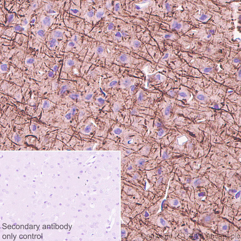
- IHC-P
Unknown
Immunohistochemistry (Formalin/PFA-fixed paraffin-embedded sections) - Anti-68kDa Neurofilament/NF-L antibody [Nfl21] - BSA and Azide free (AB273479)
This data was developed using ab273441 the same antibody clone in a different buffer formulation.
Immunohistochemical analysis of paraffin-embedded rat cerebrum tissue labeling Neurofilament light with ab273441 at 1/500 (2.116 μg/ml) dilution followed by a ready to use LeicaDS9800 (Bond® Polymer Refine Detection). Positive staining on rat cerebrum is observed. The section was incubated with ab273441 for 30 mins at room temperature and blocked mouse IgG with specific antibody ab125913 for 8min. The immunostaining was performed on a Leica Biosystems BOND® RX instrument. Counterstained with Hematoxylin.
Secondary antibody only control : Secondary antibody is a ready to use LeicaDS9800 (Bond® Polymer Refine Detection).
Heat mediated antigen retrieval with Tris-EDTA buffer (pH 9.0, epitope retrieval solution2) for 20 mins.
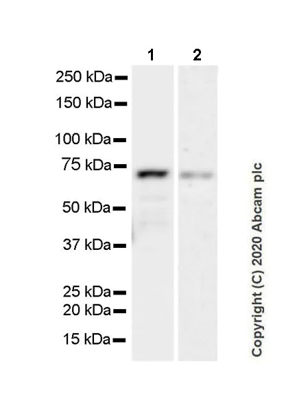
- WB
Supplier Data
Western blot - Anti-68kDa Neurofilament/NF-L antibody [Nfl21] - BSA and Azide free (AB273479)
This data was developed using ab273441 the same antibody clone in a different buffer formulation.
This blot was developed using a higher sensitivity ECL substrate.
Blocking/Dilution buffer : 5% NFDM/TBST.
Exposure times : Lane 1 : 37 seconds; Lane 2 : 3 minutes.
All lanes:
Western blot - Anti-68kDa Neurofilament/NF-L antibody [Nfl21] (<a href='/en-us/products/primary-antibodies/68kda-neurofilament-nf-l-antibody-nfl21-ab273441'>ab273441</a>) at 1/1000 dilution
Lane 1:
SK-N-BE(2) (human neuroblastoma neuroblast), whole cell lysate at 20 µg
Lane 2:
Human cerebrospinal fluid at 40 µL
Secondary
All lanes:
Peroxidase-Conjugated Goat anti-Mouse IgG (H+L) at 1/5000 dilution
Predicted band size: 62 kDa
false
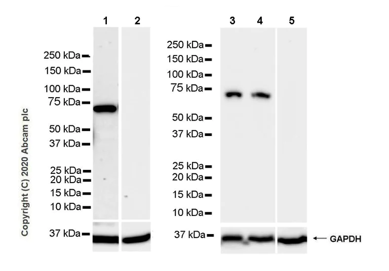
- WB
Supplier Data
Western blot - Anti-68kDa Neurofilament/NF-L antibody [Nfl21] - BSA and Azide free (AB273479)
This data was developed using ab273441 the same antibody clone in a different buffer formulation.
Negative control : liver (PMID : 2120242).
Blocking/Dilution buffer : 5% NFDM/TBST.
Exposure times : Lane 1-2 : 10 seconds; Lane 3-5 : 37 seconds.
All lanes:
Western blot - Anti-68kDa Neurofilament/NF-L antibody [Nfl21] (<a href='/en-us/products/primary-antibodies/68kda-neurofilament-nf-l-antibody-nfl21-ab273441'>ab273441</a>) at 1/1000 dilution
Lane 1:
Mouse hippocampus tissue lysate at 20 µg
Lane 2:
Mouse liver tissue lysate at 20 µg
Lane 3:
Rat brain tissue lysate at 20 µg
Lane 4:
Rat hippocampus tissue lysate at 20 µg
Lane 5:
Rat liver tissue lysate at 20 µg
Secondary
All lanes:
Western blot - Anti-mouse IgG for IP (HRP) (<a href='/en-us/products/secondary-antibodies/mouse-igg-for-ip-hrp-ab131368'>ab131368</a>) at 1/1000 dilution
Predicted band size: 62 kDa
false
Related conjugates and formulations (1)
-
Anti-68kDa Neurofilament/NF-L antibody [Nfl21]
Complementary Products


Primary Antibodies
AB278054
Anti-GFAP antibody [RM1003] - Astrocyte Marker
BOND RX™ Validated|20ul selling size|RabMAb|Recombinant

- 0
- 0

Primary Antibodies
AB52642
Anti-S100 beta antibody [EP1576Y] - Astrocyte Marker
BOND RX™ Validated|RabMAb|Recombinant|Lab Essentials|20ul selling size

- 4
- 37
Primary Antibodies
AB177487
Anti-NeuN antibody [EPR12763] - Neuronal Marker
BOND RX™ Validated|RabMAb|Recombinant|Lab Essentials|20ul selling size|Advanced Validation

- 5
- 76
Reactivity data
Product details
ab273479 is the carrier-free version of ab273441.
What are the advantages of a recombinant monoclonal antibody?
This product is a recombinant monoclonal antibody, which offers several advantages including:
- - High batch-to-batch consistency and reproducibility
- - Improved sensitivity and specificity
- - Long-term security of supply
- - Animal-free batch production
For more information, read more on recombinant antibodies.
Want a custom formulation?
This antibody clone is manufactured by Abcam. If you require a custom buffer formulation or conjugation for your experiments, please contact orders@abcam.com
Conjugation ready
Our carrier-free antibodies are typically supplied in a PBS-only formulation, purified and free of BSA, sodium azide and glycerol. This conjugation-ready format is designed for use with fluorochromes, metal isotopes, oligonucleotides, and enzymes, which makes them ideal for antibody labelling, functional and cell-based assays, flow-based assays (e.g. mass cytometry) and Multiplex Imaging applications.
Use our conjugation kits for antibody conjugates that are ready-to-use in as little as 20 minutes with 1 minute hands-on-time and 100% antibody recovery: available for fluorescent dyes, HRP, biotin and gold.
Compatibility
This product is compatible with the Maxpar® Antibody Labeling Kit from Fluidigm, without the need for antibody preparation. Maxpar® is a trademark of Fluidigm Canada Inc.
Properties and storage information
Form
Purification technique
Storage buffer
Shipped at conditions
Appropriate short-term storage conditions
Appropriate long-term storage conditions
Storage information
Supplementary information
This supplementary information is collated from multiple sources and compiled automatically.
Biological function summary
The neurofilament protein functions to stabilize the axonal diameter thereby affecting neuronal conductivity. It integrates into a complex with other neurofilament proteins and related molecules forming cross-bridges that aid in strengthening the cellular structure. This protein-protein interaction is vital for normal neuronal function and efficient nerve signal transmission.
Pathways
Neurofilament proteins contribute to cellular processes within the nervous system. It participates in the axonal transport pathway which includes the transport of synaptic vesicles and organelles along axons. This movement is facilitated by interactions with motor proteins like kinesin and dynein which help in moving these materials to their destinations. Additionally NF-L connects to actin filaments collaborating to sustain neuronal shape and function.
Product protocols
- Visit the General protocols
- Visit the Troubleshooting
Target data
Alternative Names
Product promise
Please note: All products are 'FOR RESEARCH USE ONLY. NOT FOR USE IN DIAGNOSTIC OR THERAPEUTIC PROCEDURES'.
For licensing inquiries, please contact partnerships@abcam.com