Anti-Caveolin-3 antibody - Caveolae Marker
- Lab Essentials
- What is this?
4
(11 Reviews)
|
(83 Publications )
- ICC/IF
Supplier Data
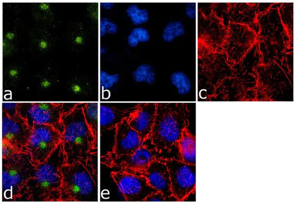
Immunocytochemistry/ Immunofluorescence - Anti-Caveolin-3 antibody - Caveolae Marker (AB2912)
Immunocytochemistry/Immunofluorescence analysis of 70% confluent log phase A-375 cells. Cells were fixed with 4% paraformaldehyde for 15 minutes, permeabilized with 0.25% Triton X-100 for 10 minutes, and blocked with 5% BSA for 1 hour at room temperature. Samples were incubated with ab2912 at 1μg/ml in 1% BSA for 3 hours at room temperature and then labelled with Alexa Fluor® 488-conjugated goat anti-rabbit IgG (H+L) at a dilution of 1/2000 for 45 minutes at room temperature (panel a : green). Nuclei (panel b : blue) were stained with DAPI. F-actin (panel c : red) was stained with Alexa Fluor® 555 Rhodamine Phalloidin (1/300). Panel d is a merged image showing cytoplasmic localization. Panel e is a no primary antibody control. The images were captured at 60X magnification.
- Flow Cyt
Supplier Data
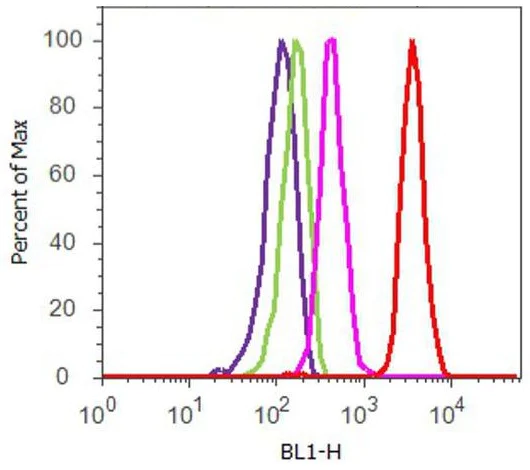
Flow Cytometry - Anti-Caveolin-3 antibody - Caveolae Marker (AB2912)
Flow cytometry analysis of U-87 MG cells. Cells were fixed with 70% ethanol for 10 minutes, permeabilized with 0.25% Triton X-100 for 20 minutes, and blocked with 5% BSA for 30 minutes at room temperature. Cells were labeled with ab2912 (red histogram) or with rabbit isotype control (pink histogram) at 3-5 ug/million cells in 2.5% BSA. After incubation at room temperature for 2 hours, the cells were labeled with Alexa Fluor® 488-conjugated goat anti-rabbit secondary antibody at a dilution of 1/400 for 30 minutes at room temperature. The purple histogram represents unstained control cells and the green histogram represents no-primary-antibody control.
- ICC/IF
Supplier Data
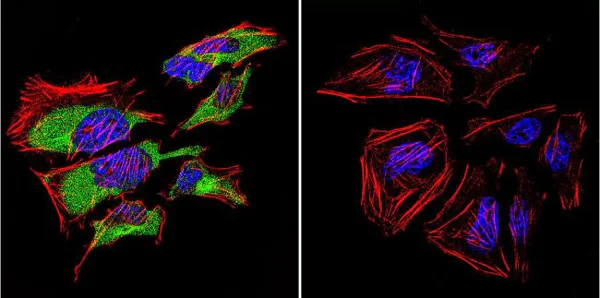
Immunocytochemistry/ Immunofluorescence - Anti-Caveolin-3 antibody - Caveolae Marker (AB2912)
Immunocytochemistry analysis of Caveolin-3 in HeLa Cells. Cells were grown on chamber slides and fixed with formaldehyde prior to staining. Cells were probed without (control) (right panel) or with ab2912 at a dilution of 1/20 overnight at 4°C, washed with PBS and incubated with a DyLight-488 conjugated secondary antibody. Caveolin-3 staining (green), F-Actin staining with Phalloidin (red) and nuclei with DAPI (blue) is shown. Images were taken at 60X magnification.
- ICC/IF
Supplier Data
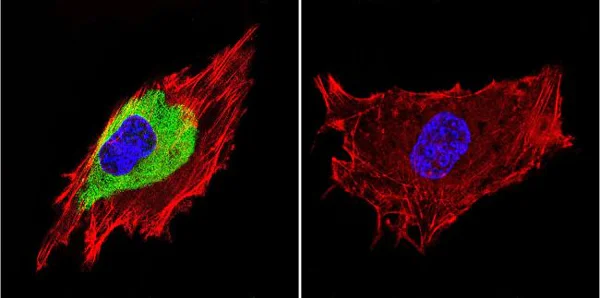
Immunocytochemistry/ Immunofluorescence - Anti-Caveolin-3 antibody - Caveolae Marker (AB2912)
Immunocytochemistry analysis of Caveolin-3 in C2C11 Cells. Cells were grown on chamber slides and fixed with formaldehyde prior to staining. Cells were probed without (control) (right panel) or with ab2912 at a dilution of 1/20 overnight at 4 C, washed with PBS and incubated with a DyLight-488 conjugated secondary antibody. Caveolin-3 staining (green), F-Actin staining with Phalloidin (red) and nuclei with DAPI (blue) is shown. Images were taken at 60X magnification.

- IP
Supplier Data
Immunoprecipitation - Anti-Caveolin-3 antibody - Caveolae Marker (AB2912)
Caveolin-3 was immunoprecipitated using 5 μg of ab2912 from mouse heart tissue lysate (Lane 3) using the protein A beads. Normal rabbit IgG was used as a isotype control (Lane 2). 10% input represents the cell extract used for immunoprecipitation (Lane 1). Western blot analysis was performed using ab2912 and HRP-conjugated goat anti-rabbit IgG (H+L) at a dilution of 1/2500. Chemiluminescent detection was performed.
All lanes:
Immunoprecipitation - Anti-Caveolin-3 antibody - Caveolae Marker (ab2912)
Predicted band size: 17 kDa
false
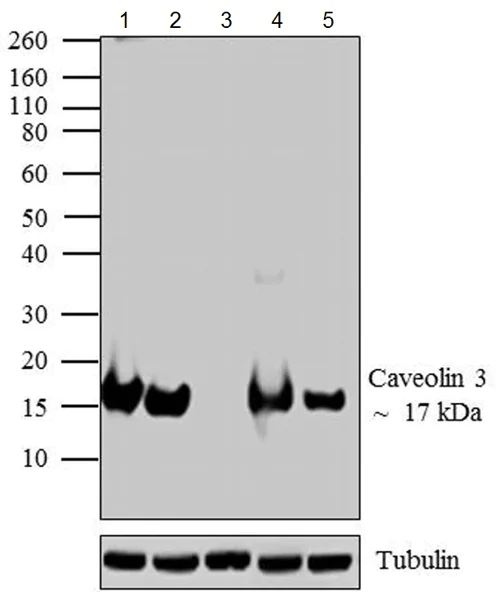
- WB
Supplier Data
Western blot - Anti-Caveolin-3 antibody - Caveolae Marker (AB2912)
Blocked with 5% skimmed milk.
All lanes:
Western blot - Anti-Caveolin-3 antibody - Caveolae Marker (ab2912)
Lane 1:
Rat heart tissue lysate at 20 µg
Lane 2:
Mouse heart tissue lysate at 20 µg
Lane 3:
HEK293 cell lysate at 20 µg
Lane 4:
Rat skeletal muscle tissue lysate at 20 µg
Lane 5:
Mouse muscle tissue lysate at 20 µg
Secondary
All lanes:
HRP-conjugated goat anti-rabbit IgG (H+L) at 1/2500 dilution
Predicted band size: 17 kDa
Observed band size: 17 kDa
true
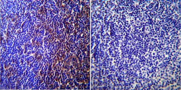
- IHC-P
Supplier Data
Immunohistochemistry (Formalin/PFA-fixed paraffin-embedded sections) - Anti-Caveolin-3 antibody - Caveolae Marker (AB2912)
Immunohistochemistry was performed on normal biopsies of deparaffinized mouse lymph node tissue. To expose target proteins heat induced antigen retrieval was performed using 10mM sodium citrate (pH6.0) buffer microwaved for 8-15 minutes. Following antigen retrieval tissues were blocked in 3% BSA-PBS for 30 minutes at room temperature. Tissues were then probed at a dilution of 1/200 with a rabbit polyclonal antibody recognizing Caveolin-3 ab2912 or without primary antibody (negative control) overnight at 4°C in a humidified chamber. Tissues were washed extensively with PBST and endogenous peroxidase activity was quenched with a peroxidase suppressor. Detection was performed using a biotin-conjugated secondary antibody and SA-HRP followed by colorimetric detection using DAB. Tissues were counterstained with hematoxylin and prepped for mounting.
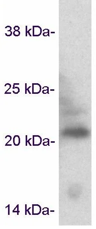
- WB
Supplier Data
Western blot - Anti-Caveolin-3 antibody - Caveolae Marker (AB2912)
All lanes:
Western blot - Anti-Caveolin-3 antibody - Caveolae Marker (ab2912)
All lanes:
Rat cardiac muscle tissue lysate
Predicted band size: 17 kDa
false
- WB
CiteAb
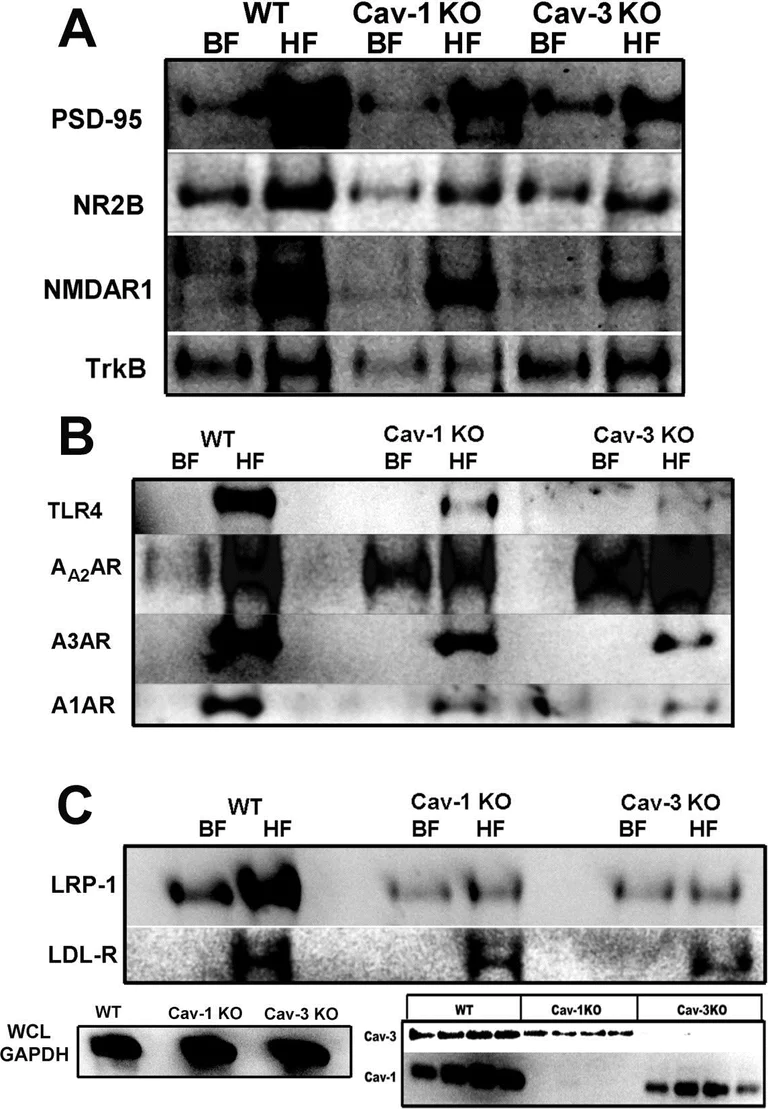
Western blot - Anti-Caveolin-3 antibody - Caveolae Marker (AB2912)
Caveolin-3 western blot using anti-Caveolin-3 antibody - Caveolae Marker ab2912. Publication image and figure legend from Niesman, I. R., Schilling, J. M., et al., 2014, J Neuroinflammation, PubMed 24593993.
ab2912 was used in this publication in western blot. This may not be the same as the application(s) guaranteed by Abcam. For a full list of applications guaranteed by Abcam for ab2912 please see the product overview.
Caveolin (Cav)-1 knock-out (KO) and Cav-3 KO mice have altered expression and membrane/lipid raft (MLR) localization of key neuronal and glial proteins. Sucrose density fractionation (SDF) and Western blot (WB) of wild-type (WT), Cav-1 KO, and Cav-3 KO brains. Buoyant fractions (BF) contain the cholesterol and sphingolipid enriched MLR, while heavy farctions (HF) contain non-MLR cellular components. (A) WB detection of PSD-95, NR2B, NR1 and TrkB expression in BF and HF. (B) WB detection of toll-like receptor-4 (TLR4), AA2AR, A3AR, and A1AR expression in BF and HF. (C) WB detection of LRP-1 and LDL-R expression in BF and HF. Bottom left, WB analysis of GAPDH in whole cell lysates (WCL) from which SDF were generated for each group. Bottom right, WB shows loss of Cav-1 and Cav-3 protein expression in the transgenic mouse used in the present study.
false
Alternative Products
Primary Antibodies
AB289544
Anti-Caveolin-3 antibody [RM1042] - Caveolae Marker
BOND RX™ Validated|20ul selling size|RabMAb|Recombinant

- 0
- 0
Primary Antibodies
AB173575
Anti-Caveolin-3 antibody [EPR11082] - Caveolae Marker
BOND RX™ Validated|RabMAb|Recombinant|Lab Essentials

- 0
- 0
Primary Antibodies
AB171752
Anti-Caveolin-3 antibody [EPR11083] - Caveolae Marker
BOND RX™ Validated|RabMAb|Recombinant

- 0
- 0

Primary Antibodies
AB182739
Anti-Caveolin-3 antibody [EPR18975] - Caveolae Marker
RabMAb|Recombinant

- 0
- 0

Complementary Products

Primary Antibodies
AB124684
Anti-Dysferlin antibody [JAI-1-49-3]
20ul selling size|RabMAb|Recombinant

- 4
- 4
Primary Antibodies
AB189254
Anti-alpha Sarcoglycan antibody [EPR14773]
RabMAb|Recombinant|20ul selling size|Trial Size

- 0
- 0
Primary Antibodies
AB281583
BOND RX™ Validated|RabMAb|Recombinant|Lab Essentials|20ul selling size

- 5
- 5
Reactivity data
Properties and storage information
Form
Purification technique
Purification notes
Storage buffer
Shipped at conditions
Appropriate short-term storage duration
Appropriate short-term storage conditions
Appropriate long-term storage conditions
Aliquoting information
Storage information
Supplementary information
This supplementary information is collated from multiple sources and compiled automatically.
Biological function summary
Caveolae perform vital functions in cellular signaling and endocytosis. Caveolin-3 assists in various signaling pathways within muscle cells and interacts with other caveolae markers to facilitate signal transduction. It often forms a complex with other caveolin proteins such as caveolin-1 creating an environment conducive for the embedding of signaling molecules. This protein also modulates the trafficking and function of certain ion channels and receptors impacting muscle physiology.
Pathways
Caveolin-3 significantly influences pathways concerned with muscle function and signal transduction. In particular it involves itself with the nitric oxide (NO) signaling pathway and beta-adrenergic signaling both critical to the contractile functions of muscle tissues. It closely interacts with proteins such as endothelial nitric oxide synthase (eNOS) and components of the G-protein coupled receptor family indicating its pivotal role in mediating physiological responses to stimuli.
Product protocols
- Visit the General protocols
- Visit the Troubleshooting
Target data
Alternative Names
Publications (83)
Recent publications for all applications. Explore the full listand refine your search
CNS neuroscience & therapeutics 31:e70367 PubMed40406932
2025
Applications
Unspecified application
Species
Unspecified reactive species
Nature communications 16:3730 PubMed40253375
2025
Applications
Unspecified application
Species
Unspecified reactive species
Skeletal muscle 15:1 PubMed39789642
2025
Applications
Unspecified application
Species
Unspecified reactive species
Proceedings of the National Academy of Sciences of the United States of America 121:e2402890121 PubMed38771868
2024
Applications
Unspecified application
Species
Unspecified reactive species
The Journal of physiology 602:4487-4510 PubMed38686538
2024
Applications
Unspecified application
Species
Unspecified reactive species
JCI insight 8: PubMed37639557
2023
Applications
Unspecified application
Species
Unspecified reactive species
eLife 12: PubMed37083699
2023
Applications
Unspecified application
Species
Unspecified reactive species
Journal of molecular and cellular cardiology 177:38-49 PubMed36842733
2023
Applications
Unspecified application
Species
Unspecified reactive species
Cells 12: PubMed36766835
2023
Applications
Unspecified application
Species
Unspecified reactive species
Journal of molecular histology 54:67-75 PubMed36719565
2023
Applications
Unspecified application
Species
Unspecified reactive species
Product promise
Please note: All products are 'FOR RESEARCH USE ONLY. NOT FOR USE IN DIAGNOSTIC OR THERAPEUTIC PROCEDURES'.
For licensing inquiries, please contact partnerships@abcam.com