Anti-CD80 antibody [RM1133]
- BOND RX™ Validated
- 20ul selling size
- RabMAb
- Recombinant
- KO Validated
- What is this?
Be the first to review this product! Submit a review
|
(3 Publications )
- IHC-P
Supplier Data
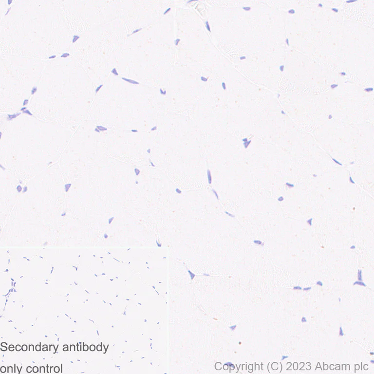
Immunohistochemistry (Formalin/PFA-fixed paraffin-embedded sections) - Anti-CD80 antibody [RM1133] (AB315832)
Immunohistochemical analysis of paraffin-embedded Human skeletal muscle tissue labeling CD80 with ab315832 at 1/200 (2.635 ug/ml) dilution, followed by a ready to use LeicaDS9800 (Bond™ Polymer Refine Detection).
Low expression tissue : nearly no staining on human skeletal muscle. The section was incubated with ab315832 for 30 mins at room temperature.
The immunostaining was performed on a Leica Biosystems BOND® RX instrument.
Counterstained with Hematoxylin.
Secondary antibody only control : Secondary antibody is a ready to use LeicaDS9800 (Bond™ Polymer Refine Detection).
Heat mediated antigen retrieval was performed with Tris-EDTA buffer (pH 9.0, Epitope Retrieval Solution2) for 20 mins.

- IHC-P
Supplier Data
Immunohistochemistry (Formalin/PFA-fixed paraffin-embedded sections) - Anti-CD80 antibody [RM1133] (AB315832)
Immunohistochemical analysis of paraffin-embedded Human tonsil tissue labeling CD80 with ab315832 at 1/200 (2.635 ug/ml) dilution, followed by a ready to use LeicaDS9800 (Bond™ Polymer Refine Detection).
Positive staining on the human tonsil. The section was incubated with ab315832 for 30 mins at room temperature.
The immunostaining was performed on a Leica Biosystems BOND® RX instrument.
Counterstained with Hematoxylin.
Secondary antibody only control : Secondary antibody is a ready to use LeicaDS9800 (Bond™ Polymer Refine Detection).
Heat mediated antigen retrieval was performed with Tris-EDTA buffer (pH 9.0, Epitope Retrieval Solution2) for 20 mins.
- IHC-P
Supplier Data
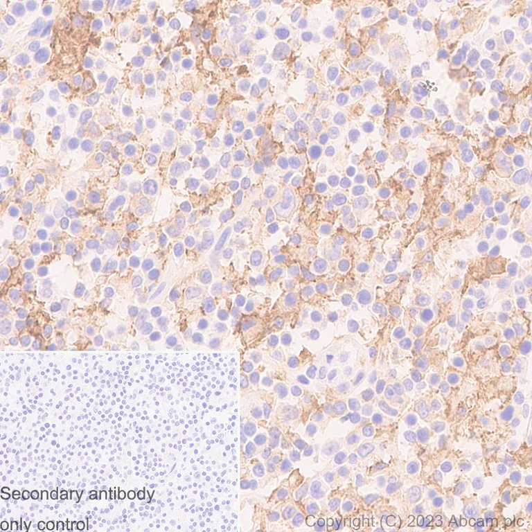
Immunohistochemistry (Formalin/PFA-fixed paraffin-embedded sections) - Anti-CD80 antibody [RM1133] (AB315832)
Immunohistochemical analysis of paraffin-embedded Human hodgkin's lymphoma tissue labeling CD80 with ab315832 at 1/200 (2.635 ug/ml) dilution, followed by a ready to use LeicaDS9800 (Bond™ Polymer Refine Detection).
Positive staining on the human hodgkin's lymphoma. The section was incubated with ab315832 for 30 mins at room temperature.
The immunostaining was performed on a Leica Biosystems BOND® RX instrument.
Counterstained with Hematoxylin.
Secondary antibody only control : Secondary antibody is a ready to use LeicaDS9800 (Bond™ Polymer Refine Detection).
Heat mediated antigen retrieval was performed with Tris-EDTA buffer (pH 9.0, Epitope Retrieval Solution2) for 20 mins.

- ICC/IF
Supplier Data
Immunocytochemistry/ Immunofluorescence - Anti-CD80 antibody [RM1133] (AB315832)
Immunofluorescent analysis of 4% Paraformaldehyde-fixed, 0.1% TritonX-100 permeabilized CD80 knock out Raji (CD80 knockout human Burkitt's lymphoma B lymphocyte cell line) cells labelling CD80 with ab315832 at 1/200 (2.635 ug/ml) dilution, followed by ab150081 Goat Anti-Rabbit IgG H&L (Alexa Fluor® 488) preadsorbed antibody at 1/1000 (2 ug/mL) dilution (Green).
Confocal image showing membranous staining in wild-type Raji cell line, and negative staining in CD80 knock out Raji cell line.
Image was taken with a confocal microscope(Leica-Microsystems, TCS SP8).
ab195889 Anti-alpha Tubulin mouse monoclonal antibody - Microtubule Marker (Alexa Fluor® 594) was used to counterstain tubulin at 1/200 (2.5ug/ml) dilution (Red). The Nuclear counterstain was DAPI (Blue).
Secondary antibody only control : Secondary antibody is ab150081 Goat Anti-Rabbit IgG H&L (Alexa Fluor® 488) preadsorbed at 1/1000 (2 ug/mL) dilution.
- Flow Cyt
Supplier Data
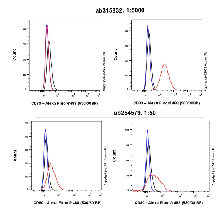
Flow Cytometry - Anti-CD80 antibody [RM1133] (AB315832)
Flow cytometric analysis of CD80 KO Raji (human CD80 knockout Burkitt's lymphoma B lymphocyte, Left) / Parental Raji (Right) cells labelling CD80 with ab315832 at 1/5000 dilution (0.01 ug)/Red compared with a Rabbit monoclonal IgG (ab172730) (Black) isotype control and an unlabelled control (cells without incubation with primary antibody and secondary antibody) (Blue).
Goat Anti-Rabbit IgG (Alexa Fluor® 488, ab150081) at 1/5000 dilution was used as the secondary antibody.
In flow cytometry, ab315832 (upper) shows positive staining on Raji and no staining on CD80 KO Raji, while ab254579 (lower) was shown non-specific binding CD80.
The upper images are applied with Anti-CD80 antibody (ab315832) at 1/5000 dilution and the lower images are applied with Anti-CD80 antibody (ab254579) at 1/50 dilution.
- IP
Supplier Data
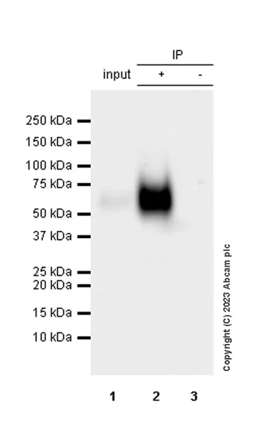
Immunoprecipitation - Anti-CD80 antibody [RM1133] (AB315832)
CD80 was immunoprecipitated from 0.35 mg Raji (human Burkitt's lymphoma B lymphocyte) whole cell lysate with ab315832 at 1/30 dilution (2ug in 0.35mg lysates). Western blot was performed on the immunoprecipitate using ab315832 at 1/1000 dilution. VeriBlot for IP secondary antibody(HRP)(ab131366) was used at 1/5000 dilution.
Blocking and diluting buffer and concentration : 5% NFDM/TBST.
All lanes:
Immunoprecipitation - Anti-CD80 antibody [RM1133] (ab315832) at 1/1000 dilution
Lane 1:
Raji (human Burkitt's lymphoma B lymphocyte) whole cell lysate
Lane 2:
ab315832 at 1/30 IP in Raji (human Burkitt's lymphoma B lymphocyte) whole cell lysate
Lane 3:
Rabbit monoclonal IgG (<a href='/en-us/products/primary-antibodies/rabbit-igg-monoclonal-epr25a-isotype-control-ab172730'>ab172730</a>) instead of ab315832 in Raji whole cell lysate
Secondary
All lanes:
Immunoprecipitation - VeriBlot for IP Detection Reagent (HRP) (<a href='/en-us/products/reagents/veriblot-for-ip-detection-reagent-hrp-ab131366'>ab131366</a>) at 1/5000 dilution
false
Exposure time: 180s

- WB
Supplier Data
Western blot - Anti-CD80 antibody [RM1133] (AB315832)
Blocking and diluting buffer and concentration : 5% NFDM/TBST.
Negative control : HUVEC (PMID : 15064090), 293T.
The molecular mass observed is consistent with what has been described in the literature (PMID : 7544014, PMID : 9079798); 55-60 kDa is due to glycosylation (PMID : 9654121).
This blot was developed using a high sensitivity ECL substrate (Lanes 6-7). The high-sensitivity ECL substrate used allows for the detection of proteins in the mid-femtogram range.
In Western blot, Anti-GAPDH antibody [EPR16891] - Loading Control (ab181602) staining at 1/200000 dilution.
All lanes:
Western blot - Anti-CD80 antibody [RM1133] (ab315832) at 1/1000 dilution
Lane 1:
Raji (human Burkitts lymphoma B lymphocyte) whole cell lysate at 20 µg
Lane 2:
Daudi (human Burkitts lymphoma lymphoblast) whole cell lysate at 20 µg
Lane 3:
HDLM-2 (human Hodgkin lymphoma cell) whole cell lysate at 20 µg
Lane 4:
HUVEC (human umbilical vein endothelial cell) whole cell lysate at 20 µg
Lane 5:
293T (human embryonic kidney epithelial cell) whole cell lysate at 20 µg
Secondary
All lanes:
Western blot - Goat Anti-Rabbit IgG H&L (HRP) (<a href='/en-us/products/secondary-antibodies/goat-rabbit-igg-h-l-hrp-ab97051'>ab97051</a>) at 1/20000 dilution
Observed band size: 55-60 kDa,36 kDa
true
Exposure time: 26s
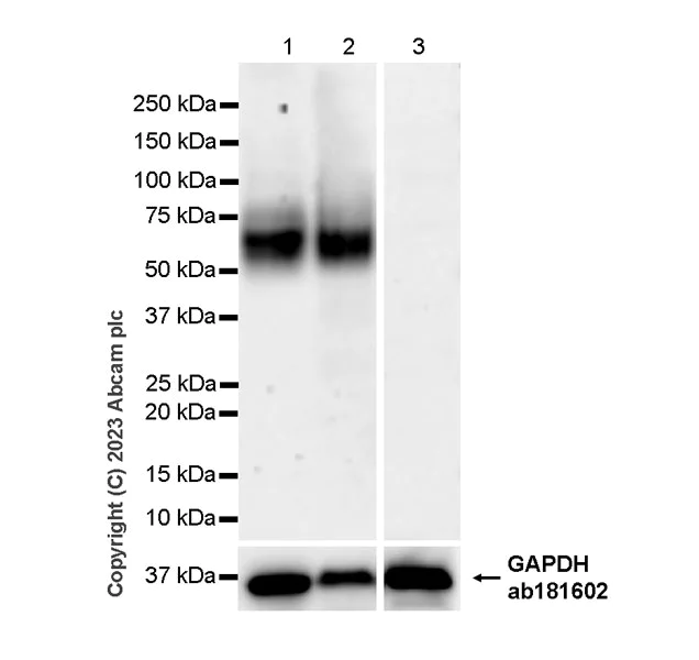
- WB
Supplier Data
Western blot - Anti-CD80 antibody [RM1133] (AB315832)
Low expression tissue : skeletal muscle.
Blocking and diluting buffer and concentration : 5% NFDM/TBST.
In Western blot, Anti-GAPDH antibody [EPR16891] - Loading Control (ab181602) staining at 1/200000 dilution.
All lanes:
Western blot - Anti-CD80 antibody [RM1133] (ab315832) at 1/1000 dilution
Lane 1:
Human tonsil tissue lysate at 20 µg
Lane 2:
Human spleen tissue lysate at 20 µg
Lane 3:
Human skeletal muscle tissue lysate at 20 µg
Secondary
All lanes:
Goat Anti-Rabbit IgG (HRP) with minimal cross-reactivity with human IgG at 1/1000 dilution
Observed band size: 55-60 kDa,36 kDa
false
Exposure time: 180s

- WB
Supplier Data
Western blot - Anti-CD80 antibody [RM1133] (AB315832)
Western blot : Anti-CD80 antibody (panel A : ab315832, panel B : ab254579) staining at 1/1000 dilution, shown in green; Mouse anti-GAPDH antibody [6C5](ab8245) loading control staining at 1/20000 dilution, shown in red.
In Western blot, ab315832 (panel A) was shown to bind specifically to CD80, while ab254579 (panel B) was shown non-specific binding CD80. Target of interest was observed at 55-60 kDa in wild-type Raji cell lysates (lane 1) with no signal observed at this size in CD80 knockout cell line (lane 2, knockout cell line ab273873/knockout cell lysate ab273827). To generate this image, samples were first run on an SDS-PAGE gel then transferred onto an immobilon-FL PVDF membrane. Membranes were blocked in a fluorescent western blot (TBS-based) blocking solution before incubation with primary antibodies overnight at 4 °C. Blots were washed in TBS-T, incubated with secondary antibodies Goat anti-Rabbit IgG H&L 800CW and Goat anti-Mouse IgG H&L 680RD at 1/20000 dilution for 1 h at room temperature, washed again then imaged.
All lanes:
Western blot - Anti-CD80 antibody [RM1133] (ab315832) at 1/1000 dilution
Lane 1:
Wild-type Raji (human Burkitts lymphoma B lymphocyte) whole cell lysate at 20 µg
Lane 2:
CD80 knockout Raji whole cell lysate at 20 µg
Secondary
All lanes:
Goat Anti-Rabbit IgG H&L (800CW) and Goat Anti-Mouse IgG H&L (680RD) at 1/20000 dilution
Observed band size: 55-60 kDa
false
Related conjugates and formulations (1)
-
Anti-CD80 antibody [RM1133] - BSA and Azide free
Complementary Products

Primary Antibodies
AB239075
20ul selling size|RabMAb|Recombinant|KO Validated

- 0
- 0

Primary Antibodies
AB182422
Anti-CD163 antibody [EPR19518]
BOND RX™ Validated|20ul selling size|RabMAb|Advanced Validation|Recombinant

- 5
- 17

Reactivity data
Product details
What are recombinant multiclonals?
Recombinant multiclonals are a mixture of recombinant antibodies co-expressed from a library of heavy and light chains. They offer several advantages including:
- - The sensitivity of polyclonal antibodies by recognising multiple epitopes
- - High batch-to-batch consistency and reproducibility
- - Improved sensitivity and specificity
- - Long-term security of supply
- - Animal-free batch production
View our range of recombinant multiclonal antibodies.
Patented technology
Our RabMAb® technology is a patented hybridoma-based technology for making rabbit monoclonal antibodies. For details on our patents, please refer to RabMAb® patents.
Properties and storage information
Form
Purification technique
Storage buffer
Shipped at conditions
Appropriate short-term storage duration
Appropriate short-term storage conditions
Appropriate long-term storage conditions
Aliquoting information
Storage information
Supplementary information
This supplementary information is collated from multiple sources and compiled automatically.
Biological function summary
CD80 plays a significant role in the immune system's ability to distinguish between self and non-self. It belongs to the B7 family and participates in the formation of immunological synapses between T cells and antigen-presenting cells. This interaction is essential for triggering T cell proliferation and cytokine production forming a part of the adaptive immune response. CD80's expression increases in response to pathogen recognition enhancing the immune system's efficiency.
Pathways
CD80 is integral to the regulation of T cell-mediated immune responses. It is involved in the CD28 co-stimulatory pathway which is vital for T cell activation and function. In this pathway CD80 interacts with proteins like CD28 and CTLA-4 to modulate immune activation and regulate immune tolerance. The proper function of CD80 within this pathway ensures a balanced immune reaction preventing autoimmunity while allowing effective defense against pathogens.
Product protocols
- Visit the General protocols
- Visit the Troubleshooting
Target data
Alternative Names
Publications (3)
Recent publications for all applications. Explore the full listand refine your search
Communications biology 8:1134 PubMed40739377
2025
Applications
Unspecified application
Species
Unspecified reactive species
Journal of nanobiotechnology 23:175 PubMed40050923
2025
Applications
Unspecified application
Species
Unspecified reactive species
Molecular medicine reports 31: PubMed39981936
2025
Applications
Unspecified application
Species
Unspecified reactive species
Product promise
Please note: All products are 'FOR RESEARCH USE ONLY. NOT FOR USE IN DIAGNOSTIC OR THERAPEUTIC PROCEDURES'.
For licensing inquiries, please contact partnerships@abcam.com