Anti-ESM1 antibody [RM1338]
- RabMAb
- Recombinant
- What is this?
Be the first to review this product! Submit a review
|
(0 Publication)
- ICC/IF
Supplier Data
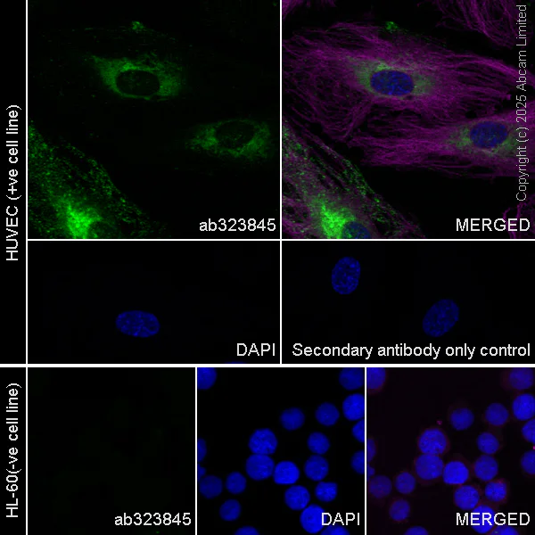
Immunocytochemistry/ Immunofluorescence - Anti-ESM1 antibody [RM1338] (AB323845)
Immunofluorescent analysis of 4% Paraformaldehyde-fixed, 0.1% TritonX-100 permeabilized HUVEC (human umbilical vein endothelial cell) cells labelling ESM1 with ab323845 at 1/500 (0.998 ug/ml) dilution, followed by ab150081 Goat Anti-Rabbit IgG H&L (Alexa Fluor® 488) preadsorbed antibody at 1/ab150081 1000 2ug/ml dilution (Green).
Confocal image showing cytoplasmic staining in HUVEC cell line (shown in green). The counterstain was observed in magenta. Nuclear DNA was labelled with DAPI (shown in blue).
Negative control : HL-60 (PMID : 8702785).
Image was taken with a confocal microscope (Leica-Microsystems, TCS SP8).
ab195889 Anti-alpha Tubulin mouse monoclonal antibody - Microtubule Marker (Alexa Fluor® 594) was used to counterstain tubulin at 1/ab195889 200 2.5ug/ml dilution (Magenta). The Nuclear counterstain was DAPI (Blue).
Secondary antibody only control : Secondary antibody is ab150081 Goat Anti-Rabbit IgG H&L (Alexa Fluor® 488) preadsorbed at 1/ab150081 1000 2ug/ml dilution.
- IP
Supplier Data
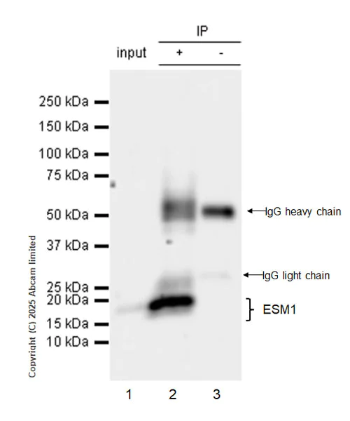
Immunoprecipitation - Anti-ESM1 antibody [RM1338] (AB323845)
ESM1 was immunoprecipitated from 0.35 mg HUVEC (human umbilical vein endothelial cell) whole cell lysate with ab323845 at 1/30 dilution (2ug in 0.35mg lysates). Western blot was performed on the immunoprecipitate using ab323845 at 1/1000 dilution.
Blocking and dilution buffer and concentration : 5% NFDM/TBST
All lanes:
Immunoprecipitation - Anti-ESM1 antibody [RM1338] (ab323845) at 1/1000 dilution
Lane 1:
HUVEC (human umbilical vein endothelial cell) whole cell lysate at 10 µg
Lane 2:
ab323845 at 1/30 IP in HUVEC (human umbilical vein endothelial cell) whole cell lysate
Lane 3:
Rabbit monoclonal IgG (<a href='/en-us/products/primary-antibodies/rabbit-igg-monoclonal-epr25a-isotype-control-ab172730'>ab172730</a>) instead of ab323845 in HUVEC whole cell lysate
Secondary
All lanes:
Immunoprecipitation - VeriBlot for IP Detection Reagent (HRP) (<a href='/en-us/products/reagents/veriblot-for-ip-detection-reagent-hrp-ab131366'>ab131366</a>) at 1/5000 dilution
Observed band size: 20 kDa
false
Exposure time: 15s
- ICC/IF
Supplier Data
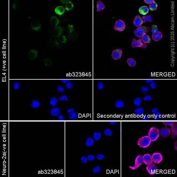
Immunocytochemistry/ Immunofluorescence - Anti-ESM1 antibody [RM1338] (AB323845)
Immunofluorescent analysis of 4% Paraformaldehyde-fixed, 0.1% TritonX-100 permeabilized EL4 (mouse lymphoma T lymphocyte) cells labelling ESM1 with ab323845 at 1/500 (0.998 ug/ml) dilution, followed by ab150081 Goat Anti-Rabbit IgG H&L (Alexa Fluor® 488) preadsorbed antibody at 1/ab150081 1000 2ug/ml dilution (Green).
Confocal image showing cytoplasmic staining in EL4 cell line (shown in green). The counterstain was observed in magenta. Nuclear DNA was labelled with DAPI (shown in blue).
Negative control : Neuro-2a.
Image was taken with a confocal microscope (Leica-Microsystems, TCS SP8).
ab195889 Anti-alpha Tubulin mouse monoclonal antibody - Microtubule Marker (Alexa Fluor® 594) was used to counterstain tubulin at 1/ab195889 200 2.5ug/ml dilution (Magenta). The Nuclear counterstain was DAPI (Blue).
Secondary antibody only control : Secondary antibody is ab150081 Goat Anti-Rabbit IgG H&L (Alexa Fluor® 488) preadsorbed at 1/ab150081 1000 2ug/ml dilution.
- IP
Supplier Data
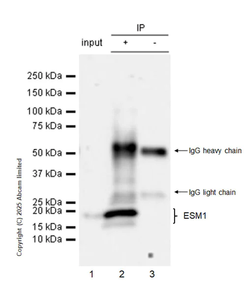
Immunoprecipitation - Anti-ESM1 antibody [RM1338] (AB323845)
ESM1 was immunoprecipitated from 0.35 mg EL4 (mouse lymphoma T lymphocyte) whole cell lysate with ab323845 at 1/30 dilution (2ug in 0.35mg lysates). Western blot was performed on the immunoprecipitate using ab323845 at 1/1000 dilution.
Blocking and dilution buffer and concentration : 5% NFDM/TBST
All lanes:
Immunoprecipitation - Anti-ESM1 antibody [RM1338] (ab323845) at 1/1000 dilution
Lane 1:
EL4 (mouse lymphoma T lymphocyte) whole cell lysate at 10 µg
Lane 2:
ab323845 at 1/30 IP in EL4 (mouse lymphoma T lymphocyte) whole cell lysate
Lane 3:
Rabbit monoclonal IgG (<a href='/en-us/products/primary-antibodies/rabbit-igg-monoclonal-epr25a-isotype-control-ab172730'>ab172730</a>) instead of ab323845 in EL4 whole cell lysate
Secondary
All lanes:
Immunoprecipitation - VeriBlot for IP Detection Reagent (HRP) (<a href='/en-us/products/reagents/veriblot-for-ip-detection-reagent-hrp-ab131366'>ab131366</a>) at 1/5000 dilution
Observed band size: 14 kDa,20 kDa
false
Exposure time: 32s
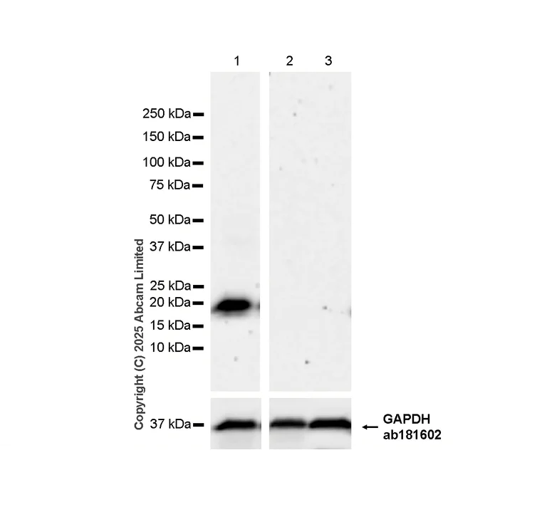
- WB
Supplier Data
Western blot - Anti-ESM1 antibody [RM1338] (AB323845)
Blocking and diluting buffer and concentration : 5% NFDM/TBST.
Negative control : A549, HL-60 (PMID : 8702785)
In Western blot, Anti-GAPDH antibody [EPR16891] - Loading Control (ab181602) staining at 1/200000 dilution.
All lanes:
Western blot - Anti-ESM1 antibody [RM1338] (ab323845) at 1/1000 dilution
Lane 1:
HUVEC (human umbilical vein endothelial cell) whole cell lysate at 40 µg
Lane 2:
A549 (human lung carcinoma epithelial cell) whole cell lysate at 40 µg
Lane 3:
HL-60 (human acute promyelocytic leukemia promyeloblast) whole cell lysate at 40 µg
Secondary
All lanes:
Western blot - Goat Anti-Rabbit IgG H&L (HRP) (<a href='/en-us/products/secondary-antibodies/goat-rabbit-igg-h-l-hrp-ab97051'>ab97051</a>) at 1/100000 dilution
Observed band size: 20 kDa,36 kDa
false
Exposure time: 103s
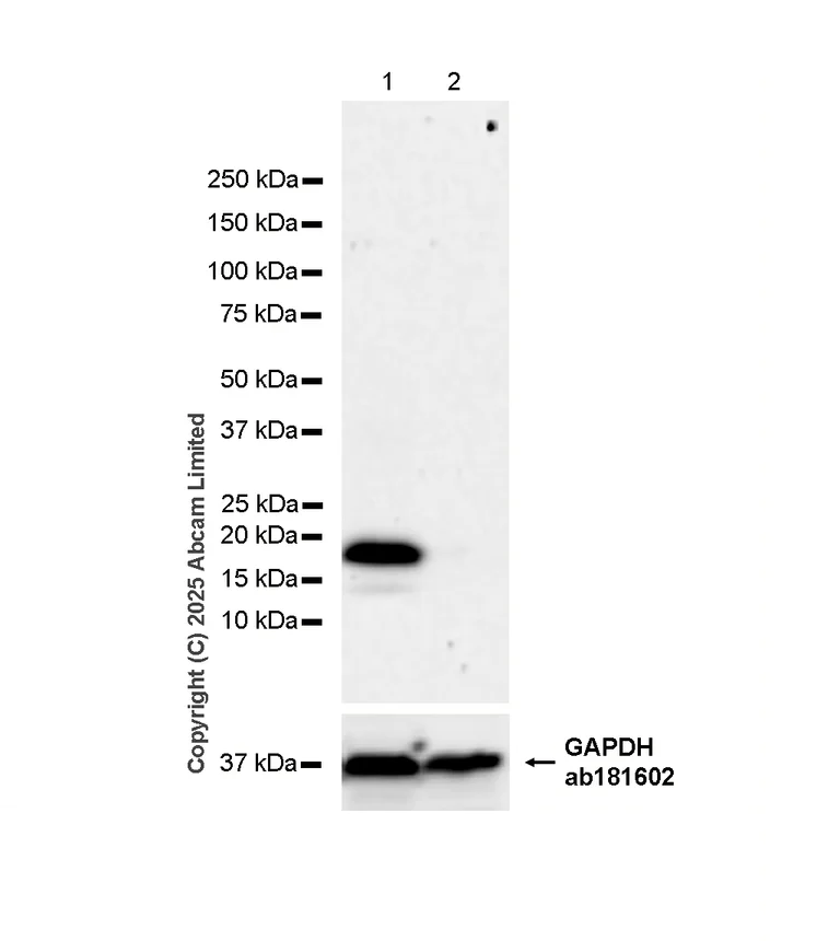
- WB
Supplier Data
Western blot - Anti-ESM1 antibody [RM1338] (AB323845)
Blocking and diluting buffer and concentration : 5% NFDM/TBST.
Negative control : Neuro-2a.
The expression profile/ molecular weight observed is consistent with what has been described in the literature (PMID : 8702785).
In Western blot, Anti-GAPDH antibody [EPR16891] - Loading Control (ab181602) staining at 1/200000 dilution.
All lanes:
Western blot - Anti-ESM1 antibody [RM1338] (ab323845) at 1/1000 dilution
Lane 1:
EL4 (mouse lymphoma T lymphocyte) whole cell lysate at 40 µg
Lane 2:
Neuro-2a (mouse neuroblastoma neuroblast) whole cell lysate at 40 µg
Secondary
All lanes:
Western blot - Goat Anti-Rabbit IgG H&L (HRP) (<a href='/en-us/products/secondary-antibodies/goat-rabbit-igg-h-l-hrp-ab97051'>ab97051</a>) at 1/100000 dilution
Observed band size: 14 kDa,20 kDa,36 kDa
false
Exposure time: 103s
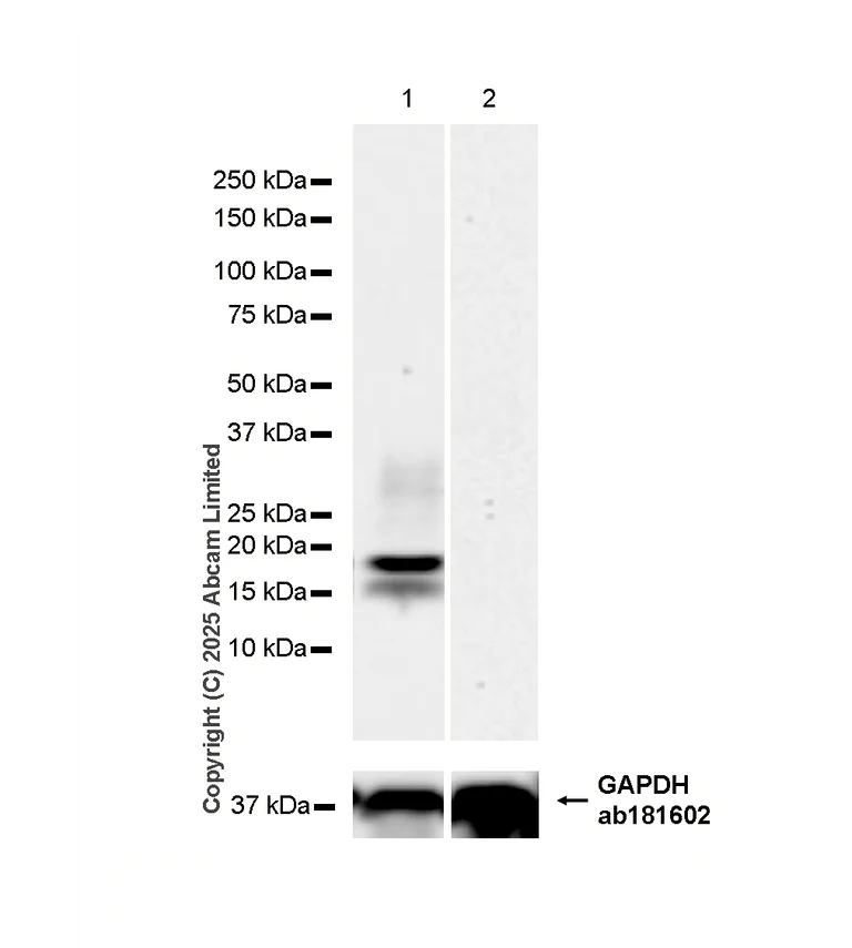
- WB
Supplier Data
Western blot - Anti-ESM1 antibody [RM1338] (AB323845)
Blocking and diluting buffer and concentration : 5% NFDM/TBST.
Negative control : skeletal muscle.
The expression profile/ molecular weight observed is consistent with what has been described in the literature (PMID : 8702785).
In Western blot, Anti-GAPDH antibody [EPR16891] - Loading Control (ab181602) staining at 1/200000 dilution.
All lanes:
Western blot - Anti-ESM1 antibody [RM1338] (ab323845) at 1/1000 dilution
Lane 1:
Mouse pituitary tissue lysate at 40 µg
Lane 2:
Mouse skeletal muscle tissue lysate at 40 µg
Secondary
All lanes:
Western blot - Goat Anti-Rabbit IgG H&L (HRP) (<a href='/en-us/products/secondary-antibodies/goat-rabbit-igg-h-l-hrp-ab97051'>ab97051</a>) at 1/100000 dilution
Observed band size: 14 kDa,20 kDa,36 kDa
false
Exposure time: 59s
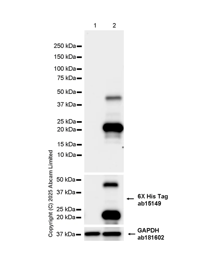
- WB
Supplier Data
Western blot - Anti-ESM1 antibody [RM1338] (AB323845)
Blocking and diluting buffer and concentration : 5% NFDM/TBST.
In Western blot, Anti-GAPDH antibody [EPR16891] - Loading Control (ab181602) staining at 1/200000 dilution.
In Western blot, Anti-6X His tag® antibody [AD1.1.10] (ab15149) staining at 1/1000 dilution.
All lanes:
Western blot - Anti-ESM1 antibody [RM1338] (ab323845) at 1/1000 dilution
Lane 1:
293T (human embryonic kidney epithelial cell) cells transfected with an empty vector containing a myc-His-tag®, whole cell lysate at 10 µg
Lane 2:
293T cells transfected with a human ESM1 expression vector containing a myc-His-tag®, whole cell lysate at 10 µg
Secondary
All lanes:
Western blot - Goat Anti-Rabbit IgG H&L (HRP) (<a href='/en-us/products/secondary-antibodies/goat-rabbit-igg-h-l-hrp-ab97051'>ab97051</a>) at 1/100000 dilution
Observed band size: 20 kDa,36 kDa
false
Exposure time: 26s
Complementary Products

Primary Antibodies
AB315238
Anti-VEGF Receptor 2 antibody [EPR28328-80]
BOND RX™ Validated|20ul selling size|RabMAb|Recombinant

- 5
- 2


Primary Antibodies
AB124964
Anti-alpha smooth muscle Actin antibody [EPR5368]
BOND RX™ Validated|RabMAb|Recombinant|KO Validated|20ul selling size

- 5
- 19
Reactivity data
Product details
Patented technology
Our RabMAb® technology is a patented hybridoma-based technology for making rabbit monoclonal antibodies. For details on our patents, please refer to RabMAb® patents.
What are recombinant multiclonals?
Recombinant multiclonals are a mixture of recombinant antibodies co-expressed from a library of heavy and light chains. They offer several advantages including:
- - The sensitivity of polyclonal antibodies by recognising multiple epitopes
- - High batch-to-batch consistency and reproducibility
- - Improved sensitivity and specificity
- - Long-term security of supply
- - Animal-free batch production
View our range of recombinant multiclonal antibodies.
Properties and storage information
Form
Purification technique
Storage buffer
Shipped at conditions
Appropriate short-term storage duration
Appropriate short-term storage conditions
Appropriate long-term storage conditions
Aliquoting information
Product protocols
- Visit the General protocols
- Visit the Troubleshooting
Target data
Alternative Names
Product promise
Please note: All products are 'FOR RESEARCH USE ONLY. NOT FOR USE IN DIAGNOSTIC OR THERAPEUTIC PROCEDURES'.
For licensing inquiries, please contact partnerships@abcam.com