Anti-GABA A Receptor alpha 6 antibody [EPR25322-129]
- BOND RX™ Validated
- 20ul selling size
- RabMAb
- Advanced Validation
- Recombinant
- What is this?
Be the first to review this product! Submit a review
|
(2 Publications )

- mIHC
Lab
Multiplex immunohistochemistry - Anti-GABA A Receptor alpha 6 antibody [EPR25322-129] (AB300069)
Multiplex immunohistochemistry analysis of formalin/PFA-fixed paraffin-embedded Human cerebellum staining of Calbindin with ab108404 at a 1/1000 dilution, DGKZ/DGK-zeta with ab239081 at 1/5000 dilution, and GABA A Receptor alpha 6 with ab300069 at a 1/500 dilution, followed by a ready to use secondary antibody Opal Polymer HRP Ms + Rb.
Panel A : merged staining of anti-GABA A Receptor alpha 6 (gray; Opal™690), anti-DGKZ/DGK-zeta (green; Opal™520) and anti-Calbindin (magenta; Opal™570) on human cerebellum.
Panel B : anti-Calbindin stained on all layers of cerebellum.
Panel C : anti-DGKZ/DGK-zeta stained on Purkinje cells.
Panel D : anti-GABA A Receptor alpha 6 stained on the granular layer.
Nuclear DNA was labeled with DAPI (shown in blue).
The section was incubated in three rounds of staining : in the order of ab300069, ab239081, and ab108404 for 30 mins at room temperature. Each round was followed by a separate fluorescent tyramide signal amplification system.
Heat mediated antigen retrieval with Tris-EDTA buffer (pH 9.0, epitope retrieval solution2) for 20 mins.
The immunostaining was performed on a Leica Biosystems BOND® RX instrument with an Opal™ 4-color kit. Image acquisition was performed with Leica SP8 confocal microscope.
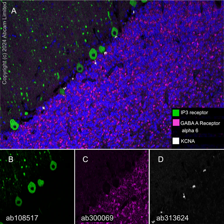
- mIHC
Lab
Multiplex immunohistochemistry - Anti-GABA A Receptor alpha 6 antibody [EPR25322-129] (AB300069)
Multiplex immunohistochemistry analysis of formalin/PFA-fixed paraffin-embedded human cerebellum tissue staining IP3 receptor with ab108517 at a 1/1000 dilution.
Panel A : merged staining of anti-IP3 receptor (green; Opal™520), anti-GABA A Receptor alpha 6 (magenta; Opal™690) and anti-KCNA (gray; Opal™570) on human cerebellum.
Panel B : anti-IP3 receptor staining Purkinje cells in human cerebellum.
Panel C : anti-GABA A Receptor alpha 6 staining granule cells in human cerebellum.
Panel D : anti-KCNA staining basket cells in human cerebellum.
Nuclear DNA was labeled with DAPI (shown in blue).
The section was incubated in three rounds of staining : in the order of ab108517, ab300069 and ab313624 for 30 mins at room temperature. Each round was followed by a separate fluorescent tyramide signal amplification system.
The immunostaining was performed on a Leica Biosystems BOND® RX instrument with an Opal™ 4-color kit using an Opal Polymer HRP Ms + Rb secondary antibody (ready to use). Image acquisition was performed with Leica SP8 confocal microscope.
Heat mediated antigen retrieval was performed with Tris-EDTA buffer (pH 9.0, epitope retrieval solution2) for 20 mins.

- IHC-P
Supplier Data
Immunohistochemistry (Formalin/PFA-fixed paraffin-embedded sections) - Anti-GABA A Receptor alpha 6 antibody [EPR25322-129] (AB300069)
Immunohistochemical analysis of paraffin-embedded Human cerebellum tissue labelling GABA A Receptor alpha 6 with ab300069 at 1/500 dilution (1.244 μg/ml) followed by a ready to use LeicaDS9800 (BOND™ Polymer Refine Detection) was used. Positive staining on human cerebellum. The section was incubated with ab300069 for 30 mins at room temperature. The immunostaining was performed on a Leica Biosystems BOND® RX instrument. Counterstained with Hematoxylin.
Secondary antibody only control : Secondary antibody is a ready to use LeicaDS9800 (BOND™ Polymer Refine Detection).
Heat mediated antigen retrieval with Tris-EDTA buffer (pH 9.0, epitope retrieval solution2) for 20 mins.

- mIHC
Lab
Multiplex immunohistochemistry - Anti-GABA A Receptor alpha 6 antibody [EPR25322-129] (AB300069)
Fluorescence multiplex immunohistochemical analysis of mouse cerebellum (formalin-fixed paraffin-embedded section).
Panel A : merged staining of anti-GABRA6 (magenta; Opal™690), anti-KCNA (green; Opal™520) and anti-DGKZ (gray; Opal™570) on mouse cerebellum.
Panel B : anti-GABRA6 staining granule cells in mouse cerebellum.
Panel C : anti-KCNA staining basket cells in mouse cerebellum.
Panel D : anti-DGKZ staining Purkinje cells in mouse cerebellum.
Nuclear DNA was labeled with DAPI (shown in blue).
The section was incubated in three rounds of staining : in the order of ab300069 at a 1/500 dilution, ab313624 at a 1/5000 and ab239080 at a 1/5000 dilution for 30 mins at room temperature. Each round was followed by a separate fluorescent tyramide signal amplification system.
Heat mediated antigen retrieval was performed with Tris-EDTA buffer (pH 9.0, epitope retrieval solution2) for 20 mins.
The immunostaining was performed on a Leica Biosystems BOND® RX instrument with an Opal™ 4-color kit. Image acquisition was performed with Leica SP8 confocal microscope.
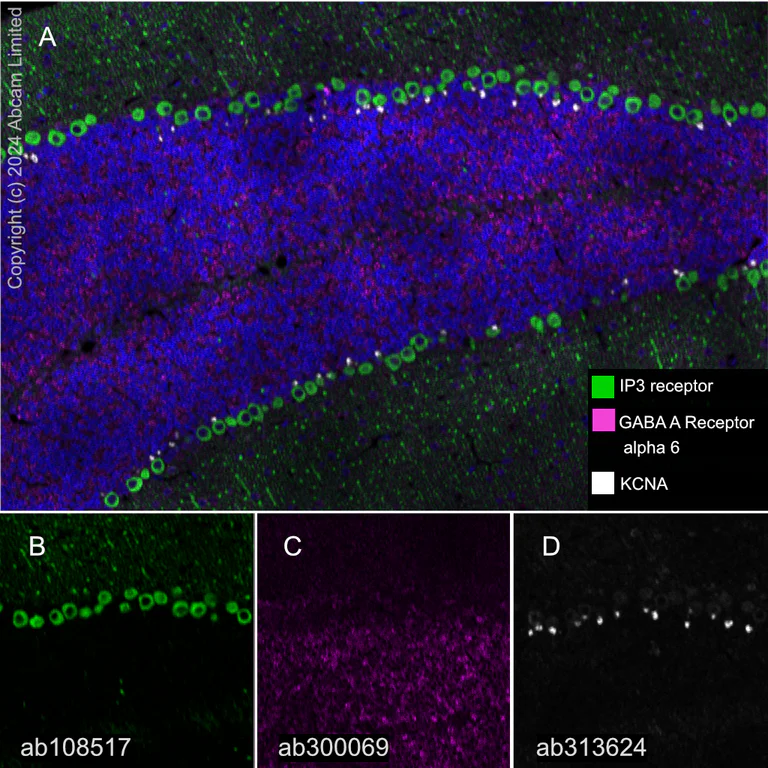
- mIHC
Lab
Multiplex immunohistochemistry - Anti-GABA A Receptor alpha 6 antibody [EPR25322-129] (AB300069)
Multiplex immunohistochemistry analysis of formalin/PFA-fixed paraffin-embedded mouse cerebellum tissue staining IP3 receptor with ab108517 at a 1/1000 dilution.
Panel A : merged staining of anti-IP3 receptor (green; Opal™520), anti-GABA A Receptor alpha 6 (magenta; Opal™690) and anti-KCNA (gray; Opal™570) on mouse cerebellum.
Panel B : anti-IP3 receptor staining Purkinje cells in mouse cerebellum.
Panel C : anti-GABA A Receptor alpha 6 staining granule cells in mouse cerebellum.
Panel D : anti-KCNA staining basket cells in mouse cerebellum.
Nuclear DNA was labeled with DAPI (shown in blue).
The section was incubated in three rounds of staining : in the order of ab108517, ab300069 and ab313624 for 30 mins at room temperature. Each round was followed by a separate fluorescent tyramide signal amplification system.
The immunostaining was performed on a Leica Biosystems BOND® RX instrument with an Opal™ 4-color kit using an Opal Polymer HRP Ms + Rb secondary antibody (ready to use). Image acquisition was performed with Leica SP8 confocal microscope.
Heat mediated antigen retrieval was performed with Tris-EDTA buffer (pH 9.0, epitope retrieval solution2) for 20 mins.
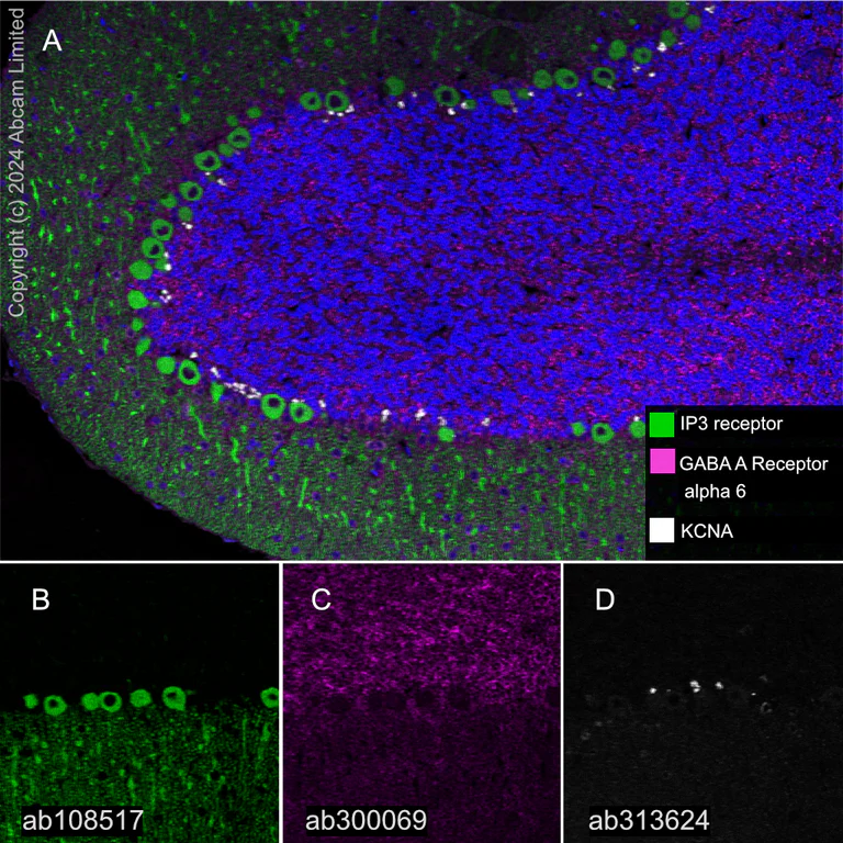
- mIHC
Lab
Multiplex immunohistochemistry - Anti-GABA A Receptor alpha 6 antibody [EPR25322-129] (AB300069)
Multiplex immunohistochemistry analysis of formalin/PFA-fixed paraffin-embedded rat cerebellum tissue staining IP3 receptor with ab108517 at a 1/1000 dilution.
Panel A : merged staining of anti-IP3 receptor (green; Opal™520), anti-GABA A Receptor alpha 6 (magenta; Opal™690) and anti-KCNA (gray; Opal™570) on rat cerebellum.
Panel B : anti-IP3 receptor staining Purkinje cells in rat cerebellum.
Panel C : anti-GABA A Receptor alpha 6 staining granule cells in rat cerebellum.
Panel D : anti-KCNA staining basket cells in rat cerebellum.
Nuclear DNA was labeled with DAPI (shown in blue).
The section was incubated in three rounds of staining : in the order of ab108517, ab300069 and ab313624 for 30 mins at room temperature. Each round was followed by a separate fluorescent tyramide signal amplification system.
The immunostaining was performed on a Leica Biosystems BOND® RX instrument with an Opal™ 4-color kit using an Opal Polymer HRP Ms + Rb secondary antibody (ready to use). Image acquisition was performed with Leica SP8 confocal microscope.
Heat mediated antigen retrieval was performed with Tris-EDTA buffer (pH 9.0, epitope retrieval solution2) for 20 mins.
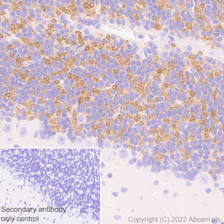
- IHC-P
Supplier Data
Immunohistochemistry (Formalin/PFA-fixed paraffin-embedded sections) - Anti-GABA A Receptor alpha 6 antibody [EPR25322-129] (AB300069)
Immunohistochemical analysis of paraffin-embedded Mouse cerebellum tissue labelling GABA A Receptor alpha 6 with ab300069 at 1/500 dilution (1.244 μg/ml) followed by a ready to use LeicaDS9800 (BOND™ Polymer Refine Detection). Positive staining on mouse cerebellum. The section was incubated with ab300069 for 30 mins at room temperature. The immunostaining was performed on a Leica Biosystems BOND® RX instrument. Counterstained with Hematoxylin.
Secondary antibody only control : Secondary antibody is a ready to use LeicaDS9800 (BOND™ Polymer Refine Detection).
Heat mediated antigen retrieval with Tris-EDTA buffer (pH 9.0, epitope retrieval solution2) for 20 mins.
- IHC-P
Supplier Data
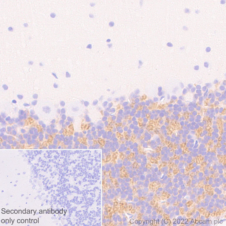
Immunohistochemistry (Formalin/PFA-fixed paraffin-embedded sections) - Anti-GABA A Receptor alpha 6 antibody [EPR25322-129] (AB300069)
Immunohistochemical analysis of paraffin-embedded Rat cerebellum tissue labelling GABA A Receptor alpha 6 with ab300069 at 1/500 dilution (1.244 μg/ml) followed by a ready to use LeicaDS9800 (BOND™ Polymer Refine Detection). Positive staining on rat cerebellum. The section was incubated with ab300069 for 30 mins at room temperature. The immunostaining was performed on a Leica Biosystems BOND® RX instrument. Counterstained with Hematoxylin.
Secondary antibody only control : Secondary antibody is a ready to use LeicaDS9800 (BOND™ Polymer Refine Detection).
Heat mediated antigen retrieval with Tris-EDTA buffer (pH 9.0, epitope retrieval solution2) for 20 mins.
- IHC-Fr
Supplier Data
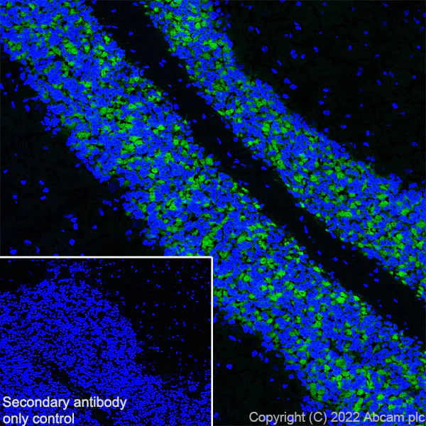
Immunohistochemistry (Frozen sections) - Anti-GABA A Receptor alpha 6 antibody [EPR25322-129] (AB300069)
Immunohistochemical analysis of 4% PFA-fixed, 0.2% Triton X-100 permeabilized frozen Rat cerebellum (fresh) tissue labeling GABA A Receptor alpha 6 with ab300069 at 1/500 (1.244 μg/ml) dilution followed by ab150081 Goat Anti-Rabbit IgG H&L (Alexa Fluor® 488) preadsorbed at 1/1000 (2 μg/mL) dilution (Green). Positive staining on rat cerebellum is observed. The nuclear counterstain was DAPI (Blue).
Secondary antibody control : Secondary antibody is ab150081 Goat Anti-Rabbit IgG H&L (Alexa Fluor® 488) preadsorbedat 1/1000 (2 μ/mL) dilution.
- IHC-Fr
Supplier Data
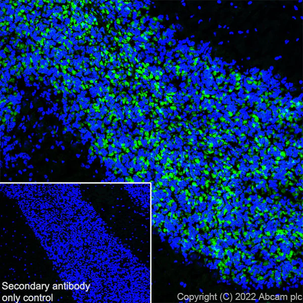
Immunohistochemistry (Frozen sections) - Anti-GABA A Receptor alpha 6 antibody [EPR25322-129] (AB300069)
Immunohistochemical analysis of 4% PFA-fixed, 0.2% Triton X-100 permeabilized frozen Mouse cerebellum (fresh) tissue labeling GABA A Receptor alpha 6 with ab300069 at 1/500 (1.244 μg/ml) dilution followed by ab150081 Goat Anti-Rabbit IgG H&L (Alexa Fluor® 488) preadsorbed at 1/1000 2 μg/mL dilution (Green). Positive staining on mouse cerebellum is observed. The nuclear counterstain was DAPI (Blue).
Secondary antibody control : Secondary antibody is ab150081 Goat Anti-Rabbit IgG H&L (Alexa Fluor® 488) preadsorbedat 1/1000 2 μ/mL dilution.
- WB
Supplier Data
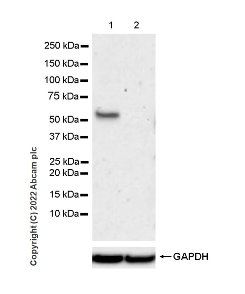
Western blot - Anti-GABA A Receptor alpha 6 antibody [EPR25322-129] (AB300069)
5% NFDM/TBST was used as blocking and diluting buffer.
Low expression:Heart (PMID : 29467616).
All lanes:
Western blot - Anti-GABA A Receptor alpha 6 antibody [EPR25322-129] (ab300069) at 1/1000 dilution
Lane 1:
Human hypothalamus tissue lysate at 20 µg
Lane 2:
Human heart tissue lysate at 20 µg
Secondary
All lanes:
Western blot - VeriBlot for IP Detection Reagent (HRP) (<a href='/en-us/products/reagents/veriblot-for-ip-detection-reagent-hrp-ab131366'>ab131366</a>) at 1/1000 dilution
Predicted band size: 51 kDa
Observed band size: 57 kDa
false
Exposure time: 180s
- WB
Lab
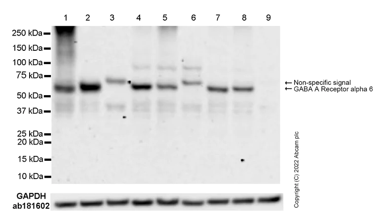
Western blot - Anti-GABA A Receptor alpha 6 antibody [EPR25322-129] (AB300069)
Blocking and diluting buffer and concentration : 5% NFDM/TBST.
Low expression tissue : heart (PMID : 29467616).
This blot was developed using a high ECL substrate.
Sometimes a non-specific band near the target signal can be observed.
All lanes:
Western blot - Anti-GABA A Receptor alpha 6 antibody [EPR25322-129] (ab300069) at 1/1000 dilution
Lane 1:
Human cerebellum tissue lysate at 20 µg
Lane 2:
Human hypothalamus tissue lysate at 20 µg
Lane 3:
Human heart tissue lysate at 20 µg
Lane 4:
Mouse brain tissue lysate at 20 µg
Lane 5:
Mouse cerebellum tissue lysate at 20 µg
Lane 6:
Mouse heart tissue lysate at 20 µg
Lane 7:
Rat brain tissue lysate at 20 µg
Lane 8:
Rat cerebellum tissue lysate at 20 µg
Lane 9:
Rat heart tissue lysate at 20 µg
Secondary
All lanes:
Goat Anti-Rabbit IgG (HRP), minimal cross-reactivity with human IgG at 1/2000 dilution
Predicted band size: 51 kDa
Observed band size: 57 kDa
false
Exposure time: 180s
- WB
Supplier Data
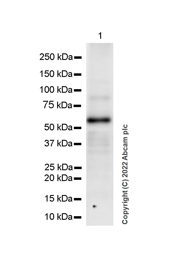
Western blot - Anti-GABA A Receptor alpha 6 antibody [EPR25322-129] (AB300069)
This data was developed using ab300069, the same antibody clone in a different buffer formulation.
5% NFDM/TBST was used as blocking and diluting buffer.
All lanes:
Western blot - Anti-GABA A Receptor alpha 6 antibody [EPR25322-129] (ab300069) at 1/1000 dilution
All lanes:
Mouse brain tissue lysate at 20 µg
Secondary
All lanes:
Western blot - Goat Anti-Rabbit IgG H&L (HRP) (<a href='/en-us/products/secondary-antibodies/goat-rabbit-igg-h-l-hrp-ab97051'>ab97051</a>) at 1/20000 dilution
Predicted band size: 51 kDa
Observed band size: 57 kDa
false
Exposure time: 59s
- WB
Supplier Data
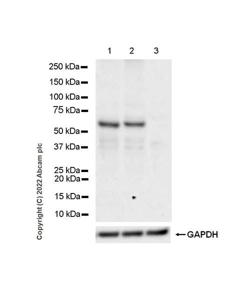
Western blot - Anti-GABA A Receptor alpha 6 antibody [EPR25322-129] (AB300069)
Blocking and diluting buffer and concentration : 5% NFDM/TBST.
Low expression : Heart (PMID : 29467616)
This blot was developed using a high sensitivity ECL substrate. The high-sensitivity ECL substrate used allows for the detection of proteins in the mid-femtogram range.
All lanes:
Western blot - Anti-GABA A Receptor alpha 6 antibody [EPR25322-129] (ab300069) at 1/1000 dilution
Lane 1:
Rat brain tissue lysate at 20 µg
Lane 2:
Rat cerebellum tissue lysate at 20 µg
Lane 3:
Rat heart tissue lysate at 20 µg
Secondary
All lanes:
Goat Anti-Rabbit IgG (HRP) with minimal cross-reactivity with human IgG (Merck DC03L) at 1/2000 dilution
Predicted band size: 51 kDa
Observed band size: 57 kDa
true
Exposure time: 180s
Related conjugates and formulations (1)
-
Anti-GABA A Receptor alpha 6 antibody [EPR25322-129] - BSA and Azide free
Complementary Products

Primary Antibodies
AB75838
Anti-GABA B Receptor 2/GABBR2 antibody [EP2411Y]
RabMAb|Recombinant|20ul selling size

- 5
- 4


Reactivity data
Product details
Patented technology
Our RabMAb® technology is a patented hybridoma-based technology for making rabbit monoclonal antibodies. For details on our patents, please refer to RabMAb® patents.
What are the advantages of a recombinant monoclonal antibody?
This product is a recombinant monoclonal antibody, which offers several advantages including:
- - High batch-to-batch consistency and reproducibility
- - Improved sensitivity and specificity
- - Long-term security of supply
- - Animal-free batch production
For more information, read more on recombinant antibodies.
Properties and storage information
Form
Purification technique
Storage buffer
Shipped at conditions
Appropriate short-term storage duration
Appropriate short-term storage conditions
Appropriate long-term storage conditions
Aliquoting information
Storage information
Product protocols
- Visit the General protocols
- Visit the Troubleshooting
Target data
Alternative Names
Publications (2)
Recent publications for all applications. Explore the full listand refine your search
Molecular medicine (Cambridge, Mass.) 31:242 PubMed40537751
2025
Applications
Unspecified application
Species
Unspecified reactive species
International journal of molecular sciences 24: PubMed38069205
2023
Applications
Unspecified application
Species
Unspecified reactive species
Product promise
Please note: All products are 'FOR RESEARCH USE ONLY. NOT FOR USE IN DIAGNOSTIC OR THERAPEUTIC PROCEDURES'.
For licensing inquiries, please contact partnerships@abcam.com