Anti-GIRK2 antibody [EPR23841-83] - BSA and Azide free
- BOND RX™ Validated
- RabMAb
- Recombinant
- What is this?
Be the first to review this product! Submit a review
|
(0 Publication)
- IHC-P
Supplier Data
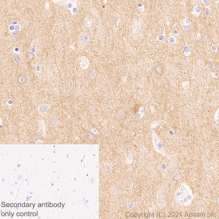
Immunohistochemistry (Formalin/PFA-fixed paraffin-embedded sections) - Anti-GIRK2 antibody [EPR23841-83] - BSA and Azide free (AB281831)
This data was developed using ab259909, the same antibody clone in a different buffer formulation.
Immunohistochemical analysis of paraffin-embedded human cerebrum tissue labelling GIRK2 with ab259909 at 1/200 (2.345 μg/ml) dilution followed by ready to use LeicaDS9800 (Bond™ Polymer Refine Detection). The section was incubated with ab259909 for 30 mins at room temperature. The immunostaining was performed on a Leica Biosystems BOND® RX instrument. Counterstained with Hematoxylin.
Cytoplasmic staining on human cerebrum (PMID : 8642402).
Secondary antibody only control : Secondary antibody is ready to use LeicaDS9800 (Bond™ Polymer Refine Detection).
Heat mediated antigen retrieval with Tris-EDTA buffer (pH 9.0, epitope retrieval solution2) for 20 mins.
- IHC-Fr
Lab
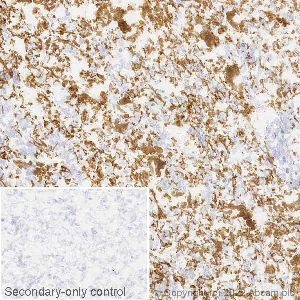
Immunohistochemistry (Frozen sections) - Anti-GIRK2 antibody [EPR23841-83] - BSA and Azide free (AB281831)
IHC image of GIRK2 staining in a section of frozen human normal substantia nigra performed on a Leica Biosystems BOND® RX instrument using the standard protocol. The section was fixed in 10% paraformaldehyde (10 min) prior to staining. The section was incubated with ab259909, 0.05 μg/ml, for 15 mins at room temperature and detected using an HRP conjugated compact polymer system. DAB was used as the chromogen. The section was then counterstained with haematoxylin and mounted with DPX. The inset secondary-only control image is taken from an identical assay without primary antibody. For other IHC staining systems (automated and non-automated) customers should optimize variable parameters such as antigen retrieval conditions, primary antibody concentration and antibody incubation times. Tissue obtained from the Human Research Tissue Bank, supported by the NIHR Cambridge Biomedical Research Centre. This data was developed using ab259909, the same antibody clone in a different buffer formulation.
- IHC-P
Supplier Data
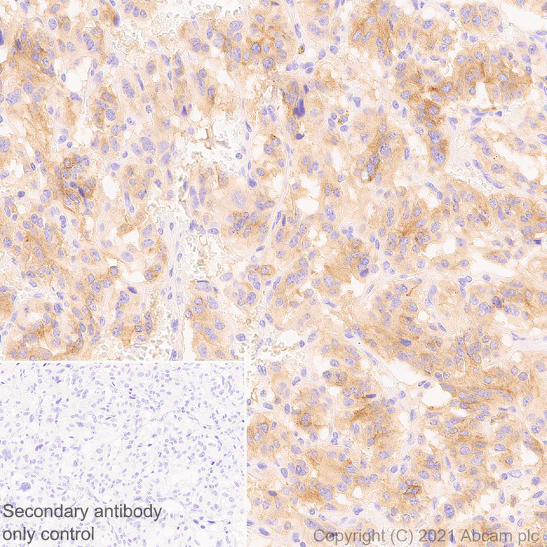
Immunohistochemistry (Formalin/PFA-fixed paraffin-embedded sections) - Anti-GIRK2 antibody [EPR23841-83] - BSA and Azide free (AB281831)
This data was developed using ab259909, the same antibody clone in a different buffer formulation.
Immunohistochemical analysis of paraffin-embedded human insulinoma tissue labelling GIRK2 with ab259909 at 1/200 (2.345 μg/ml) dilution followed by ready to use LeicaDS9800 (Bond™ Polymer Refine Detection). The section was incubated with ab259909 for 30 mins at room temperature. The immunostaining was performed on a Leica Biosystems BOND® RX instrument. Counterstained with Hematoxylin.
Cytoplasmic staining on human insulinoma (PMID : 7592809).
Secondary antibody only control : Secondary antibody is ready to use LeicaDS9800 (Bond™ Polymer Refine Detection).
Heat mediated antigen retrieval with Tris-EDTA buffer (pH 9.0, epitope retrieval solution2) for 20 mins.
- IHC-Fr
Lab
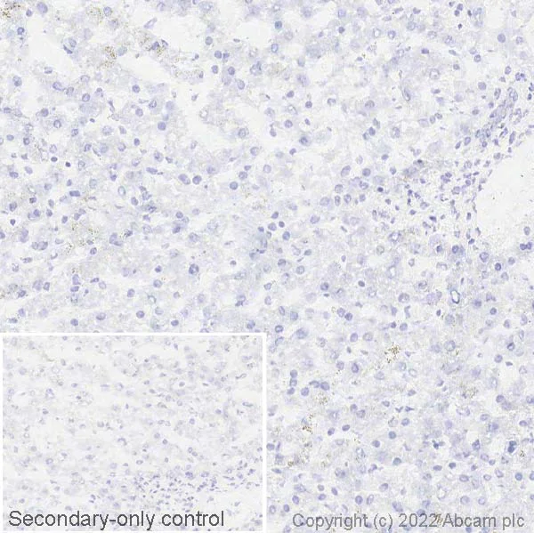
Immunohistochemistry (Frozen sections) - Anti-GIRK2 antibody [EPR23841-83] - BSA and Azide free (AB281831)
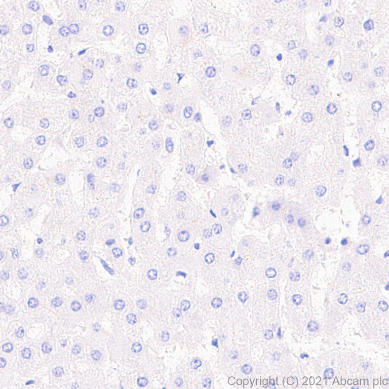
Negative tissue control image : IHC image of GIRK2 staining in a section of frozen human normal liver performed on a Leica Biosystems BOND® RX instrument using the standard protocol. The section was fixed in 10% paraformaldehyde (10 min) prior to staining. The section was incubated with ab259909, 0.05 μg/ml, for 15 mins at room temperature and detected using an HRP conjugated compact polymer system. DAB was used as the chromogen. The section was then counterstained with haematoxylin and mounted with DPX. The inset secondary-only control image is taken from an identical assay without primary antibody. For other IHC staining systems (automated and non-automated) customers should optimize variable parameters such as antigen retrieval conditions, primary antibody concentration and antibody incubation times. Tissue obtained from the Human Research Tissue Bank, supported by the NIHR Cambridge Biomedical Research Centre. This data was developed using ab259909, the same antibody clone in a different buffer formulation.
- IHC-P
Supplier Data
Immunohistochemistry (Formalin/PFA-fixed paraffin-embedded sections) - Anti-GIRK2 antibody [EPR23841-83] - BSA and Azide free (AB281831)
This data was developed using ab259909, the same antibody clone in a different buffer formulation.
Immunohistochemical analysis of paraffin-embedded human liver tissue labelling GIRK2 with ab259909 at 1/200 (2.345 μg/ml) dilution followed by ready to use LeicaDS9800 (Bond™ Polymer Refine Detection). The section was incubated with ab259909 for 30 mins at room temperature. The immunostaining was performed on a Leica Biosystems BOND® RX instrument. Counterstained with Hematoxylin.
Negative control : No staining on human liver (PMID : 7592809).
Secondary antibody only control : Secondary antibody is ready to use LeicaDS9800 (Bond™ Polymer Refine Detection).
Heat mediated antigen retrieval with Tris-EDTA buffer (pH 9.0, epitope retrieval solution2) for 20 mins.
- IHC-Fr
Supplier Data
Immunohistochemistry (Frozen sections) - Anti-GIRK2 antibody [EPR23841-83] - BSA and Azide free (AB281831)
This data was developed using ab259909, the same antibody clone in a different buffer formulation.
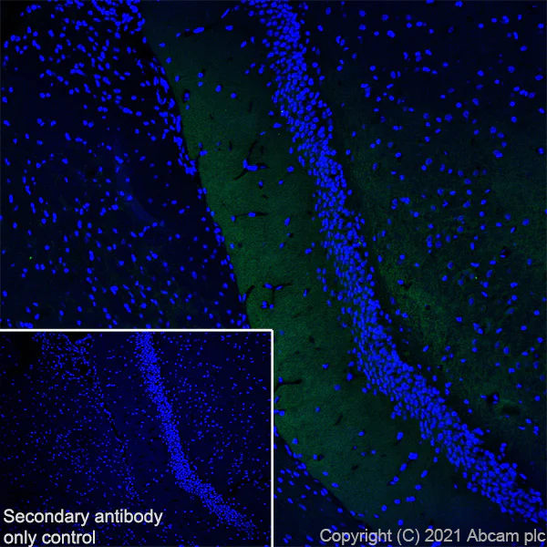
Immunohistochemical analysis of 4% PFA-fixed, 0.2% Triton X-100 permeabilized frozen rat hippocampus tissue labeling GIRK2 with ab259909 at 1/100 (4.69 μg/ml) dilution followed by ab150077 Goat Anti-Rabbit IgG H&L (Alexa Fluor® 488) at 1/1000 dilution (Green). The nuclear counterstain was DAPI (Blue).
Cytoplasmic staining on rat hippocampus (PMID : 8929423) is observed.
Secondary antibody control : Secondary antibody is ab150077 Goat Anti-Rabbit IgG H&L (Alexa Fluor® 488) at 1/1000 dilution.
Heat mediated antigen retrieval using sodium citrate buffer (10mM citrate pH 6.0 + 0.05% Tween-20).
- IHC-P
Supplier Data
Immunohistochemistry (Formalin/PFA-fixed paraffin-embedded sections) - Anti-GIRK2 antibody [EPR23841-83] - BSA and Azide free (AB281831)
This data was developed using ab259909, the same antibody clone in a different buffer formulation.
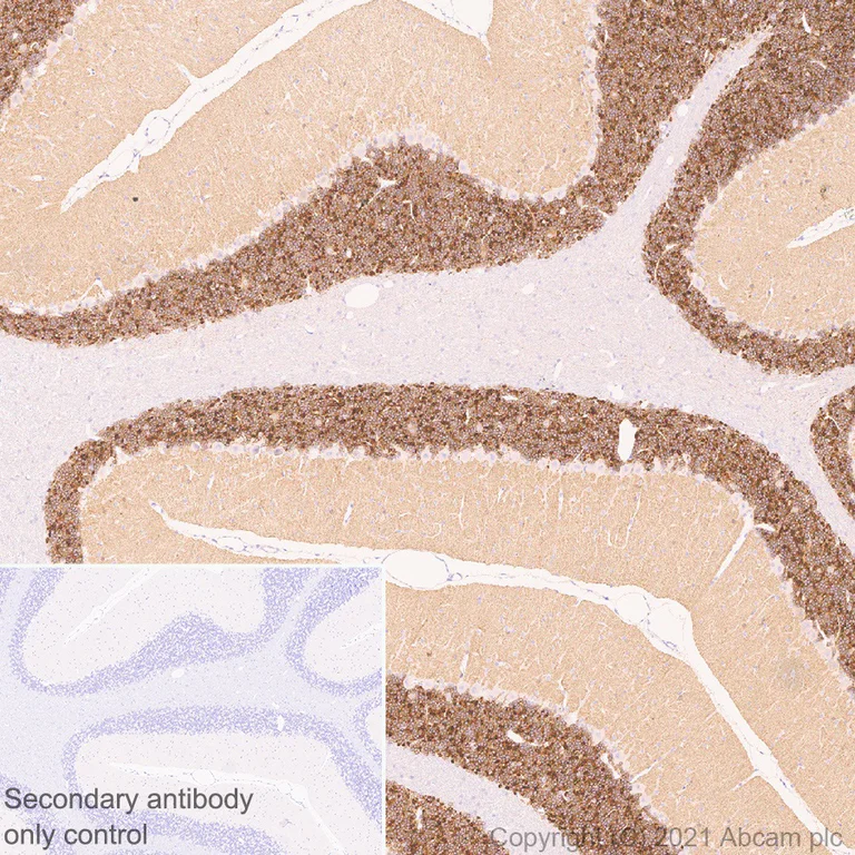
Immunohistochemical analysis of paraffin-embedded rat cerebellum tissue labelling GIRK2 with ab259909 at 1/200 (2.345 μg/ml) dilution followed by ready to use LeicaDS9800 (Bond™ Polymer Refine Detection). The section was incubated with ab259909 for 30 mins at room temperature. The immunostaining was performed on a Leica Biosystems BOND® RX instrument. Counterstained with Hematoxylin.
Postive staining predominantly on granular layer of rat cerebellum (PMID : 8642402, PMID : 9023358).
Secondary antibody only control : Secondary antibody is ready to use LeicaDS9800 (Bond™ Polymer Refine Detection).
Heat mediated antigen retrieval with Tris-EDTA buffer (pH 9.0, epitope retrieval solution2) for 20 mins.
- IHC-Fr
Supplier Data
Immunohistochemistry (Frozen sections) - Anti-GIRK2 antibody [EPR23841-83] - BSA and Azide free (AB281831)
This data was developed using ab259909, the same antibody clone in a different buffer formulation.
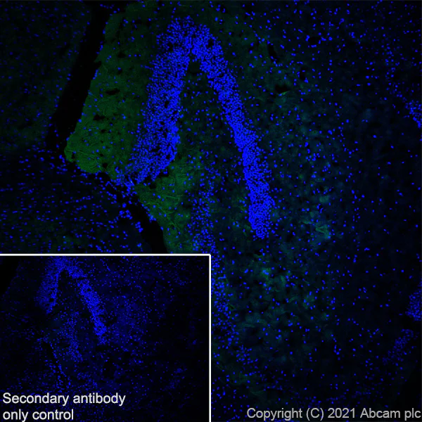
Immunohistochemical analysis of 4% PFA-fixed, 0.2% Triton X-100 permeabilized frozen mouse hippocampus tissue labeling GIRK2 with ab259909 at 1/100 (4.69 μg/ml) dilution followed by ab150077 Goat Anti-Rabbit IgG H&L (Alexa Fluor® 488) at 1/1000 dilution (Green). The nuclear counterstain was DAPI (Blue).
Cytoplasmic staining on mouse hippocampus (PMID : 8929423) is observed.
Secondary antibody control : Secondary antibody is ab150077 Goat Anti-Rabbit IgG H&L (Alexa Fluor® 488) at 1/1000 dilution.
Heat mediated antigen retrieval using sodium citrate buffer (10mM citrate pH 6.0 + 0.05% Tween-20).
- IHC-Fr
Supplier Data
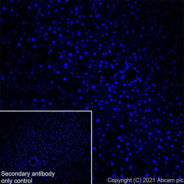
Immunohistochemistry (Frozen sections) - Anti-GIRK2 antibody [EPR23841-83] - BSA and Azide free (AB281831)
This data was developed using ab259909, the same antibody clone in a different buffer formulation.
Immunohistochemical analysis of 4% PFA-fixed, 0.2% Triton X-100 permeabilized frozen rat liver tissue labeling GIRK2 with ab259909 at 1/100 (4.69 μg/ml) dilution followed by ab150077 Goat Anti-Rabbit IgG H&L (Alexa Fluor® 488) at 1/1000 dilution (Green). The nuclear counterstain was DAPI (Blue).
Negative control : No staining on rat liver (PMID : 7592809) is observed.
Secondary antibody control : Secondary antibody is ab150077 Goat Anti-Rabbit IgG H&L (Alexa Fluor® 488)at 1/1000 dilution.
Heat mediated antigen retrieval using sodium citrate buffer (10mM citrate pH 6.0 + 0.05% Tween-20).
- IHC-Fr
Supplier Data
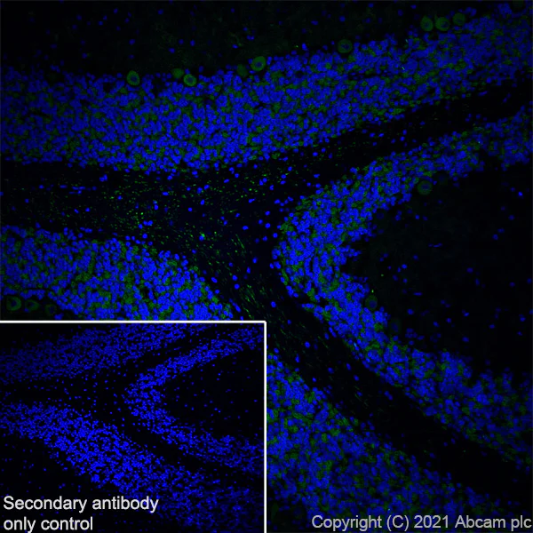
Immunohistochemistry (Frozen sections) - Anti-GIRK2 antibody [EPR23841-83] - BSA and Azide free (AB281831)
This data was developed using ab259909, the same antibody clone in a different buffer formulation.
Immunohistochemical analysis of 4% PFA-fixed, 0.2% Triton X-100 permeabilized frozen rat cerebellum tissue labeling GIRK2 with ab259909 at 1/100 (4.69 μg/ml) dilution followed by ab150077 Goat Anti-Rabbit IgG H&L (Alexa Fluor® 488) at 1/1000 dilution (Green). The nuclear counterstain was DAPI (Blue).
Cytoplasmic staining on rat cerebellum (PMID : 8929423) is observed.
Secondary antibody control : Secondary antibody is ab150077 Goat Anti-Rabbit IgG H&L (Alexa Fluor® 488) at 1/1000 dilution.
Heat mediated antigen retrieval using sodium citrate buffer (10mM citrate pH 6.0 + 0.05% Tween-20).
- IHC-Fr
Supplier Data
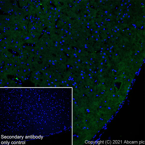
Immunohistochemistry (Frozen sections) - Anti-GIRK2 antibody [EPR23841-83] - BSA and Azide free (AB281831)
This data was developed using ab259909, the same antibody clone in a different buffer formulation.
Immunohistochemical analysis of 4% PFA-fixed, 0.2% Triton X-100 permeabilized frozen rat cerebral cortex tissue labeling GIRK2 with ab259909 at 1/100 (4.69 μg/ml) dilution followed by ab150077 Goat Anti-Rabbit IgG H&L (Alexa Fluor® 488) at 1/1000 dilution (Green). The nuclear counterstain was DAPI (Blue).
Cytoplasmic staining on rat cerebral cortex (PMID : 8929423) is observed.
Secondary antibody control : Secondary antibody is ab150077 Goat Anti-Rabbit IgG H&L (Alexa Fluor® 488) at 1/1000 dilution.
Heat mediated antigen retrieval using sodium citrate buffer (10mM citrate pH 6.0 + 0.05% Tween-20).
- IHC-Fr
Supplier Data
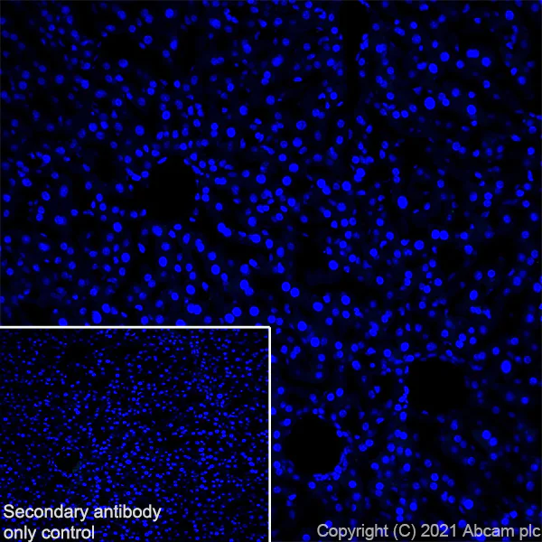
Immunohistochemistry (Frozen sections) - Anti-GIRK2 antibody [EPR23841-83] - BSA and Azide free (AB281831)
This data was developed using ab259909, the same antibody clone in a different buffer formulation.
Immunohistochemical analysis of 4% PFA-fixed, 0.2% Triton X-100 permeabilized frozen mouse liver tissue labeling GIRK2 with ab259909 at 1/100 (4.69 μg/ml) dilution followed by ab150077 Goat Anti-Rabbit IgG H&L (Alexa Fluor® 488) at 1/1000 dilution (Green). The nuclear counterstain was DAPI (Blue).
Negative control : No staining on mouse liver (PMID : 7592809) is observed.
Secondary antibody control : Secondary antibody is ab150077 Goat Anti-Rabbit IgG H&L (Alexa Fluor® 488)at 1/1000 dilution.
Heat mediated antigen retrieval using sodium citrate buffer (10mM citrate pH 6.0 + 0.05% Tween-20).
- IHC-Fr
Supplier Data
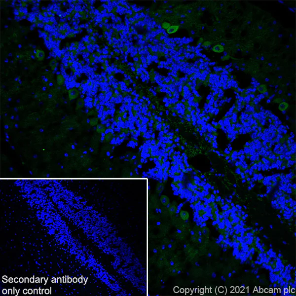
Immunohistochemistry (Frozen sections) - Anti-GIRK2 antibody [EPR23841-83] - BSA and Azide free (AB281831)
This data was developed using ab259909, the same antibody clone in a different buffer formulation.
Immunohistochemical analysis of 4% PFA-fixed, 0.2% Triton X-100 permeabilized frozen mouse cerebellum tissue labeling GIRK2 with ab259909 at 1/100 (4.69 μg/ml) dilution followed by ab150077 Goat Anti-Rabbit IgG H&L (Alexa Fluor® 488) at 1/1000 dilution (Green). The nuclear counterstain was DAPI (Blue).
Cytoplasmic staining on mouse cerebellum (PMID : 8929423) is observed.
Secondary antibody control : Secondary antibody is ab150077 Goat Anti-Rabbit IgG H&L (Alexa Fluor® 488) at 1/1000 dilution.
Heat mediated antigen retrieval using sodium citrate buffer (10mM citrate pH 6.0 + 0.05% Tween-20).
- IHC-Fr
Supplier Data
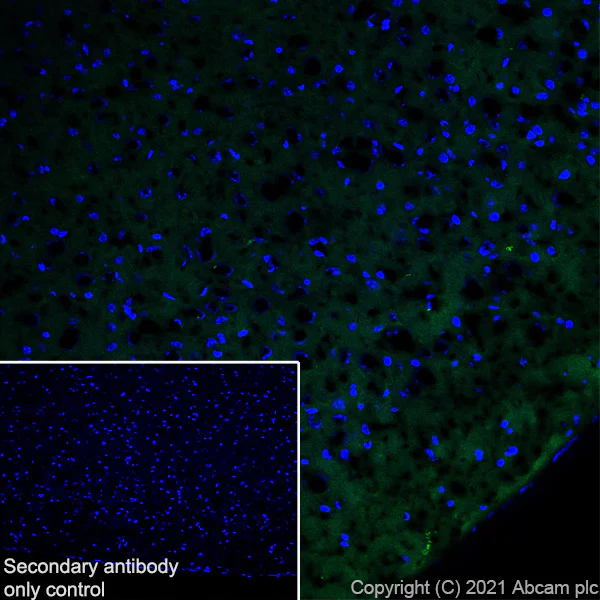
Immunohistochemistry (Frozen sections) - Anti-GIRK2 antibody [EPR23841-83] - BSA and Azide free (AB281831)
This data was developed using ab259909, the same antibody clone in a different buffer formulation.
Immunohistochemical analysis of 4% PFA-fixed, 0.2% Triton X-100 permeabilized frozen mouse cerebral cortex tissue labeling GIRK2 with ab259909 at 1/100 (4.69 μg/ml) dilution followed by ab150077 Goat Anti-Rabbit IgG H&L (Alexa Fluor® 488) at 1/1000 dilution (Green). The nuclear counterstain was DAPI (Blue).
Cytoplasmic staining on mouse cerebral cortex (PMID : 8929423) is observed.
Secondary antibody control : Secondary antibody is ab150077 Goat Anti-Rabbit IgG H&L (Alexa Fluor® 488) at 1/1000 dilution.
Heat mediated antigen retrieval using sodium citrate buffer (10mM citrate pH 6.0 + 0.05% Tween-20).
- IHC-P
Supplier Data
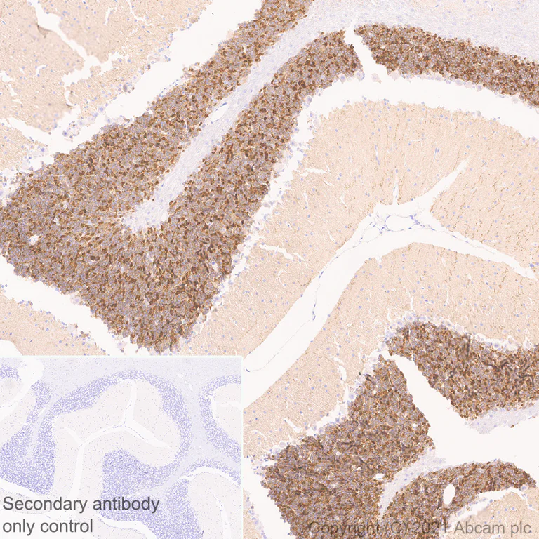
Immunohistochemistry (Formalin/PFA-fixed paraffin-embedded sections) - Anti-GIRK2 antibody [EPR23841-83] - BSA and Azide free (AB281831)
This data was developed using ab259909, the same antibody clone in a different buffer formulation.
Immunohistochemical analysis of paraffin-embedded mouse cerebellum tissue labelling GIRK2 with ab259909 at 1/200 (2.345 μg/ml) dilution followed by ready to use LeicaDS9800 (Bond™ Polymer Refine Detection). The section was incubated with ab259909 for 30 mins at room temperature. The immunostaining was performed on a Leica Biosystems BOND® RX instrument. Counterstained with Hematoxylin.
Postive staining predominantly on granular layer of mouse cerebellum (PMID : 8642402, PMID : 9023358 ).
Secondary antibody only control : Secondary antibody is ready to use LeicaDS9800 (Bond™ Polymer Refine Detection).
Heat mediated antigen retrieval with Tris-EDTA buffer (pH 9.0, epitope retrieval solution2) for 20 mins.
- IP
Supplier Data
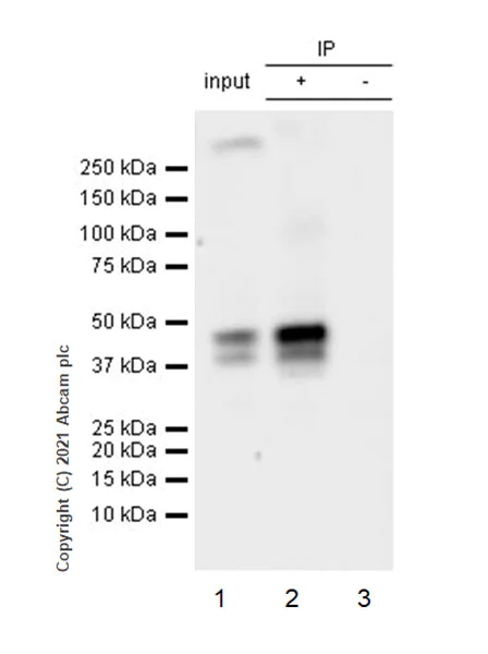
Immunoprecipitation - Anti-GIRK2 antibody [EPR23841-83] - BSA and Azide free (AB281831)
This data was developed using ab259909, the same antibody clone in a different buffer formulation.
GIRK2 was immunoprecipitated from 0.35 mg mouse brain tissue lysate 10 μg with ab259909 at 1/30 dilution (2 μg in 0.35 mg lysates). Western blot was performed on the immunoprecipitate using ab259909 at 1/1000 dilution. VeriBlot for IP secondary antibody(HRP)(ab131366) was used at 1/5000 dilution.
Lane 1 : Mouse brain tissue lysate 10 μg.
Lane 2 : ab259909 IP in mouse brain tissue lysate.
Lane 3 : Rabbit monoclonal IgG (ab172730) instead of ab259909 in mouse brain tissue lysate.
Blocking and dilution buffer and concentration : 5% NFDM/TBST.
Exposure time : 23 seconds.
Input sample is non-boiled as boiling may cause protein aggregates.The expression of truncated GIRK2 proteins observed around 40KD is consistent with what has been described in the literature (PMID : 28951616).
All lanes:
Immunoprecipitation - Anti-GIRK2 antibody [EPR23841-83] (<a href='/en-us/products/primary-antibodies/girk2-antibody-epr23841-83-ab259909'>ab259909</a>)
Predicted band size: 48 kDa
Observed band size: 40 kDa,48 kDa
false
- WB
Lab
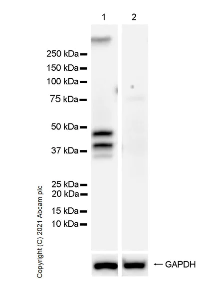
Western blot - Anti-GIRK2 antibody [EPR23841-83] - BSA and Azide free (AB281831)
This data was developed using ab259909, the same antibody clone in a different buffer formulation.
Blocking and diluting buffer and concentration : 5% NFDM/TBST.
Molecular weight around 40kDa, cleaved forms. (PMID : 28951616).
Samples are non-boiled as boiling may cause protein aggregates.
Negative control : liver (PMID : 8929423).
Exposure time : 5.5 seconds.
All lanes:
Western blot - Anti-GIRK2 antibody [EPR23841-83] (<a href='/en-us/products/primary-antibodies/girk2-antibody-epr23841-83-ab259909'>ab259909</a>) at 1/1000 dilution
Lane 1:
Human brain tissue lysate at 20 µg
Lane 2:
Human liver tissue lysate at 40 µg
Secondary
All lanes:
Goat Anti-Rabbit IgG (HRP) with minimal cross-reactivity with human IgG at 1/2000 dilution
Predicted band size: 48 kDa
Observed band size: 35 kDa,40 kDa,48 kDa
false
- WB
Lab
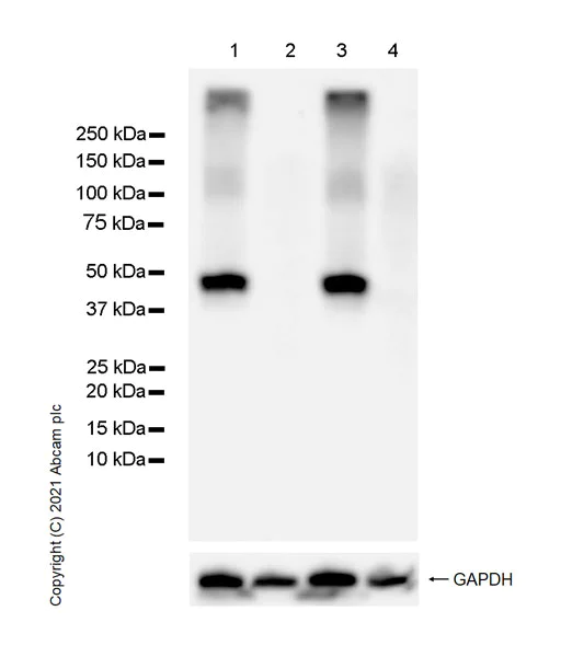
Western blot - Anti-GIRK2 antibody [EPR23841-83] - BSA and Azide free (AB281831)
This data was developed using ab259909, the same antibody clone in a different buffer formulation.
Blocking and diluting buffer and concentration : 5% NFDM/TBST.
Samples are non-boiled as boiling may cause protein aggregates.
Negative control : liver (PMID : 8929423).
Exposure time : 5.5 seconds.
All lanes:
Western blot - Anti-GIRK2 antibody [EPR23841-83] (<a href='/en-us/products/primary-antibodies/girk2-antibody-epr23841-83-ab259909'>ab259909</a>) at 1/1000 dilution
Lane 1:
Mouse brain tissue lysate at 20 µg
Lane 2:
Mouse liver tissue lysate at 40 µg
Lane 3:
Rat brain tissue lysate at 20 µg
Lane 4:
Rat liver tissue lysate at 40 µg
Secondary
All lanes:
Goat Anti-Rabbit IgG (HRP) with minimal cross-reactivity with human IgG at 1/2000 dilution
Predicted band size: 48 kDa
false
Related conjugates and formulations (1)
-
Anti-GIRK2 antibody [EPR23841-83]
Complementary Products


Primary Antibodies
AB108404
Anti-Calbindin antibody [EP3478]
BOND RX™ Validated|20ul selling size|RabMAb|Advanced Validation|Recombinant

- 4
- 3
Primary Antibodies
AB184451
Anti-Dopamine Transporter antibody [EPR19695]
BOND RX™ Validated|20ul selling size|RabMAb|Recombinant

- 0
- 0

Reactivity data
Product details
ab281831 is the carrier-free version of ab259909.
Patented technology
Our RabMAb® technology is a patented hybridoma-based technology for making rabbit monoclonal antibodies. For details on our patents, please refer to RabMAb® patents.
What are the advantages of a recombinant monoclonal antibody?
This product is a recombinant monoclonal antibody, which offers several advantages including:
- - High batch-to-batch consistency and reproducibility
- - Improved sensitivity and specificity
- - Long-term security of supply
- - Animal-free batch production
For more information, read more on recombinant antibodies.
Conjugation ready
Our carrier-free antibodies are typically supplied in a PBS-only formulation, purified and free of BSA, sodium azide and glycerol. This conjugation-ready format is designed for use with fluorochromes, metal isotopes, oligonucleotides, and enzymes, which makes them ideal for antibody labelling, functional and cell-based assays, flow-based assays (e.g. mass cytometry) and Multiplex Imaging applications.
Use our conjugation kits for antibody conjugates that are ready-to-use in as little as 20 minutes with 1 minute hands-on-time and 100% antibody recovery: available for fluorescent dyes, HRP, biotin and gold.
Compatibility
This product is compatible with the Maxpar® Antibody Labeling Kit from Fluidigm, without the need for antibody preparation. Maxpar® is a trademark of Fluidigm Canada Inc.
Properties and storage information
Form
Purification technique
Storage buffer
Shipped at conditions
Appropriate short-term storage conditions
Appropriate long-term storage conditions
Supplementary information
This supplementary information is collated from multiple sources and compiled automatically.
Biological function summary
The GIRK2 channel contributes to maintaining resting membrane potentials and controlling neuronal excitability. GIRK2 forms heteromultimeric complexes with other GIRK subunits such as GIRK1 to fully function. These complexes are essential for the proper regulation of synaptic transmission and responsiveness to neurotransmitters particularly in areas of the brain like the cerebellum and hippocampus which are critical for cognitive and motor functions.
Pathways
GIRK2 plays a significant role in the GABAergic neurotransmission and dopaminergic pathways. In the GABAergic pathway GIRK2 aids in mediating inhibitory signals by enhancing the hyperpolarization of neurons therefore dampening neurotransmission. In dopaminergic pathways GIRK2 interacts closely with dopamine receptors to modulate dopamine-induced potassium conductance impacting processes related to mood and reward. These interactions implicate GIRK2 in the regulation of important signaling pathways related to neuronal function and mood regulation.
Product protocols
- Visit the General protocols
- Visit the Troubleshooting
Target data
Alternative Names
Product promise
Please note: All products are 'FOR RESEARCH USE ONLY. NOT FOR USE IN DIAGNOSTIC OR THERAPEUTIC PROCEDURES'.
For licensing inquiries, please contact partnerships@abcam.com