Anti-Histone H3 (acetyl K18) antibody [EPR16595] - BSA and Azide free
- RabMAb
- Advanced Validation
- Recombinant
- What is this?
Be the first to review this product! Submit a review
|
(1 Publication)
- IHC-P
Supplier Data
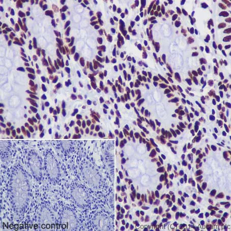
Immunohistochemistry (Formalin/PFA-fixed paraffin-embedded sections) - Anti-Histone H3 (acetyl K18) antibody [EPR16595] - BSA and Azide free (AB239414)
Immunohistochemical analysis of paraffin-embedded Human colon tissue labeling Histone H3 (acetyl K18) with ab177870 at 1/1000 dilution, followed by Goat Anti-Rabbit IgG H&L (HRP) (ab97051) secondary antibody at 1/500 dilution. Nucleus staining on glandular epithelium of colon tissue is observed. Counter stained with Hematoxylin.
Negative control : Using PBS instead of primary antibody, secondary antibody is Goat Anti-Rabbit IgG H&L (HRP) (ab97051) at 1/500 dilution.
This data was developed using the same antibody clone in a different buffer formulation containing PBS, BSA, glycerol, and sodium azide (ab177870).
Perform heat mediated antigen retrieval with Tris/EDTA buffer pH 9.0 before commencing with IHC staining protocol.
- ICC/IF
Supplier Data
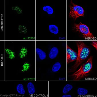
Immunocytochemistry/ Immunofluorescence - Anti-Histone H3 (acetyl K18) antibody [EPR16595] - BSA and Azide free (AB239414)
Immunofluorescent analysis of 4% paraformaldehyde-fixed, 0.1% Triton X-100 permeabilized HeLa (Human epithelial cells from cervix adenocarcinoma) cells labeling Histone H3 (acetyl K18) with ab177870 at 1/2000 dilution, followed by Goat Anti-Rabbit IgG H&L (Alexa Fluor® 488) (ab150077) secondary antibody at 1/400 dilution (green). Confocal image showing nuclear staining on HeLa cell line. Acetylation levels increased after treatment with Trichostatin A (500 ng/ml) for 4 hours. The nuclear counter stain is DAPI (blue). Tubulin is detected with ab7291 (Anti-alpha Tubulin antibody [DM1A] - Loading Control) at 1/500 dilution and Goat Anti-Mouse IgG H&L (Alexa Fluor® 594) preadsorbed (ab150120) at 1/500 dilution (red).
The negative controls are as follows;
-ve control 1 : ab177870 at 1/2000 dilution followed by ab150120 at 1/500 dilution.
-ve control 2 : ab7291 at 1/500 dilution followed by ab150077 at 1/400 dilution.
This data was developed using the same antibody clone in a different buffer formulation containing PBS, BSA, glycerol, and sodium azide (ab177870).
- ChIP
Supplier Data
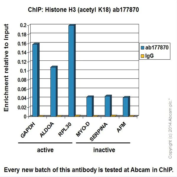
ChIP - Anti-Histone H3 (acetyl K18) antibody [EPR16595] - BSA and Azide free (AB239414)
Chromatin was prepared from HeLa (Human epithelial cells from cervix adenocarcinoma) cells according to the Abcam X-ChIP protocol. Cells were fixed with formaldehyde for 10min. The ChIP was performed with 25μg of chromatin, 2μg of ab177870 (blue), and 20μl of Anti rabbit IgG sepharose beads. 2μg of Rabbit normal IgG was added to the beads control (yellow). The immunoprecipitated DNA was quantified by real time PCR (SYBR approach). Primers and probes are located in the first kb of the transcribed region.This data was developed using the same antibody clone in a different buffer formulation containing PBS, BSA, glycerol, and sodium azide (ab177870).
- IHC-P
Supplier Data
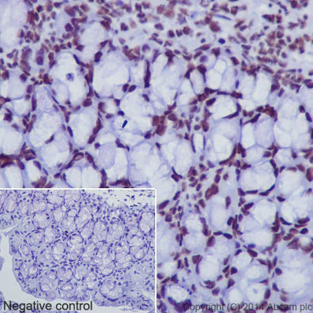
Immunohistochemistry (Formalin/PFA-fixed paraffin-embedded sections) - Anti-Histone H3 (acetyl K18) antibody [EPR16595] - BSA and Azide free (AB239414)
Immunohistochemical analysis of paraffin-embedded Mouse colon tissue labeling Histone H3 (acetyl K18) with ab177870 at 1/2000 dilution, followed by Goat Anti-Rabbit IgG H&L (HRP) (ab97051) secondary antibody at 1/500 dilution. Nuclear staining on glandular epithelium of mouse colon tissue is observed. Counter stained with Hematoxylin.
Negative control : Using PBS instead of primary antibody, secondary antibody is Goat Anti-Rabbit IgG H&L (HRP) (ab97051) at 1/500 dilution.
This data was developed using the same antibody clone in a different buffer formulation containing PBS, BSA, glycerol, and sodium azide (ab177870).
Perform heat mediated antigen retrieval with Tris/EDTA buffer pH 9.0 before commencing with IHC staining protocol.
- IHC-P
Supplier Data
Immunohistochemistry (Formalin/PFA-fixed paraffin-embedded sections) - Anti-Histone H3 (acetyl K18) antibody [EPR16595] - BSA and Azide free (AB239414)
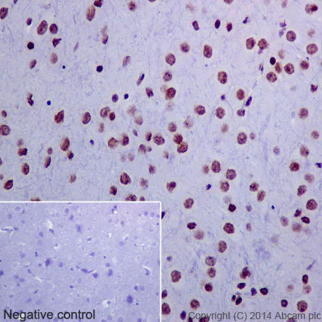
Immunohistochemical analysis of paraffin-embedded Rat cerebral cortex tissue labeling Histone H3 (acetyl K18) with ab177870 at 1/500 dilution, followed by prediluted Goat Anti-Rabbit IgG H&L (HRP). Nuclear staining on Rat cerebral cortex tissue is observed. Counter stained with Hematoxylin.
Negative control : Using PBS instead of primary antibody, secondary antibody is prediluted Goat Anti-Rabbit IgG H&L (HRP).
This data was developed using the same antibody clone in a different buffer formulation containing PBS, BSA, glycerol, and sodium azide (ab177870).
Perform heat mediated antigen retrieval with Tris/EDTA buffer pH 9.0 before commencing with IHC staining protocol.
- ChIC/CUT&RUN-seq
Lab
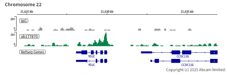
ChIC/CUT&RUN sequencing - Anti-Histone H3 (acetyl K18) antibody [EPR16595] - BSA and Azide free (AB239414)
This data was developed using the same antibody clone in a different buffer formulation containing PBS, BSA, glycerol, and sodium azide (,ab177870).
ChIC/CUT&RUN was performed using a pAG-MNase at a final concentration of 700 ng/mL, 2.5 x 10^5 HeLa (human cervical adenocarcinoma epithelial cell) cells and 5 µg of ab177870 [EPR16595]. The resulting DNA was sequenced on the Illumina NovaSeq 6000 to a depth of 10 million reads. The negative IgG control ab172730 is also shown.
The University of Geneva owns patents relevant to ChIC (Chromatin Immuno-Cleavage) methods.
- ChIC/CUT&RUN-seq
Lab
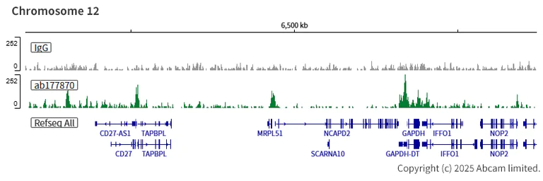
ChIC/CUT&RUN sequencing - Anti-Histone H3 (acetyl K18) antibody [EPR16595] - BSA and Azide free (AB239414)
This data was developed using the same antibody clone in a different buffer formulation containing PBS, BSA, glycerol, and sodium azide (,ab177870).
ChIC/CUT&RUN was performed using a pAG-MNase at a final concentration of 700 ng/mL, 2.5 x 10^5 HeLa (human cervical adenocarcinoma epithelial cell) cells and 5 µg of ab177870 [EPR16595]. The resulting DNA was sequenced on the Illumina NovaSeq 6000 to a depth of 10 million reads. The negative IgG control ab172730 is also shown.
The University of Geneva owns patents relevant to ChIC (Chromatin Immuno-Cleavage) methods.
- ChIC/CUT&RUN-seq
Lab
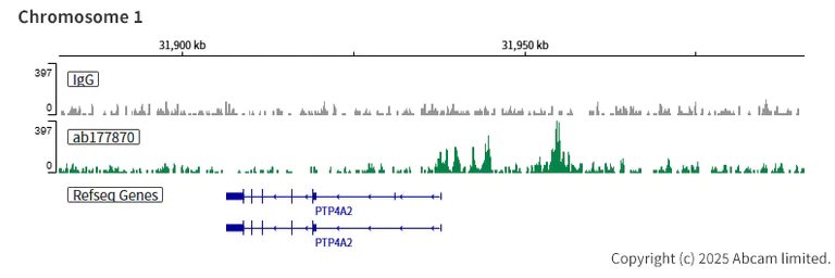
ChIC/CUT&RUN sequencing - Anti-Histone H3 (acetyl K18) antibody [EPR16595] - BSA and Azide free (AB239414)
This data was developed using the same antibody clone in a different buffer formulation containing PBS, BSA, glycerol, and sodium azide (,ab177870).
ChIC/CUT&RUN was performed using a pAG-MNase at a final concentration of 700 ng/mL, 2.5 x 10^5 HeLa (human cervical adenocarcinoma epithelial cell) cells and 5 µg of ab177870 [EPR16595]. The resulting DNA was sequenced on the Illumina NovaSeq 6000 to a depth of 10 million reads. The negative IgG control ab172730 is also shown.
The University of Geneva owns patents relevant to ChIC (Chromatin Immuno-Cleavage) methods.
- PepArr
Lab
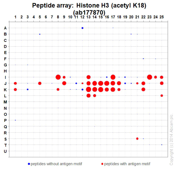
Peptide Array - Anti-Histone H3 (acetyl K18) antibody [EPR16595] - BSA and Azide free (AB239414)
ab177870 at 0.02 μg/mL was tested in Peptide Array against 501 different modified and unmodified histone peptides; each peptide is printed on the array at six concentrations (each in triplicate). A Goat Anti-Rabbit IgG, (H+L), Fluo 647nm conjugated at 1/50000 dilution was used as the secondary antibody. Washing buffer and time was protein Array Washing Buffer, 2*10mins and 2*5mins and blocking buffer was 5% BSA in TBST. Circle area represents affinity between the antibody and a peptide : all antigen-containing peptides are displayed as red circles, all other peptides as blue circles. The affinity is calculated as the area under the curve when antibody binding values are plotted against the corresponding peptide concentration. Each circle area is normalized to the peptide with the strongest affinity. Scanner details : Innoscan 710 This data was developed using the same antibody clone in a different buffer formulation containing PBS, BSA, glycerol, and sodium azide (ab177870).

- Dot
Supplier Data
Dot Blot - Anti-Histone H3 (acetyl K18) antibody [EPR16595] - BSA and Azide free (AB239414)
Dot blot analysis using ab177870 at 1/1000 dilution and Goat Anti-Rabbit IgG H&L (HRP) (ab97051) as secondary antibody at 1/100,000 dilution.
Lane 1 : Histone H3(acetyl K4 + K9 + K14 + K18) peptide
Lane 2 : Histone H3(acetyl K4) peptide
Lane 3 : Histone H3(acetyl K9) peptide
Lane 4 : Histone H3(acetyl K14) peptide
Lane 5 : Histone H3(acetyl K18) peptide
Lane 6 : Histone H3 unmodified peptide
Blocking and Diluting buffer and concentration : 5% NFDM/TBST
Exposure time : 10 seconds
This data was developed using the same antibody clone in a different buffer formulation containing PBS, BSA, glycerol, and sodium azide (ab177870).
Related conjugates and formulations (1)
-
Anti-Histone H3 (acetyl K18) antibody [EPR16595] - ChIP Grade
Alternative Products
Primary Antibodies
AB176916
Anti-Histone H3 (tri methyl K9) antibody [EPR16601] - ChIP Grade
20ul selling size|RabMAb|Advanced Validation|Recombinant

- 4
- 14
Primary Antibodies
AB32356
Anti-Histone H3 (di methyl K4) antibody [Y47] - ChIP Grade
20ul selling size|RabMAb|Advanced Validation|Recombinant

- 5
- 14
Primary Antibodies
AB52946
Anti-Histone H3 (acetyl K14) antibody [EP964Y] - ChIP Grade
20ul selling size|BOND RX™ Validated|RabMAb|Recombinant|Advanced Validation

- 5
- 12
Primary Antibodies
AB176843
Anti-Histone H3 (citrulline R2) antibody [EPR17703]
20ul selling size|RabMAb|Advanced Validation|Recombinant

- 5
- 1
Primary Antibodies
AB219407
Anti-Histone H3 (citrulline R17) antibody [EPR20358-120]
RabMAb|Recombinant|20ul selling size

- 5
- 1
Primary Antibodies
AB277918
Alexa Fluor® 488 Anti-Histone H3 (acetyl K14) antibody [EP964Y]
RabMAb|Recombinant

- 0
- 0
Primary Antibodies
AB277919
Alexa Fluor® 647 Anti-Histone H3 (acetyl K14) antibody [EP964Y]
RabMAb|Recombinant

- 0
- 0
Primary Antibodies
AB192985
Anti-Histone H3 (tri methyl K27) antibody [EPR18607] - ChIP Grade
20ul selling size|RabMAb|Advanced Validation|Recombinant

- 4
- 14
Complementary Products

Sample Prep & Detection Kits
AB270703
5’ Feature Barcode Antibody Conjugation Kit - Lightning-Link® - Oligos 1-10

- 0
- 0
Primary Antibodies
AB1791
Anti-Histone H3 antibody - Nuclear Marker and ChIP Grade
Lab Essentials

- 5
- 233
Primary Antibodies
AB4729
Anti-Histone H3 (acetyl K27) antibody - ChIP Grade
BOND RX™ Validated

- 5
- 85
Primary Antibodies
AB8898
Anti-Histone H3 (tri methyl K9) antibody - ChIP Grade
BOND RX™ Validated

- 5
- 92
Primary Antibodies
AB109463
Anti-Histone H4 (acetyl K16) antibody [EPR1004]
20ul selling size|RabMAb|Advanced Validation|Recombinant

- 5
- 15
Reactivity data
Product details
ab239414 is the carrier-free version of ab177870.
Patented technology
Our RabMAb® technology is a patented hybridoma-based technology for making rabbit monoclonal antibodies. For details on our patents, please refer to RabMAb® patents.
What are the advantages of a recombinant monoclonal antibody?
This product is a recombinant monoclonal antibody, which offers several advantages including:
- - High batch-to-batch consistency and reproducibility
- - Improved sensitivity and specificity
- - Long-term security of supply
- - Animal-free batch production
For more information, read more on recombinant antibodies.
Conjugation ready
Our carrier-free antibodies are typically supplied in a PBS-only formulation, purified and free of BSA, sodium azide and glycerol. This conjugation-ready format is designed for use with fluorochromes, metal isotopes, oligonucleotides, and enzymes, which makes them ideal for antibody labelling, functional and cell-based assays, flow-based assays (e.g. mass cytometry) and Multiplex Imaging applications.
Use our conjugation kits for antibody conjugates that are ready-to-use in as little as 20 minutes with 1 minute hands-on-time and 100% antibody recovery: available for fluorescent dyes, HRP, biotin and gold.
Compatibility
This product is compatible with the Maxpar® Antibody Labeling Kit from Fluidigm, without the need for antibody preparation. Maxpar® is a trademark of Fluidigm Canada Inc.
Properties and storage information
Form
Purification technique
Storage buffer
Shipped at conditions
Appropriate short-term storage conditions
Appropriate long-term storage conditions
Storage information
Product protocols
- Visit the General protocols
- Visit the Troubleshooting
Target data
Alternative Names
Publications (1)
Recent publications for all applications. Explore the full list and refine your search
Synapse (New York, N.Y.) 75:e22221 PubMed34958692
2021
Applications
Unspecified application
Species
Unspecified reactive species
Product promise
Please note: All products are 'FOR RESEARCH USE ONLY. NOT FOR USE IN DIAGNOSTIC OR THERAPEUTIC PROCEDURES'.
For licensing inquiries, please contact partnerships@abcam.com