Anti-Histone H3 (phospho S28) antibody [E191] - ChIP Grade
- BOND RX™ Validated
- 20ul selling size
- RabMAb
- Advanced Validation
- Recombinant
- What is this?
4
(4 Reviews)
|
(16 Publications )
- IHC-P
Lab
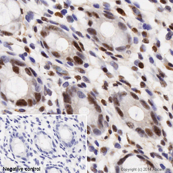
Immunohistochemistry (Formalin/PFA-fixed paraffin-embedded sections) - Anti-Histone H3 (phospho S28) antibody [E191] - ChIP Grade (AB32388)
IHC image of ab32388 staining Histone H3 in Human normal colon formalin fixed paraffin embedded tissue* sections, performed on a Leica Bond. The section was pre-treated using heat mediated antigen retrieval with sodium citrate buffer (pH6, epitope retrieval solution 1) for 20 mins. The section was then incubated with ab32388, 0.1ug/ml, for 15 mins at room temperature and detected using an HRP conjugated compact polymer system. DAB was used as the chromogen. The section was then counterstained with haematoxylin and mounted with DPX. No primary antibody was used in the negative control (shown on the inset).
For other IHC staining systems (automated and non-automated) customers should optimize variable parameters such as antigen retrieval conditions, primary antibody concentration and antibody incubation times.
*Tissue obtained from the Human Research Tissue Bank, supported by the NIHR Cambridge Biomedical Research Centre
- Flow Cyt (Intra)
Lab
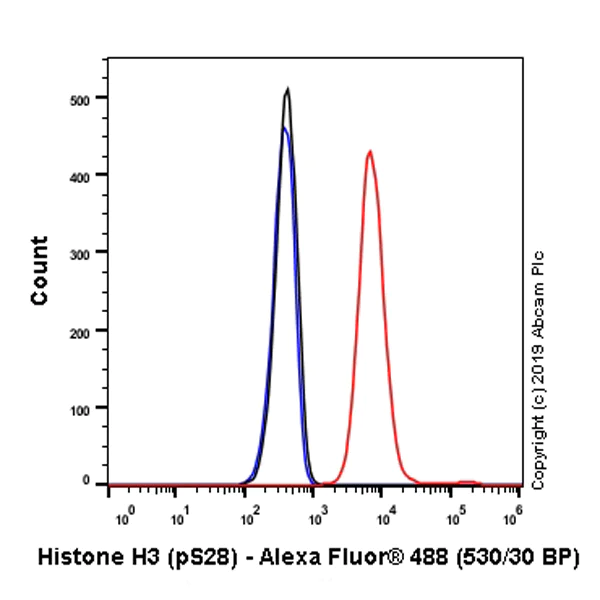
Flow Cytometry (Intracellular) - Anti-Histone H3 (phospho S28) antibody [E191] - ChIP Grade (AB32388)
Overlay histogram showing HeLa cells stained with ab32388 (red line). The cells were fixed with 4% paraformaldehyde and then permeabilized with 90% methanol. The cells were then incubated in 1x PBS / 10% normal goat serum / 0.3M glycine to block non-specific protein-protein interactions followed by the antibody (ab32388, 1/1200 dilution, 1.02 μg/ml) for 30 min at 22°C. The secondary antibody used was Alexa Fluor® 488 goat anti-rabbit IgG (H+L) (ab150077) at 1/2000 dilution for 30 min at 22°C. Isotype control antibody (black line) was rabbit IgG (monoclonal) (ab172730) (1μg/1x106 cells) used under the same conditions. Unlabelled sample (blue line) was also used as a control. Acquisition of >5,000 events were collected using a 20mW Argon ion laser (488nm) and 525/30 bandpass filter.
- Flow Cyt (Intra)
Unknown
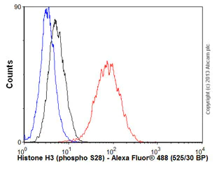
Flow Cytometry (Intracellular) - Anti-Histone H3 (phospho S28) antibody [E191] - ChIP Grade (AB32388)
Overlay histogram showing HeLa cells stained with ab32388 (red line). The cells were fixed with 80% methanol (5 min) and then permeabilized with 0.1% PBS-Tween for 20 min. The cells were then incubated in 1x PBS / 10% normal goat serum / 0.3M glycine to block non-specific protein-protein interactions followed by the antibody (ab32388, 1/100 dilution) for 30 min at 22°C. The secondary antibody used was Alexa Fluor® 488 goat anti-rabbit IgG (H+L) (ab150077) at 1/2000 dilution for 30 min at 22°C. Isotype control antibody (black line) was rabbit IgG (monoclonal) (1μg/1x106 cells) used under the same conditions. Unlabelled sample (blue line) was also used as a control. Acquisition of >5,000 events were collected using a 20mW Argon ion laser (488nm) and 525/30 bandpass filter.
- ICC/IF
Unknown
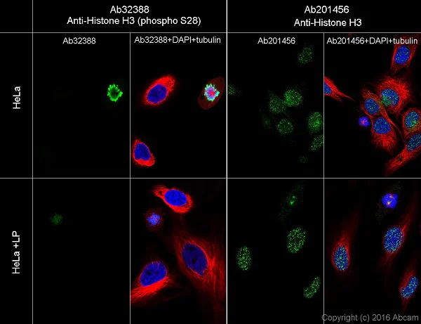
Immunocytochemistry/ Immunofluorescence - Anti-Histone H3 (phospho S28) antibody [E191] - ChIP Grade (AB32388)
Immunofluorescent analysis of 4% Paraformaldehyde-fixed, 0.1% tritonX-100 permeabilized HeLa (Human epithelial cell line from cervix adenocarcinoma) cells, LP starved and non-starved, labeling anti-Histone H3 (phospho S28) with ab32388 at 1/100 dilution followed by Goat anti-Rabbit secondary IgG AlexaFluor®488 (ab150077) secondary antibody at 1/1000 dilution (green).
Confocal image showing nuclear staining on M phase of HeLa cells, then the signal decreased after LP treatment.
For the pan antibody, there was no great difference after LP treatment. The data showed mostly nuclear staining

- IP
Lab
Immunoprecipitation - Anti-Histone H3 (phospho S28) antibody [E191] - ChIP Grade (AB32388)
Immunoprecipitation dilution was 1/60.
Western blot was performed on the immunoprecipitate using ab32388 at 1/500 dilution (2.444 μg/ml). VeriBlot for IP secondary antibody (HRP) (ab131366) at 1 : 1000 dilution.
Blocking/Diluting buffer and concentration : 5% NFDM/TBST.
All lanes:
Immunoprecipitation - Anti-Histone H3 (phospho S28) antibody [E191] - ChIP Grade (ab32388)
Lane 1:
HeLa (human cervix adenocarcinoma epithelial cell) treated with 0.5µM Nocodazole for 24h whole cell lysate at 10 µg
Lane 2:
ab32388 IP in Nocodazole treated HeLa whole cell lysate
Lane 3:
Rabbit monoclonal IgG (<a href='/en-us/products/primary-antibodies/rabbit-igg-monoclonal-epr25a-isotype-control-ab172730'>ab172730</a>) instead of ab32388 in nocodazole treated HeLa whole cell lysate
Predicted band size: 15 kDa
Observed band size: 17 kDa
false
- ChIP
Unknown
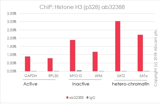
ChIP - Anti-Histone H3 (phospho S28) antibody [E191] - ChIP Grade (AB32388)
Chromatin was prepared from HeLa cells according to the Abcam X-ChIP protocol. Cells were fixed with formaldehyde for 10min.
The ChIP was performed with 25 μg of chromatin, 5 μg of ab32388 (red), and 20 μl of Protein A/G sepharose beads. Rabbit normal IgG was added to the beads control (gray). The immunoprecipitated DNA was quantified by real time PCR (Sybr green approach).
Primers and probes are located in the first kb of the transcribed region.
- WB
Supplier Data
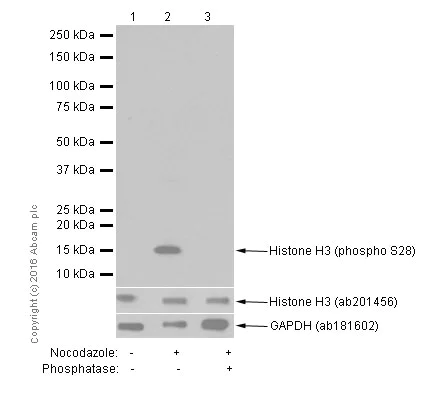
Western blot - Anti-Histone H3 (phospho S28) antibody [E191] - ChIP Grade (AB32388)
Blocking/dilution buffer : 2% BSA/TBST
All lanes:
Western blot - Anti-Histone H3 (phospho S28) antibody [E191] - ChIP Grade (ab32388) at 1/1000 dilution
Lane 1:
HeLa (Human epithelial cell line from cervix adenocarcinoma) whole cell lysate at 15 µg
Lane 2:
Whole cell lysate from HeLa (Human epithelial cell line from cervix adenocarcinoma) cells, treated with 100ng/ml nocodazole for 18 hours at 15 µg
Lane 3:
Whole cell lysate from HeLa (Human epithelial cell line from cervix adenocarcinoma) cells, treated with 100ng/ml nocodazole for 18 hours. Membrane incubated with phosphatase at 15 µg
Secondary
All lanes:
Western blot - Goat Anti-Rabbit IgG H&L (HRP) (<a href='/en-us/products/secondary-antibodies/goat-rabbit-igg-h-l-hrp-ab97051'>ab97051</a>) at 1/20000 dilution
Predicted band size: 15 kDa
Observed band size: 17 kDa
false
Exposure time: 15s
- WB
Unknown
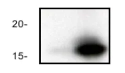
Western blot - Anti-Histone H3 (phospho S28) antibody [E191] - ChIP Grade (AB32388)
All lanes:
Western blot - Anti-Histone H3 (phospho S28) antibody [E191] - ChIP Grade (ab32388) at 1/2000 dilution
Lane 1:
NIH 3T3 cell lysate -untreated
Lane 2:
NIH 3T3 cell lysate -treated with FBS + CalA.
Predicted band size: 15 kDa
Observed band size: 17 kDa
false
- Dot
Lab
Dot Blot - Anti-Histone H3 (phospho S28) antibody [E191] - ChIP Grade (AB32388)
Dot blot analysis of Histone H3 (phospho S28) labeled with ab32388 at 1/1000 dilution. Lane 1 : Histone H3 (phospho S28) peptide a Lane 2 : Histone H3 (phospho S28) peptide c1 Goat Anti-Rabbit IgG, (H+L), Peroxidase conjugated (ab97051) was used as secondary antobody at 1/100000 dilution. Blocking/Dilution buffer : 5% NFDM/TBST. Exposure time : 180 minutes.
- Dot
Unknown
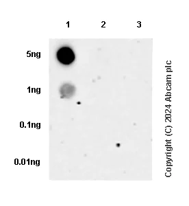
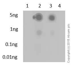
Dot Blot - Anti-Histone H3 (phospho S28) antibody [E191] - ChIP Grade (AB32388)
Dot blot performed using ab32388 at a dilution of 1/100. Lane 1 - Unmodified H3 peptide. Lane 2 - H3S28ph peptide. Lane 3 - H3.3S28ph peptide. Lane 4 - H3.3S31ph peptide. A HRP conjugated goat anti-rabbit (H+L) was used as the secondary antibody at a dilution of 1/2500. The exposure time was 3 minutes and the dilution and blocking buffer used were 5% NFDM/TBST.

- PepArr
Lab
Peptide Array - Anti-Histone H3 (phospho S28) antibody [E191] - ChIP Grade (AB32388)
ab32388 (0.02μg/mL) was tested in Peptide Array against 501 different modified and unmodified histone peptides; each peptide is printed on the array at six concentrations (each in triplicate). A Goat Anti-Rabbit IgG, (H+L), Fluo 647nm conjugated at 1/50000 was used as the secondary antibody. Protein Array Washing Buffer, 2*10 mins and 2*5 mins. Blocking buffer was 5% BSA in TBST and Innoscan 710 scanner was used. Circle area represents affinity between the antibody and a peptide : all antigen-containing peptides are displayed as red circles, all other peptides as blue circles. The affinity is calculated as the area under the curve when antibody binding values are plotted against the corresponding peptide concentration. Each circle area is normalized to the peptide with the strongest affinity.
The complete dataset, including full list of all peptides and information on the position of each peptide in the diagram, is available under the Product Protocol section.
Related conjugates and formulations (10)
-
Anti-Histone H3 (phospho S28) antibody [E191] - BSA and Azide free
-
660 APC
APC Anti-Histone H3 (phospho S28) antibody [E191] (ChIP Grade)
-
519 Alexa Fluor® 488
Alexa Fluor® 488 Anti-Histone H3 (phospho S28) antibody [E191]
-
565 Alexa Fluor® 555
Alexa Fluor® 555 Anti-Histone H3 (phospho S28) antibody [E191] - ChIP Grade
-
603 Alexa Fluor® 568
Alexa Fluor® 568 Anti-Histone H3 (phospho S28) antibody [E191] - ChIP Grade
-
617 Alexa Fluor® 594
Alexa Fluor® 594 Anti-Histone H3 (phospho S28) antibody [E191] - ChIP Grade
-
665 Alexa Fluor® 647
Alexa Fluor® 647 Anti-Histone H3 (phospho S28) antibody [E191]
-
775 Alexa Fluor® 750
Alexa Fluor® 750 Anti-Histone H3 (phospho S28) antibody [E191] - ChIP Grade
-
HRP Anti-Histone H3 (phospho S28) antibody [E191] (ChIP Grade)
-
578 PE
PE Anti-Histone H3 (phospho S28) antibody [E191] (ChIP Grade)
Alternative Products
Primary Antibodies
AB176916
Anti-Histone H3 (tri methyl K9) antibody [EPR16601] - ChIP Grade
20ul selling size|RabMAb|Advanced Validation|Recombinant

- 4
- 14
Primary Antibodies
AB32356
Anti-Histone H3 (di methyl K4) antibody [Y47] - ChIP Grade
20ul selling size|RabMAb|Advanced Validation|Recombinant

- 5
- 14
Primary Antibodies
AB52946
Anti-Histone H3 (acetyl K14) antibody [EP964Y] - ChIP Grade
20ul selling size|BOND RX™ Validated|RabMAb|Recombinant|Advanced Validation

- 5
- 12
Primary Antibodies
AB176842
Anti-Histone H3 antibody [EPR16987] - Nuclear Marker and ChIP Grade
20ul selling size|RabMAb|Recombinant|Lab Essentials|Advanced Validation

- 5
- 15
Primary Antibodies
AB176843
Anti-Histone H3 (citrulline R2) antibody [EPR17703]
20ul selling size|RabMAb|Advanced Validation|Recombinant

- 5
- 1
Primary Antibodies
AB219407
Anti-Histone H3 (citrulline R17) antibody [EPR20358-120]
RabMAb|Recombinant|20ul selling size

- 5
- 1
Primary Antibodies
AB277918
Alexa Fluor® 488 Anti-Histone H3 (acetyl K14) antibody [EP964Y]
RabMAb|Recombinant

- 0
- 0
Primary Antibodies
AB277919
Alexa Fluor® 647 Anti-Histone H3 (acetyl K14) antibody [EP964Y]
RabMAb|Recombinant

- 0
- 0
Primary Antibodies
AB192985
Anti-Histone H3 (tri methyl K27) antibody [EPR18607] - ChIP Grade
20ul selling size|RabMAb|Advanced Validation|Recombinant

- 4
- 14
Complementary Products

Primary Antibodies
AB16667
BOND RX™ Validated|KO Validated|RabMAb|Advanced Validation|Recombinant|20ul selling size

- 5
- 133
Primary Antibodies
AB32356
Anti-Histone H3 (di methyl K4) antibody [Y47] - ChIP Grade
20ul selling size|RabMAb|Advanced Validation|Recombinant
- 5
- 14

Primary Antibodies
AB32107
Anti-Histone H3 (phospho S10 + T11) antibody [E173]
20ul selling size|RabMAb|Recombinant

- 5
- 4
Reactivity data
Product details
Patented technology
Our RabMAb® technology is a patented hybridoma-based technology for making rabbit monoclonal antibodies. For details on our patents, please refer to RabMAb® patents.
What are the advantages of a recombinant monoclonal antibody?
This product is a recombinant monoclonal antibody, which offers several advantages including:
- - High batch-to-batch consistency and reproducibility
- - Improved sensitivity and specificity
- - Long-term security of supply
- - Animal-free batch production
For more information, read more on recombinant antibodies.
Properties and storage information
Form
Purification technique
Storage buffer
Shipped at conditions
Appropriate short-term storage duration
Appropriate short-term storage conditions
Appropriate long-term storage conditions
Aliquoting information
Storage information
Product protocols
- Visit the General protocols
- Visit the Troubleshooting
- Download peptideArrayWebsite|en
Target data
Alternative Names
Publications (16)
Recent publications for all applications. Explore the full listand refine your search
PloS one 19:e0309471 PubMed39570878
2024
Applications
Unspecified application
Species
Unspecified reactive species
Nature communications 14:8484 PubMed38123565
2023
Applications
Unspecified application
Species
Unspecified reactive species
Journal of advanced research 57:163-180 PubMed37182685
2023
Applications
Unspecified application
Species
Unspecified reactive species
PLoS genetics 17:e1009868 PubMed34752469
2021
Applications
Unspecified application
Species
Unspecified reactive species
Nature communications 12:896 PubMed33563994
2021
Applications
Unspecified application
Species
Unspecified reactive species
Nature 583:852-857 PubMed32699416
2020
Applications
Unspecified application
Species
Unspecified reactive species
Environmental and molecular mutagenesis 61:534-550 PubMed32297368
2020
Applications
Unspecified application
Species
Unspecified reactive species
Nature communications 11:612 PubMed32001712
2020
Applications
Unspecified application
Species
Unspecified reactive species
Theriogenology 116:83-88 PubMed29783047
2018
Applications
Unspecified application
Species
Unspecified reactive species
Developmental neurobiology 76:939-55 PubMed26585436
2015
Applications
IHC-Fr
Species
Human
Product promise
Please note: All products are 'FOR RESEARCH USE ONLY. NOT FOR USE IN DIAGNOSTIC OR THERAPEUTIC PROCEDURES'.
For licensing inquiries, please contact partnerships@abcam.com