Anti-HNF1 alpha antibody [EPR23054-108] - BSA and Azide free
- BOND RX™ Validated
- KO Validated
- Recombinant
- Advanced Validation
- RabMAb
- What is this?
Be the first to review this product! Submit a review
|
(1 Publication)
- IHC-P
Unknown
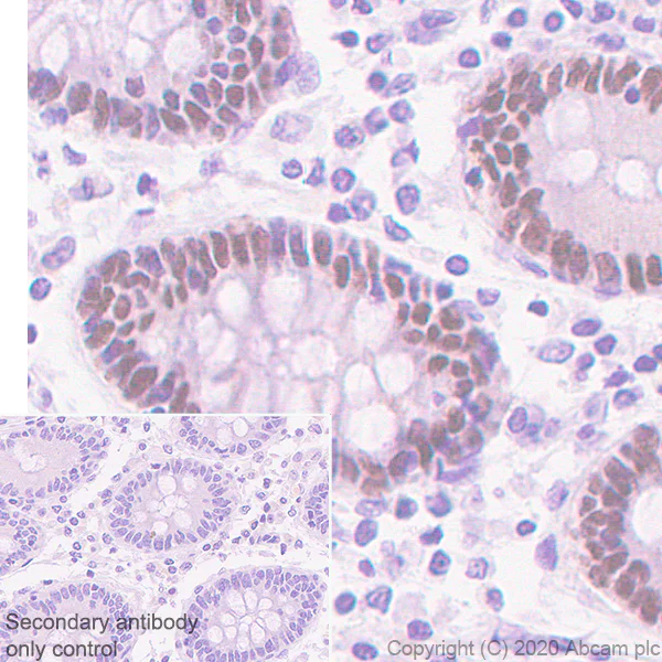
Immunohistochemistry (Formalin/PFA-fixed paraffin-embedded sections) - Anti-HNF1 alpha antibody [EPR23054-108] - BSA and Azide free (AB272708)
Immunohistochemical analysis of paraffin-embedded Human colon tissue labeling HNF1 alpha with ab272693 at 1/2000 dilution followed by a ready to use Rabbit specific IHC polymer detection kit HRP/DAB (ab209101). Nuclear staining on human colon. The section was incubated with ab272693 for 30 mins at room temperature. The immunostaining was performed on a Leica Biosystems BOND® RX instrument. Counterstained with Hematoxylin.
Secondary antibody only control : Secondary antibody is a ready to use Rabbit specific IHC polymer detection kit HRP/DAB (ab209101).
Heat mediated antigen retrieval with Tris-EDTA buffer (pH 9.0, epitope retrieval solution2) for 20 mins.
This data was developed using the same antibody clone in a different buffer formulation containing PBS, BSA, glycerol, and sodium azide (ab272693).
- IHC-P
Unknown
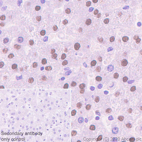
Immunohistochemistry (Formalin/PFA-fixed paraffin-embedded sections) - Anti-HNF1 alpha antibody [EPR23054-108] - BSA and Azide free (AB272708)
Immunohistochemical analysis of paraffin-embedded Human liver tissue labeling HNF1 alpha with ab272693 at 1/2000 dilution followed by a ready to use Rabbit specific IHC polymer detection kit HRP/DAB (ab209101). Nuclear staining on human liver. The section was incubated with ab272693 for 30 mins at room temperature. The immunostaining was performed on a Leica Biosystems BOND® RX instrument. Counterstained with Hematoxylin.
Secondary antibody only control : Secondary antibody is a ready to use Rabbit specific IHC polymer detection kit HRP/DAB (ab209101).
Heat mediated antigen retrieval with Tris-EDTA buffer (pH 9.0, epitope retrieval solution2) for 20 mins.
This data was developed using the same antibody clone in a different buffer formulation containing PBS, BSA, glycerol, and sodium azide (ab272693).
- Flow Cyt (Intra)
Unknown
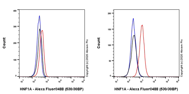
Flow Cytometry (Intracellular) - Anti-HNF1 alpha antibody [EPR23054-108] - BSA and Azide free (AB272708)
Intracellular flow cytometric analysis of 4% paraformaldehyde fixed, 90% methanol permeabilized HeLa (Human cervix adenocarcinoma epithelial cell, Left) / HepG2 (Human hepatocellular carcinoma epithelial cell, Right) cells labelling HNF1 alpha with ab272693 at 1/500 dilution (0.1ug) (Red) compared with a Rabbit monoclonal IgG (ab172730) (Black) isotype control and an unlabelled control (cells without incubation with primary antibody and secondary antibody) (Blue). A Goat anti rabbit IgG (Alexa Fluor®488, ab150077) at 1/2000 dilution was used as the secondary antibody.
Negative control : HeLa (PMID : 10677375).
This data was developed using the same antibody clone in a different buffer formulation containing PBS, BSA, glycerol, and sodium azide (ab272693).
- ICC/IF
Unknown
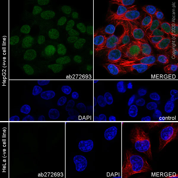
Immunocytochemistry/ Immunofluorescence - Anti-HNF1 alpha antibody [EPR23054-108] - BSA and Azide free (AB272708)
Immunofluorescent analysis of 100% methanol-fixed, 0.1% Triton X-100 permeabilized HepG2 and HeLa cells labelling HNF1 alpha with ab272693 at 1/1000 dilution, followed by ab150077 Goat Anti-Rabbit IgG H&L (Alexa Fluor® 488) antibody at 1/1000 dilution (Green). Confocal image showing nuclear staining in HepG2 cells. 100% methanol fixation is recommended. Negative control : HeLa (PMID : 10677375). ab195889 Anti-alpha Tubulin antibody (Alexa Fluor® 594) was used to counterstain tubulin at 1/200 dilution (Red). The Nuclear counterstain was DAPI (Blue).
Secondary antibody only control : Secondary antibody is ab150077 Goat Anti-Rabbit IgG H&L (Alexa Fluor® 488) at 1/1000 dilution.
This data was developed using the same antibody clone in a different buffer formulation containing PBS, BSA, glycerol, and sodium azide (ab272693).
- WB
Lab
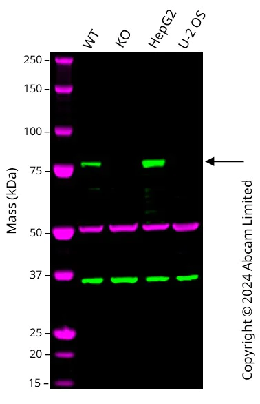
Western blot - Anti-HNF1 alpha antibody [EPR23054-108] - BSA and Azide free (AB272708)
This data was developed using the same antibody clone in a different buffer formulation (ab272693).
Western blot : Anti-HNF1A antibody [EPR23054-108] (ab272693) staining at 1/1000 dilution, shown in green; Mouse anti-Alpha Tubulin [DM1A] (ab7291) loading control staining at 1/20000 dilution, shown in magenta. In Western blot, ab272693 was shown to bind specifically to HNF1A. A band was observed at 75-80 kDa in wild-type A549 cell lysates with no signal observed at this size in HNF1A knockout cell line. To generate this image, wild-type and HNF1A knockout A549 cell lysates were analysed. First, samples were run on an SDS-PAGE gel then transferred onto a nitrocellulose membrane. Membranes were blocked in 3 % milk in TBS-0.1 % Tween® 20 (TBS-T) before incubation with primary antibodies overnight at 4 °C. Blots were washed four times in TBS-T, incubated with secondary antibodies for 1 h at room temperature, washed again four times then imaged. Secondary antibodies used were Goat anti-Rabbit IgG H&L 800CW and Goat anti-Mouse IgG H&L 680RD at 1/20000 dilution.
All lanes:
Western blot - Anti-HNF1 alpha antibody [EPR23054-108] (<a href='/en-us/products/primary-antibodies/hnf1-alpha-antibody-epr23054-108-ab272693'>ab272693</a>) at 1/1000 dilution
Lane 1:
Wild-type A549 cell lysate at 20 µg
Lane 2:
HNF1A knockout A549 cell lysate at 20 µg
Lane 3:
HepG2 cell lysate at 20 µg
Lane 4:
U-2 OS cell lysate at 20 µg
Secondary
All lanes:
Goat anti-Rabbit IgG H&L 800CW and Goat anti-Mouse IgG H&L 680RD at 1/20000 dilution
Observed band size: 75 kDa,80 kDa
false
- ChIC/CUT&RUN-seq
Supplier Data
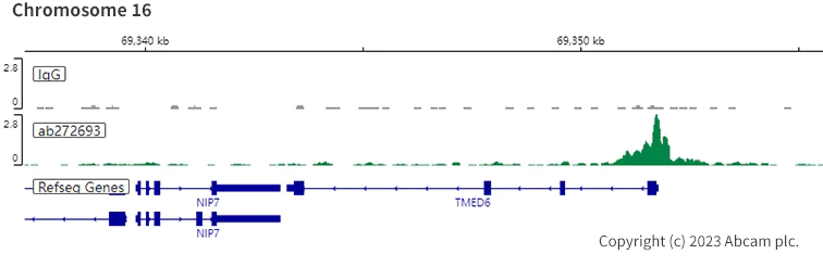
ChIC/CUT&RUN sequencing - Anti-HNF1 alpha antibody [EPR23054-108] - BSA and Azide free (AB272708)
ChIC/CUT&RUN was performed using a pAG-MNAse at a final concentration of 700 ng/mL, 2.5 x 10^5 HepG2 (Human liver hepatocellular carcinoma cell line)cells and 5µg of ab272693 [EPR23054-108]. The resulting DNA was sequenced on the Illumina NovaSeq 6000 to a depth of 10 million reads. The negative IgG control ab172730 is also shown. Additional screenshots of mapped reads can be downloaded here. The University of Geneva owns patents relevant to ChIC (Chromatin Immuno-Cleavage) methods. This data was developed using the same antibody clone in a different buffer formulation containing PBS, BSA, glycerol, and sodium azide (ab272693).
Related conjugates and formulations (1)
-
Anti-HNF1 alpha antibody [EPR23054-108]
Complementary Products

Primary Antibodies
AB181604
Anti-HNF-4-alpha antibody [EPR16885] - ChIP Grade
20ul selling size|RabMAb|Advanced Validation|Recombinant

- 5
- 4


Primary Antibodies
AB170933
Anti-FOXA1 + FOXA2 + FOXA3 antibody [EPR10881]
RabMAb|Recombinant|Advanced Validation|20ul selling size

- 5
- 1
Reactivity data
Product details
ab272708 is the carrier-free version of ab272693.
Patented technology
Our RabMAb® technology is a patented hybridoma-based technology for making rabbit monoclonal antibodies. For details on our patents, please refer to RabMAb® patents.
What are the advantages of a recombinant monoclonal antibody?
This product is a recombinant monoclonal antibody, which offers several advantages including:
- - High batch-to-batch consistency and reproducibility
- - Improved sensitivity and specificity
- - Long-term security of supply
- - Animal-free batch production
For more information, read more on recombinant antibodies.
Conjugation ready
Our carrier-free antibodies are typically supplied in a PBS-only formulation, purified and free of BSA, sodium azide and glycerol. This conjugation-ready format is designed for use with fluorochromes, metal isotopes, oligonucleotides, and enzymes, which makes them ideal for antibody labelling, functional and cell-based assays, flow-based assays (e.g. mass cytometry) and Multiplex Imaging applications.
Use our conjugation kits for antibody conjugates that are ready-to-use in as little as 20 minutes with 1 minute hands-on-time and 100% antibody recovery: available for fluorescent dyes, HRP, biotin and gold.
Compatibility
This product is compatible with the Maxpar® Antibody Labeling Kit from Fluidigm, without the need for antibody preparation. Maxpar® is a trademark of Fluidigm Canada Inc.
Properties and storage information
Form
Purification technique
Storage buffer
Shipped at conditions
Appropriate short-term storage conditions
Appropriate long-term storage conditions
Storage information
Supplementary information
This supplementary information is collated from multiple sources and compiled automatically.
Biological function summary
The HNF1 alpha functions as part of the dimeric HNF1 protein complex often partnering with HNF1 beta. It governs a wide array of genes including those involved in carbohydrate and lipid metabolism. By influencing the gene expression HNF1 alpha assists in maintaining normal metabolic processes. Its activity is critical for proper liver function and insulin secretion from pancreatic beta cells.
Pathways
HNF1 alpha actively integrates into key metabolic pathways such as the gluconeogenesis and the cholesterol biosynthesis pathways. In these pathways it works alongside other proteins like HNF4 alpha and PGC-1 alpha contributing significantly to glucose and lipid homeostasis. The transcription factor impacts the metabolic pathway reinforcing its integral role in maintaining energy balance and metabolic health.
Product protocols
- Visit the General protocols
- Visit the Troubleshooting
- Download chicCutRunSequencingBooklet|en
Target data
Alternative Names
Publications (1)
Recent publications for all applications. Explore the full listand refine your search
Scientific reports 12:11602 PubMed35804081
2022
Applications
Unspecified application
Species
Unspecified reactive species
Product promise
Please note: All products are 'FOR RESEARCH USE ONLY. NOT FOR USE IN DIAGNOSTIC OR THERAPEUTIC PROCEDURES'.
For licensing inquiries, please contact partnerships@abcam.com