Anti-Influenza A virus PB1 antibody [EPR28248-26]
- BOND RX™ Validated
- 20ul selling size
- RabMAb
- Recombinant
- What is this?
Be the first to review this product! Submit a review
|
(0 Publication)
- IHC-P
Supplier Data
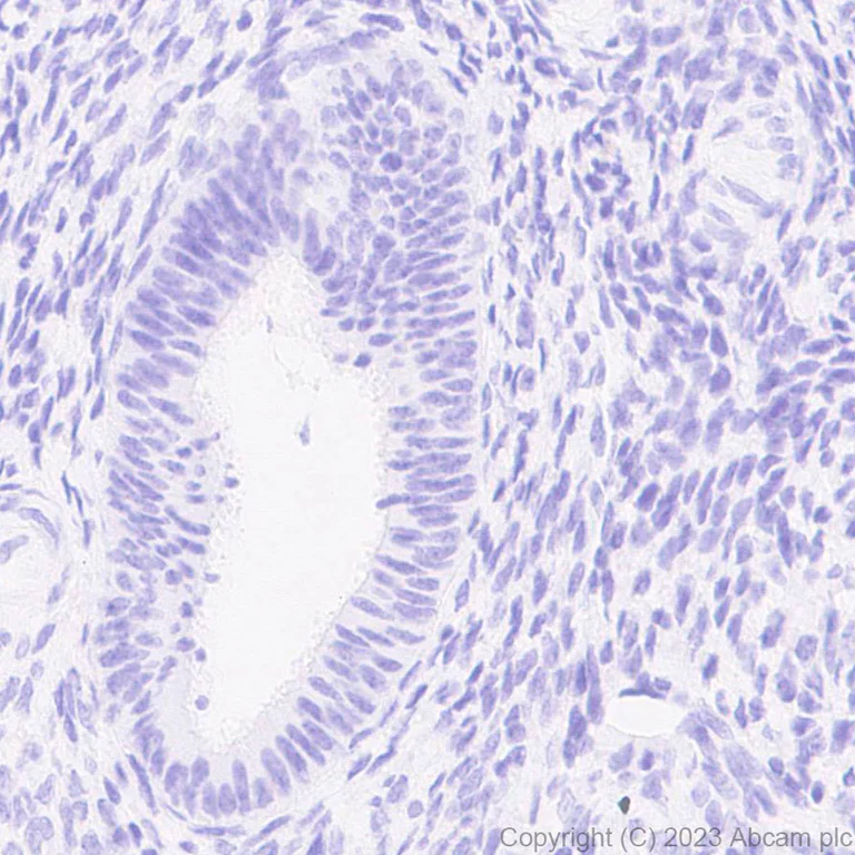
Immunohistochemistry (Formalin/PFA-fixed paraffin-embedded sections) - Anti-Influenza A virus PB1 antibody [EPR28248-26] (AB313887)
Immunohistochemical analysis of paraffin-embedded human endometrium tissue labeling Influenza A virus PB1 protein with ab313887 at 1/1000 (0.5 µg/ml) followed by a ready to use Goat Anti-Rabbit IgG H&L (HRP). Negative control : No staining on human endometrium. The section was incubated with ab313887 for 30 mins at room temperature. The immunostaining was performed on a Leica Biosystems BOND® RX instrument. Counterstained with Hematoxylin. Secondary antibody only control : Secondary antibody is a ready to use Goat Anti-Rabbit IgG H&L (HRP). Heat mediated antigen retrieval was performed with Tris-EDTA buffer (pH 9.0, Epitope Retrieval Solution2) for 20 mins.
- IHC-P
Supplier Data
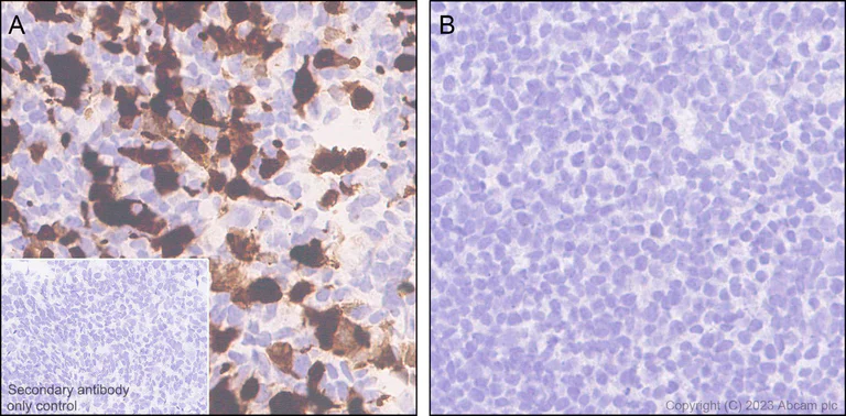
Immunohistochemistry (Formalin/PFA-fixed paraffin-embedded sections) - Anti-Influenza A virus PB1 antibody [EPR28248-26] (AB313887)
Immunohistochemical analysis of paraffin-embedded (A) 293T (human epithelial cell line from embryonic kidney) transfected with an Influenza A virus PB1 expression vector containing a Myc-his tag and (B) 293T transfected with empty vector containing a Myc-his tag with ab313887 at 1/1000 (0.5 µg/ml) followed by a ready to use Goat Anti-Rabbit IgG H&L (HRP). Positive staining on (A) 293T (human epithelial cell line from embryonic kidney) transfected with an Influenza A virus PB1 expression vector containing a Myc-his tag, no staining on (B) 293T transfected with empty vector containing a Myc-his tag. The section was incubated with ab313887 for 30 mins at room temperature. The immunostaining was performed on a Leica Biosystems BOND® RX instrument. Counterstained with Hematoxylin. Secondary antibody only control : Secondary antibody is a ready to use Goat Anti-Rabbit IgG H&L (HRP). Heat mediated antigen retrieval was performed with Tris-EDTA buffer (pH 9.0, Epitope Retrieval Solution2) for 20 mins
- Flow Cyt (Intra)
Supplier Data
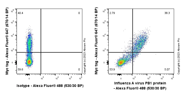
Flow Cytometry (Intracellular) - Anti-Influenza A virus PB1 antibody [EPR28248-26] (AB313887)
Flow cytometric analysis of 4% paraformaldehyde fixed 90% methanol permeabilized 293T (human embryonic kidney epithelial cell) transfected with a Influenza A virus PB1 protein expression vector containing a myc tag cells labelling Influenza A virus PB1 protein with ab313887 at 1/500 dilution (0.1 ug)/Right (Red) compared with a Rabbit monoclonal IgG (ab172730) / Left isotype control and an unlabelled control (cells without incubation with primary antibody and secondary antibody) (Blue). A Goat Anti-Rabbit IgG (Alexa Fluor® 488, ab150081) at 1/5000 dilution was used as the secondary antibody. Cells are co-stained with Myc tag conjugated to Alexa Fluor®647.

- ICC/IF
Supplier Data
Immunocytochemistry/ Immunofluorescence - Anti-Influenza A virus PB1 antibody [EPR28248-26] (AB313887)
Immunofluorescent analysis of 4% Paraformaldehyde-fixed, 0.1% TritonX-100 permeabilized 293T (human embryonic kidney epithelial cell) cells labelling Influenza A virus PB1 protein with ab313887 at 1/1000 (0.5 µg/ml) dilution, followed by ab150081 Goat Anti-Rabbit IgG H&L (Alexa Fluor® 488) preadsorbed antibody at 1/1000 (2 µg/ml) dilution (Green). Confocal image showing positive staining staining in 293T cells transfected with a Influenza A virus PB1 protein expression vector containing a myc tag. Image was taken with a confocal microscope(Leica-Microsystems, TCS SP8) Myc-Tag Mouse mAb (Alexa Fluor® 647) was used to counterstain tubulin at 1/100 (0.38 µg/ml) dilution (Red). The Nuclear counterstain was DAPI (Blue). Secondary antibody only control : Secondary antibody is ab150081 Goat Anti-Rabbit IgG H&L (Alexa Fluor® 488) preadsorbed at 1/1000 (2 µg/ml) dilution.
- WB
Supplier Data
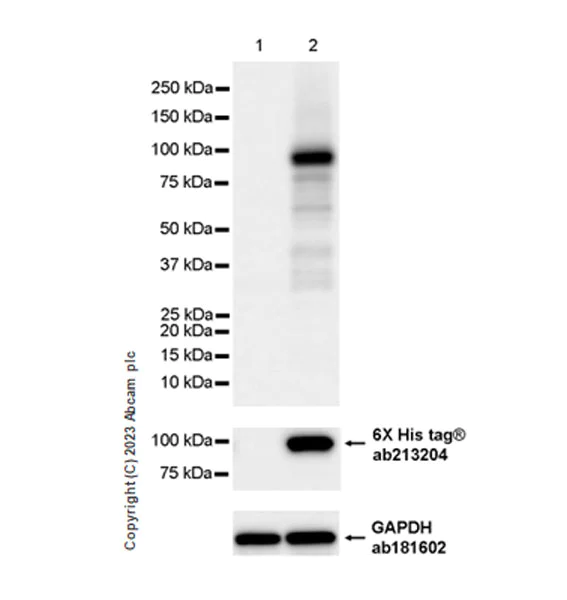
Western blot - Anti-Influenza A virus PB1 antibody [EPR28248-26] (AB313887)
Blocking and diluting buffer and concentration : 5% NFDM/TBST In Western blot, Anti-GAPDH antibody [EPR16891] - Loading Control (ab181602) staining at 1/200000 dilution. In Western blot, Anti-6X His tag® antibody [EPR20547] - ChIP Grade (ab213204) staining at 1/5000 dilution Exposure time : 8 seconds
All lanes:
Western blot - Anti-Influenza A virus PB1 antibody [EPR28248-26] (ab313887) at 1/1000 dilution
Lane 1:
293T (human embryonic kidney epithelial cell) cells transfected with an empty vector containing a myc-His-tag® whole cell lysate at 7 µg
Lane 2:
293T cells transfected with an Influenza A virus PB1 expression vector containing a myc-His-tag® whole cell lysate at 7 µg
Secondary
All lanes:
Western blot - Goat Anti-Rabbit IgG H&L (HRP) (<a href='/en-us/products/secondary-antibodies/goat-rabbit-igg-h-l-hrp-ab97051'>ab97051</a>) at 1/100000 dilution
Observed band size: 87 kDa
false
Exposure time: 8s
- I-ELISA
Supplier Data
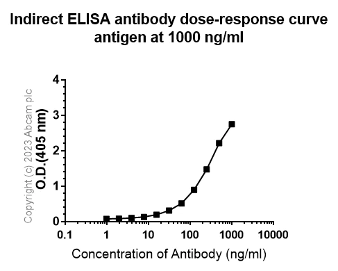
Indirect ELISA - Anti-Influenza A virus PB1 antibody [EPR28248-26] (AB313887)
Indirect ELISA analysis of ab313887 at 1000-0 ng/ml. The Secondary antibody used was Alkaline Phosphatase-conjugated AffiniPure Goat Anti-Rabbit IgG (H+L) at 1 : 2500 dilution. Antigen : Influenza A virus PB1 protein. Antigen concentration : 1000 ng/ml.
Related conjugates and formulations (1)
-
Anti-Influenza A virus PB1 antibody [EPR28248-26] - BSA and Azide free
Complementary Products
Primary Antibodies
AB313746
Anti-Influenza A Virus PA protein antibody [EPR28250-39] - BSA and Azide free
BOND RX™ Validated|RabMAb|Recombinant
- 0
- 0
Primary Antibodies
AB313343
Anti-Influenza A Virus Nucleoprotein antibody [EPR28253-17]
20ul selling size|RabMAb|Recombinant

- 5
- 1
Primary Antibodies
AB312856
Anti-Influenza A Virus PB2 protein antibody [EPR28249-46]
BOND RX™ Validated|20ul selling size|RabMAb|Recombinant
- 4
- 1
Primary Antibodies
AB280974
Mouse IgG1 monoclonal [R312-MouseIgG1]-Isotype control
BOND RX™ Validated|Recombinant|Trial Size

- 0
- 0
Reactivity data
Product details
Patented technology
Our RabMAb® technology is a patented hybridoma-based technology for making rabbit monoclonal antibodies. For details on our patents, please refer to RabMAb® patents.
What are the advantages of a recombinant monoclonal antibody?
This product is a recombinant monoclonal antibody, which offers several advantages including:
- - High batch-to-batch consistency and reproducibility
- - Improved sensitivity and specificity
- - Long-term security of supply
- - Animal-free batch production
For more information, read more on recombinant antibodies.
Properties and storage information
Form
Purification technique
Storage buffer
Shipped at conditions
Appropriate short-term storage duration
Appropriate short-term storage conditions
Appropriate long-term storage conditions
Aliquoting information
Storage information
Supplementary information
This supplementary information is collated from multiple sources and compiled automatically.
Biological function summary
The PB1 protein collaborates with two other polymerase proteins PB2 and PA in the formation of the influenza virus RNA polymerase complex. This complex is essential for catalyzing the transcription and replication of the virus's segmented negative-strand RNA genome within the host cell. The polymerase activity of PB1 drives the production of viral RNA ensuring the assembly of new viral particles for subsequent cycles of infection.
Pathways
PB1 protein functions centrally in the viral replication and transcription pathways within infected host cells. It interacts with PB2 and PA proteins to align with these pathways facilitating genomic replication and transcription of viral mRNA. The role of PB1 in catalyzing RNA synthesis also connects it to the host's cellular machinery pathways exploiting components such as the cellular nuclear export pathway to ensure efficient viral genome replication and viral propagation.
Product protocols
- Visit the General protocols
- Visit the Troubleshooting
Target data
Alternative Names
Product promise
Please note: All products are 'FOR RESEARCH USE ONLY. NOT FOR USE IN DIAGNOSTIC OR THERAPEUTIC PROCEDURES'.
For licensing inquiries, please contact partnerships@abcam.com