Anti-IRF5 antibody [RP23040171]
- Recombinant
- What is this?
Be the first to review this product! Submit a review
|
(0 Publication)
Rabbit Recombinant Multiclonal IRF5 antibody. Suitable for ICC/IF, ChIP and reacts with Human samples. Immunogen corresponding to Synthetic Peptide within Human IRF5.
View Alternative Names
Interferon regulatory factor 5, IRF-5, IRF5
- ICC/IF
Supplier Data
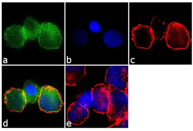
Immunocytochemistry/ Immunofluorescence - Anti-IRF5 Antibody [RP23040171] (AB313466)
Immunofluorescence was performed on fixed and permeabilized THP-1 cells for detection of endogenous IRF5 using ab313466 (2 ug/ml) and labeled with Goat anti-Rabbit IgG (H+L) Secondary Antibody, Alexa Fluor® 488 conjugate ( 1 : 2000). Panel a) shows representative cells that were stained for detection and localization of IRF5 protein (green), Panel b) is stained for nuclei (blue) using with DAPI . Panel c) represents cytoskeletal F-actin staining using Alexa Fluor® 555 Rhodamine Phalloidin ( 1 : 300). Panel d) is a composite image of Panels a, b and c clearly demonstrating cytoplasmic localization of IRF5. Panel e) represents control cells with no primary antibody to assess background. The images were captured at 60X magnification.
- ICC/IF
Supplier Data
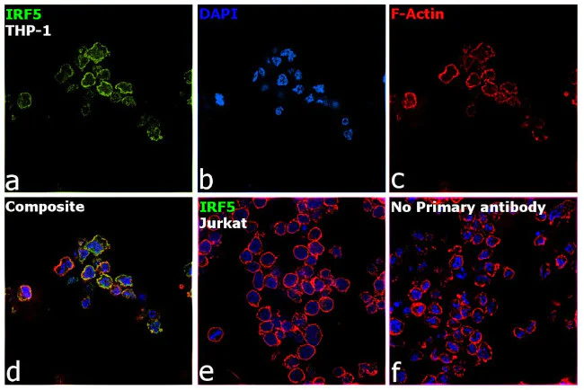
Immunocytochemistry/ Immunofluorescence - Anti-IRF5 Antibody [RP23040171] (AB313466)
Immunofluorescence analysis of IRF5 was performed using 70% confluent log phase THP-1 cells. The cells were fixed with 4% paraformaldehyde for 10 minutes, permeabilized with 0.1% Triton X-100 for 15 minutes, and blocked with 2% BSA for 1 hour at room temperature. The cells were labeled with ab313466 at 5 ug/ml n 0.1% BSA, incubated at 4 degree Celsius overnight and then with Donkey anti-Rabbit IgG (H+L) Highly Cross-Adsorbed Secondary Antibody, Alexa Fluor®Plus 488 at a dilution of 1 : 2000 for 45 minutes at room temperature (Panel a : green). Nuclei (Panel b : blue) were stained with with DAPI . F-actin (Panel c : red) was stained with Rhodamine Phalloidin ( 1 : 300). Panel d represents the merged image showing cytoplasmic and nuclear localization. Panel e represents Jurkat cells having no expression of IRF5. Panel f represents control cells with no primary antibody to assess background. The images were captured at 60X magnification.
- ChIP
Supplier Data
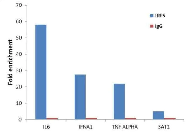
ChIP - Anti-IRF5 Antibody [RP23040171] (AB313466)
Enrichment of endogenous IRF5 protein at specific gene loci using ab313466 : Chromatin Immunoprecipitation (ChIP) was performed using ab313466,(5ug) on sheared chromatin from 2 million THP1 cells treated with 10 ng/mL of Leptomycin B for 90 minutes. Normal Rabbit IgG (1 ug) was used as a negative IP control. The purified DNA was analyzed by 7500 Fast qPCR system with optimized PCR primer pairs of the active IL6, IFNA1, TNF ALPHA promoter regions used as positive control target genes, and the region of the inactive SAT2 satellite repeat, used as negative control target gene. Data is presented as fold enrichment of the antibody signal versus the negative control IgG using the comparative CT method.
Complementary Products
Primary Antibodies
AB309088
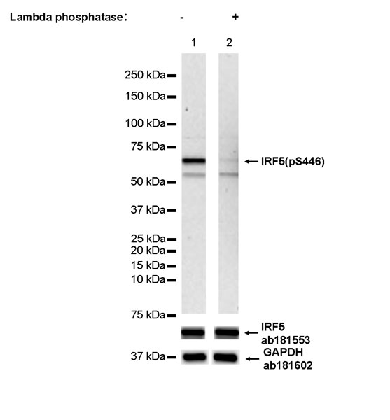
Anti-IRF5 (phospho S446) antibody [EPR26132-254]
20ul selling size|RabMAb|Recombinant|KO Validated
- 0
- 0
Primary Antibodies
AB68481
20ul selling size|RabMAb|Recombinant|KO Validated

- 5
- 2
Primary Antibodies
AB183734
RabMAb|Recombinant|Lab Essentials|20ul selling size
- 5
- 24
Primary Antibodies
AB124964
Anti-alpha smooth muscle Actin antibody [EPR5368]
BOND RX™ Validated|RabMAb|Recombinant|KO Validated|20ul selling size

- 5
- 19
Reactivity data
Product details
What are recombinant multiclonals?
Recombinant multiclonals are a mixture of recombinant antibodies co-expressed from a library of heavy and light chains. They offer several advantages including:
- - The sensitivity of polyclonal antibodies by recognising multiple epitopes
- - High batch-to-batch consistency and reproducibility
- - Improved sensitivity and specificity
- - Long-term security of supply
- - Animal-free batch production
View our range of recombinant multiclonal antibodies.
Properties and storage information
Form
Purification technique
Storage buffer
Shipped at conditions
Appropriate short-term storage duration
Appropriate short-term storage conditions
Appropriate long-term storage conditions
Aliquoting information
Storage information
Supplementary information
This supplementary information is collated from multiple sources and compiled automatically.
Biological function summary
IRF5 influences the immune response through the regulation of cytokine production. It activates genes involved in producing pro-inflammatory cytokines such as TNF-α and IL-6 which are important for clearing viral infections and activating immune cells. IRF5 does not usually form part of a protein complex but it interacts closely with TRAF6 and MyD88 for its activation. Its pivotal role in cytokine production highlights its importance in controlling inflammation.
Pathways
IRF5 is an important component of the Toll-like receptor (TLR) signaling pathway and plays a role in the MyD88-dependent pathway. In this context IRF5 gets activated by TLRs interacting with MyD88 and TAK1 to enhance the transcription of important genes for immune response. It is also closely connected to the IRF3 protein which shares similar activation pathways and can similarly initiate an antiviral state.
Product protocols
- Visit the General protocols
- Visit the Troubleshooting
Target data
Product promise
Please note: All products are 'FOR RESEARCH USE ONLY. NOT FOR USE IN DIAGNOSTIC OR THERAPEUTIC PROCEDURES'.
For licensing inquiries, please contact partnerships@abcam.com