Anti-KIF5A + KIF5B + KIF5C antibody [EPR24325-98]
- 20ul selling size
- RabMAb
- Recombinant
- What is this?
Be the first to review this product! Submit a review
|
(0 Publication)

- Flow Cyt (Intra)
Supplier Data
Flow Cytometry (Intracellular) - Anti-KIF5A + KIF5B + KIF5C antibody [EPR24325-98] (AB271049)
Flow cytometric analysis of 4% paraformaldehyde fixed 90% methanol permeabilized SH-SY5Y (Human neuroblastoma epithelial cell) cells labelling KIF5A + KIF5B + KIF5C with ab271049 at 1/500 dilution (0.1ug) (Red) compared with a Rabbit monoclonal IgG (ab172730) (Black) isotype control and an unlabelled control (cells without incubation with primary antibody and secondary antibody) (Blue). A Goat F(ab')2 Anti-Rabbit IgG(DyLight® 488, ab98507) at 1/500 dilution was used as the secondary antibody.
- ICC/IF
Supplier Data
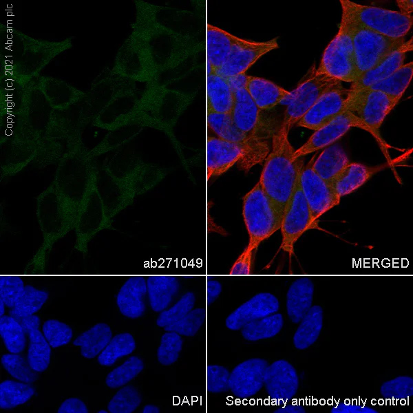
Immunocytochemistry/ Immunofluorescence - Anti-KIF5A + KIF5B + KIF5C antibody [EPR24325-98] (AB271049)
Immunofluorescent analysis of 4% Paraformaldehyde-fixed, 0.1% TritonX-100 permeabilized SH-SY5Y cells labelling KIF5A + KIF5B + KIF5C with ab271049 at 1/50 (9.72 ug/ml) dilution, followed by ab150080 Goat Anti-Rabbit IgG H&L (Alexa Fluor® 594) antibody at 1/500 (4 ug/ml) dilution (Green). Confocal image showing cytoplasmic staining in SH-SY5Y cell line. is observed. ab195889 Anti-alpha Tubulin mouse monoclonal antibody - Microtubule Marker (Alexa Fluor® 594) was used to counterstain tubulin at 1/200 (2.5 ug/ml) dilution (Red). The Nuclear counterstain was DAPI (Blue).
Secondary antibody only control : Secondary antibody is ab150080 Goat Anti-Rabbit IgG H&L (Alexa Fluor® 594) at 1/500 (4 ug/ml) dilution.
- IP
Supplier Data
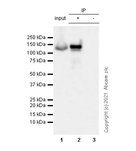
Immunoprecipitation - Anti-KIF5A + KIF5B + KIF5C antibody [EPR24325-98] (AB271049)
KIF5A + KIF5B + KIF5C was immunoprecipitated from 0.35 mg SH-SY5Y (human neuroblastoma epithelial cell) whole cell lysate 10 ug with ab271049 at 1/30 dilution (2ug in 0.35mg lysates). Western blot was performed on the immunoprecipitate using ab271049 at 1/1000 dilution. VeriBlot for IP secondary antibody(HRP)(ab131366) was used at 1/5000 dilution.
Lane 1 : SH-SY5Y (human neuroblastoma epithelial cell) whole cell lysate 10 ug
Lane 2 : ab271049 IP in SH-SY5Y whole cell lysate
Lane 3 : Rabbit monoclonal IgG (ab172730) instead of ab271049 in SH-SY5Y whole cell lysate
Blocking and dilution buffer and concentration : 5% NFDM/TBST.
Exposure time : 3 seconds
All lanes:
Immunoprecipitation - Anti-KIF5A + KIF5B + KIF5C antibody [EPR24325-98] (ab271049)
Observed band size: 117 kDa
false

- Flow Cyt (Intra)
Supplier Data
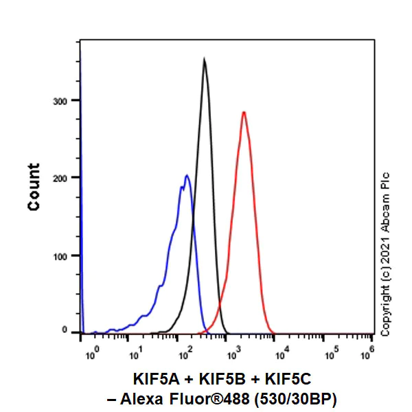
Flow Cytometry (Intracellular) - Anti-KIF5A + KIF5B + KIF5C antibody [EPR24325-98] (AB271049)
Flow cytometric analysis of 4% paraformaldehyde fixed 90% methanol permeabilized C6 (Rat glial tumor glial cell) cells labelling KIF5A + KIF5B + KIF5C with ab271049 at 1/500 dilution (0.1ug) (Red) compared with a Rabbit monoclonal IgG (ab172730) (Black) isotype control and an unlabelled control (cells without incubation with primary antibody and secondary antibody) (Blue). A Goat Anti-Rabbit IgG (Alexa Fluor® 488, ab150081) at 1/2000 dilution was used as the secondary antibody.
- ICC/IF
Supplier Data
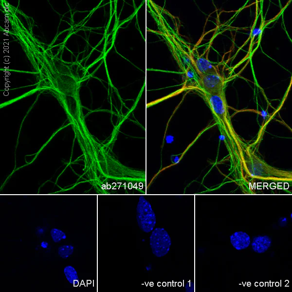
Immunocytochemistry/ Immunofluorescence - Anti-KIF5A + KIF5B + KIF5C antibody [EPR24325-98] (AB271049)
Immunofluorescent analysis of 4% Paraformaldehyde-fixed, 0.1% TritonX-100 permeabilized mouse primary neuron cells labelling KIF5A + KIF5B + KIF5C with ab271049 at 1/50 (9.72 ug/ml) dilution, followed by ab150080 Goat Anti-Rabbit IgG H&L (Alexa Fluor® 594) antibody at 1/500 (4 ug/ml) dilution (Green). Confocal image showing cytoplasmic staining in mouse primary neuron. Confocal scanning Z step was set as 0.3 μm followed by image processing with maximum Z projection. ab195889 Anti-alpha Tubulin mouse monoclonal antibody - Microtubule Marker (Alexa Fluor® 594) was used to counterstain tubulin at 1/200 (2.5 ug/ml) dilution (Red). The Nuclear counterstain was DAPI (Blue).
Secondary antibody only control : Secondary antibody is ab150080 Goat Anti-Rabbit IgG H&L (Alexa Fluor® 594) at 1/500 (4 ug/ml) dilution.
- Flow Cyt (Intra)
Supplier Data
Flow Cytometry (Intracellular) - Anti-KIF5A + KIF5B + KIF5C antibody [EPR24325-98] (AB271049)
Flow cytometric analysis of 4% paraformaldehyde fixed 90% methanol permeabilized Neuro-2a (Mouse neuroblastoma neuroblast) cells labelling KIF5A + KIF5B + KIF5C with ab271049 at 1/500 dilution (0.1ug) (Red) compared with a Rabbit monoclonal IgG (ab172730) (Black) isotype control and an unlabelled control (cells without incubation with primary antibody and secondary antibody) (Blue). A Goat Anti-Rabbit IgG (Alexa Fluor® 488, ab150081) at 1/2000 dilution was used as the secondary antibody.
- ICC/IF
Supplier Data
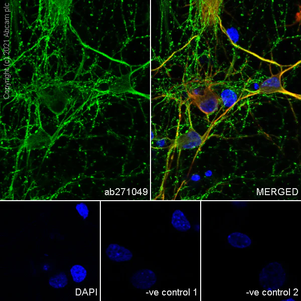
Immunocytochemistry/ Immunofluorescence - Anti-KIF5A + KIF5B + KIF5C antibody [EPR24325-98] (AB271049)
Immunofluorescent analysis of 4% Paraformaldehyde-fixed, 0.1% TritonX-100 permeabilized rat primary neuron cells labelling KIF5A + KIF5B + KIF5C with ab271049 at 1/50 (9.72 ug/ml) dilution, followed by ab150080 Goat Anti-Rabbit IgG H&L (Alexa Fluor® 594) antibody at 1/500 (4 ug/ml) dilution (Green). Confocal image showing cytoplasmic staining in rat primary neuron. Confocal scanning Z step was set as 0.3 μm followed by image processing with maximum Z projection ab11267 Anti-MAP2 mouse monoclonal antibody was used to counterstain tubulin at 1/500 (4 ug/ml) dilution (Red). The Nuclear counterstain was DAPI (Blue).
Secondary antibody only control : Secondary antibody is ab150080 Goat Anti-Rabbit IgG H&L (Alexa Fluor® 594) at 1/500 (4 ug/ml) dilution.
- IP
Supplier Data
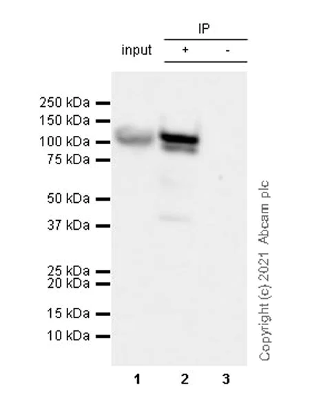
Immunoprecipitation - Anti-KIF5A + KIF5B + KIF5C antibody [EPR24325-98] (AB271049)
KIF5A + KIF5B + KIF5C was immunoprecipitated from 0.35 mg Neuro-2a (mouse neuroblastoma neuroblast) whole cell lysate 10 ug with ab271049 at 1/30 dilution (2ug in 0.35mg lysates). Western blot was performed on the immunoprecipitate using ab271049 at 1/1000 dilution. VeriBlot for IP secondary antibody(HRP)(ab131366) was used at 1/5000 dilution.
Lane 1 : Neuro-2a (mouse neuroblastoma neuroblast) whole cell lysate 10 ug
Lane 2 : ab271049 IP in Neuro-2a whole cell lysate
Lane 3 : Rabbit monoclonal IgG (ab172730) instead of ab271049 in Neuro-2a whole cell lysate
Blocking and dilution buffer and concentration : 5% NFDM/TBST.
Exposure time : 3 seconds
All lanes:
Immunoprecipitation - Anti-KIF5A + KIF5B + KIF5C antibody [EPR24325-98] (ab271049)
Observed band size: 117 kDa
false
- WB
Lab
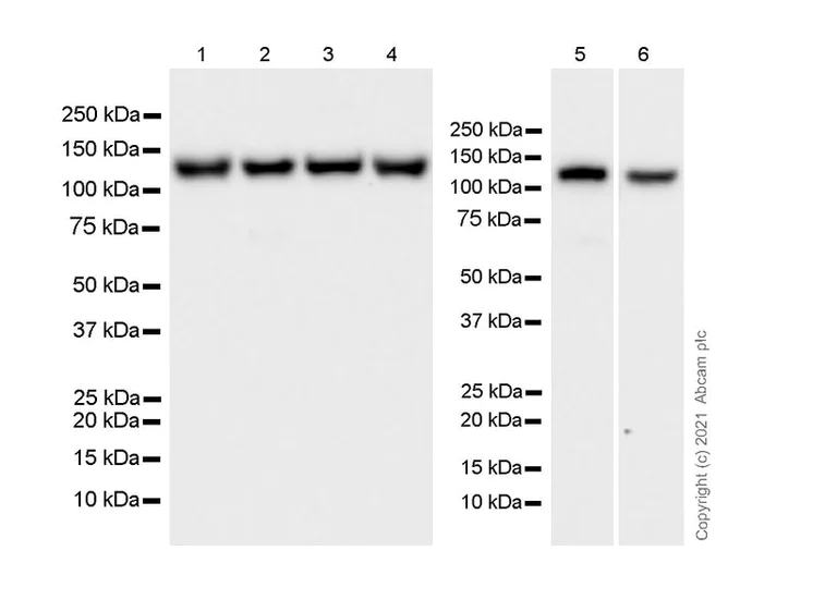
Western blot - Anti-KIF5A + KIF5B + KIF5C antibody [EPR24325-98] (AB271049)
Blocking and diluting buffer and concentration : 5% NFDM/TBST
Exposure time : 10 seconds
All lanes:
Western blot - Anti-KIF5A + KIF5B + KIF5C antibody [EPR24325-98] (ab271049) at 1/1000 dilution
Lane 1:
Neuro-2a (mouse neuroblastoma neuroblast), whole cell lysate at 20 µg
Lane 2:
C2C12 (mouse myoblasts myoblast), whole cell lysate at 20 µg
Lane 3:
U-87 MG (human glioblastoma-astrocytoma epithelial cell), whole cell lysate at 20 µg
Lane 4:
SH-SY5Y (human neuroblastoma epithelial cell), whole cell lysate at 20 µg
Lane 5:
Rat brain cortex tissue lysate at 20 µg
Lane 6:
C6 (rat glial tumor glial cell), whole cell lysate at 20 µg
Secondary
All lanes:
Goat Anti-Rabbit IgG, (H+L), Peroxidase conjugated (<a href='/en-us/products/secondary-antibodies/goat-rabbit-igg-h-l-hrp-ab97051'>ab97051</a>) at 1/100000 dilution
Predicted band size: 117 kDa
Observed band size: 117 kDa
false
- WB
Lab
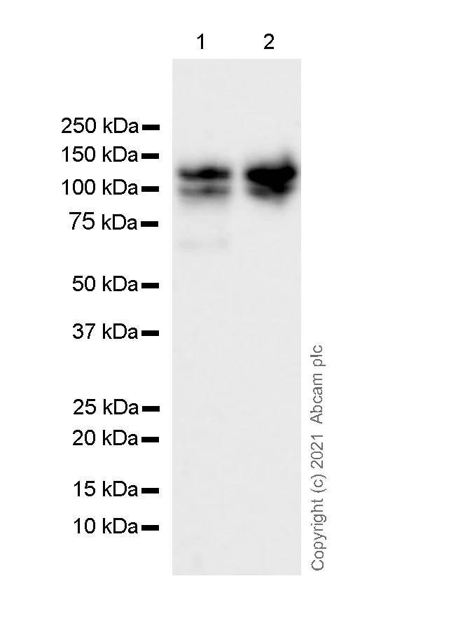
Western blot - Anti-KIF5A + KIF5B + KIF5C antibody [EPR24325-98] (AB271049)
Blocking and diluting buffer and concentration : 5% NFDM/TBST
Degraded fragment (107 KD) has also been described (PMID : 11144355)
Exposure time : 10 seconds
All lanes:
Western blot - Anti-KIF5A + KIF5B + KIF5C antibody [EPR24325-98] (ab271049) at 1/1000 dilution
Lane 1:
Human cerebellum tissue lysate at 20 µg
Lane 2:
Human hypothalamus tissue lysate at 20 µg
Secondary
All lanes:
VeriBlot for IP secondary antibody(HRP)(<a href='/en-us/products/reagents/veriblot-for-ip-detection-reagent-hrp-ab131366'>ab131366</a>) at 1/1000 dilution
Predicted band size: 117 kDa
Observed band size: 107 kDa,117 kDa
false
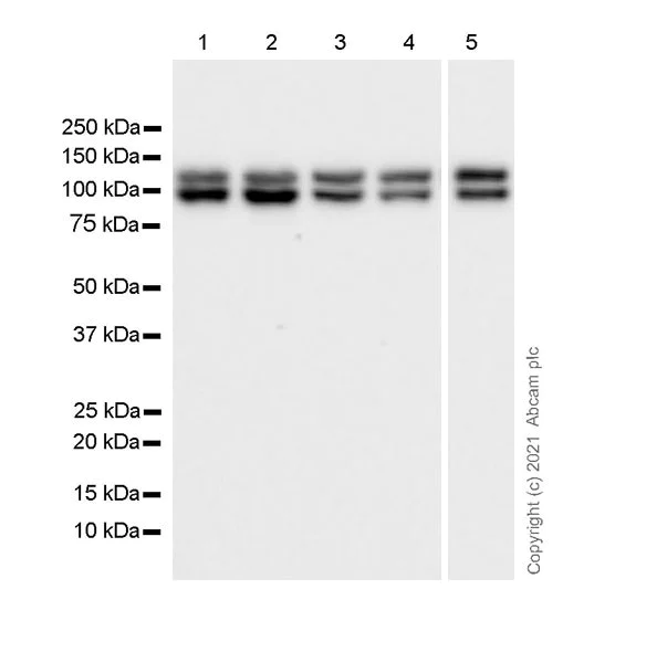
- WB
Lab
Western blot - Anti-KIF5A + KIF5B + KIF5C antibody [EPR24325-98] (AB271049)
Blocking and diluting buffer and concentration : 5% NFDM/TBST
Degraded fragment (107 KD) has also been described (PMID : 11144355)
Exposure time : 10 seconds
All lanes:
Western blot - Anti-KIF5A + KIF5B + KIF5C antibody [EPR24325-98] (ab271049) at 1/1000 dilution
Lane 1:
Mouse brain tissue lysate at 20 µg
Lane 2:
Mouse cerebral cortex tissue lysate at 20 µg
Lane 3:
Mouse cerebellum tissue lysate at 20 µg
Lane 4:
Rat brain tissue lysate at 20 µg
Lane 5:
Rat cerebellum tissue lysate at 20 µg
Secondary
All lanes:
Goat Anti-Rabbit IgG, (H+L), Peroxidase conjugated (<a href='/en-us/products/secondary-antibodies/goat-rabbit-igg-h-l-hrp-ab97051'>ab97051</a>) at 1/100000 dilution
Predicted band size: 117 kDa
Observed band size: 107 kDa,117 kDa
false
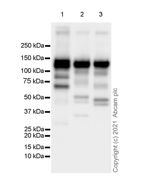
- WB
Lab
Western blot - Anti-KIF5A + KIF5B + KIF5C antibody [EPR24325-98] (AB271049)
Blocking and diluting buffer and concentration : 5% NFDM/TBST
Exposure time : 3 seconds
All lanes:
Western blot - Anti-KIF5A + KIF5B + KIF5C antibody [EPR24325-98] (ab271049) at 1/5000 dilution
Lane 1:
HEK-293T (human embryonic kidney) transfected with KIF5A expression vector containing a myc-His-tag®, whole cell lysate at 2 µg
Lane 2:
HEK-293T transfected with KIF5B expression vector containing a myc-His-tag®, whole cell lysate at 2 µg
Lane 3:
HEK-293T transfected with KIF5C expression vector containing a myc-His-tag®, whole cell lysate at 2 µg
Secondary
All lanes:
Goat Anti-Rabbit IgG, (H+L), Peroxidase conjugated (<a href='/en-us/products/secondary-antibodies/goat-rabbit-igg-h-l-hrp-ab97051'>ab97051</a>) at 1/100000 dilution
Predicted band size: 117 kDa
false
Related conjugates and formulations (1)
-
Anti-KIF5A + KIF5B + KIF5C antibody [EPR24325-98] - BSA and Azide free
Complementary Products
Primary Antibodies
AB7291
Anti-alpha Tubulin antibody [DM1A] - Loading Control
BOND RX™ Validated

- 5
- 92
Primary Antibodies
AB167429
Anti-KIF5B+KIF5C antibody [EPR10276(B)]
RabMAb|Recombinant|KO Validated|20ul selling size

- 4
- 3
Primary Antibodies
AB52866
Anti-alpha Tubulin antibody [EP1332Y] - Loading Control
BOND RX™ Validated|RabMAb|Recombinant|Lab Essentials|20ul selling size|Trial Size

- 5
- 31
Primary Antibodies
AB183734
RabMAb|Recombinant|Lab Essentials|20ul selling size

- 5
- 24
Reactivity data
Product details
Patented technology
Our RabMAb® technology is a patented hybridoma-based technology for making rabbit monoclonal antibodies. For details on our patents, please refer to RabMAb® patents.
What are the advantages of a recombinant monoclonal antibody?
This product is a recombinant monoclonal antibody, which offers several advantages including:
- - High batch-to-batch consistency and reproducibility
- - Improved sensitivity and specificity
- - Long-term security of supply
- - Animal-free batch production
For more information, read more on recombinant antibodies.
Properties and storage information
Form
Purification technique
Storage buffer
Shipped at conditions
Appropriate short-term storage duration
Appropriate short-term storage conditions
Appropriate long-term storage conditions
Aliquoting information
Storage information
Product protocols
- Visit the General protocols
- Visit the Troubleshooting
Target data
Additional targets
Alternative Names
Product promise
Please note: All products are 'FOR RESEARCH USE ONLY. NOT FOR USE IN DIAGNOSTIC OR THERAPEUTIC PROCEDURES'.
For licensing inquiries, please contact partnerships@abcam.com