Anti-MAP2 antibody [EPR19691] - Chicken IgY (Chimeric) - BSA and Azide free
- BOND RX™ Validated
- Recombinant
- What is this?
Be the first to review this product! Submit a review
|
(0 Publication)
- IHC-P
Lab
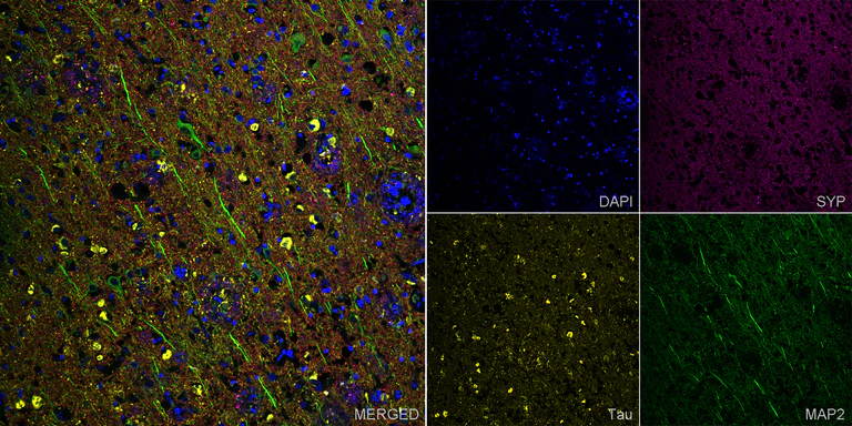
Immunohistochemistry (Formalin/PFA-fixed paraffin-embedded sections) - Anti-MAP2 antibody [EPR19691] - Chicken IgY (Chimeric) - BSA and Azide free (AB318994)
Composite multiplex immunofluorescence staining of SYP, MAP2 and Tau (MBD region) staining in a section of formalin-fixed paraffin-embedded human Alzheimer’s brain*.
Performed on a Leica BOND. The section was pre-treated using heat mediated antigen retrieval with EDTA (Ph9.0) using retrieval settings of 100°C for 40 minutes. The section was then incubated at room temperature for 1 hour with ab309493 at 1µg/ml dilution (shown in green), ab318993 at 1µg/ml (shown in magenta), and ab308439 at 1µg/ml (shown in yellow). Then incubated for 1 hour with ab150119 Goat Anti-Mouse IgG H&L (Alexa Fluor® 647) preadsorbed 1/1000, ab150086 Goat Anti-Rabbit IgG H&L (Alexa Fluor® 555) preadsorbed 1/1000, and ab150173 Goat Anti-Chicken IgY H&L (Alexa Fluor® 488) preadsorbed 1/1000. Nuclear DNA was labelled with DAPI (shown in blue). The section was then mounted using Dako Fluorescence Mounting Medium ®.
Image was taken with a confocal microscope (Leica-Microsystems, TCS SP8).
For other IHC staining systems (automated and non-automated), customers should optimize variable parameters such as antigen retrieval conditions, antibody concentrations and incubation times.
*Tissue obtained from the Human Research Tissue Bank, supported by the NIHR Cambridge Biomedical Research Centre.
- ICC/IF
Supplier Data
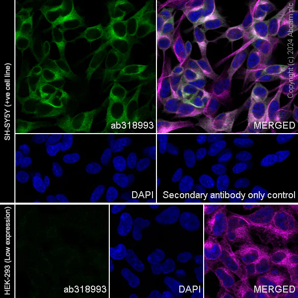
Immunocytochemistry/ Immunofluorescence - Anti-MAP2 antibody [EPR19691] - Chicken IgY (Chimeric) - BSA and Azide free (AB318994)
This data was developed using ab318993, the same antibody clone in a different buffer formulation.
Immunofluorescent analysis of 4% Paraformaldehyde-fixed, 0.1% TritonX-100 permeabilized SH-SY5Y (human neuroblastoma epithelial cell) cells labelling MAP2 with ab318993 at 1/100 (9.72 ug/ml) dilution, followed by ab150173 Goat Anti-Chicken IgY H&L (Alexa Fluor® 488) preadsorbed antibody at 1/1000 2 ug/ml dilution (Green).
Confocal image showing cytoplasmic staining in SH-SY5Y cell line (shown in green). The counterstain was observed in magenta. Nuclear DNA was labeled with DAPI (shown in blue).
Negative control : HEK-293
Image was taken with a confocal microscope (Leica-Microsystems, TCS SP8).
ab195889 Anti-alpha Tubulin mouse monoclonal antibody - Microtubule Marker (Alexa Fluor® 594) was used to counterstain tubulin at 1/200 2.5ug/ml dilution (Magenta). The Nuclear counterstain was DAPI (Blue).
Secondary antibody only control : Secondary antibody is ab150173 Goat Anti-Chicken IgY H&L (Alexa Fluor® 488) preadsorbed at 1/1000 2 ug/ml dilution.
- IHC-P
Supplier Data
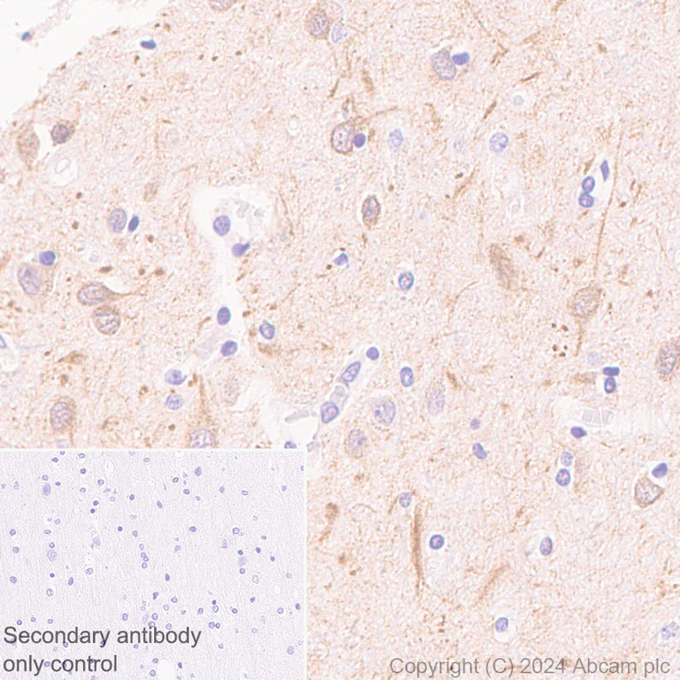
Immunohistochemistry (Formalin/PFA-fixed paraffin-embedded sections) - Anti-MAP2 antibody [EPR19691] - Chicken IgY (Chimeric) - BSA and Azide free (AB318994)
This data was developed using ab318993, the same antibody clone in a different buffer formulation.
Immunohistochemical analysis of paraffin-embedded Human cerebrum tissue labeling MAP2 with ab318993 at 1/50000 (0.019 ug/ml) dilution, followed by a ready to use LeicaDS9800 (Bond™ Polymer Refine Detection).
Positive staining on human cerebrum.
The section was incubated with ab318993 for 10 mins at room temperature and followed by anti-Chicken IgY antibody (ab97136) for 8 mins during the LeicaDS9800 kit staining procedure.
The immunostaining was performed on a Leica Biosystems BOND® RX instrument
Counterstained with Hematoxylin.
Secondary antibody only control : Secondary antibody is a ready to use LeicaDS9800 (Bond™ Polymer Refine Detection).
Heat mediated antigen retrieval was performed with Citrate buffer (pH 6.0, Epitope Retrieval Solution 1) for 10 mins
- IHC-Fr
Supplier Data
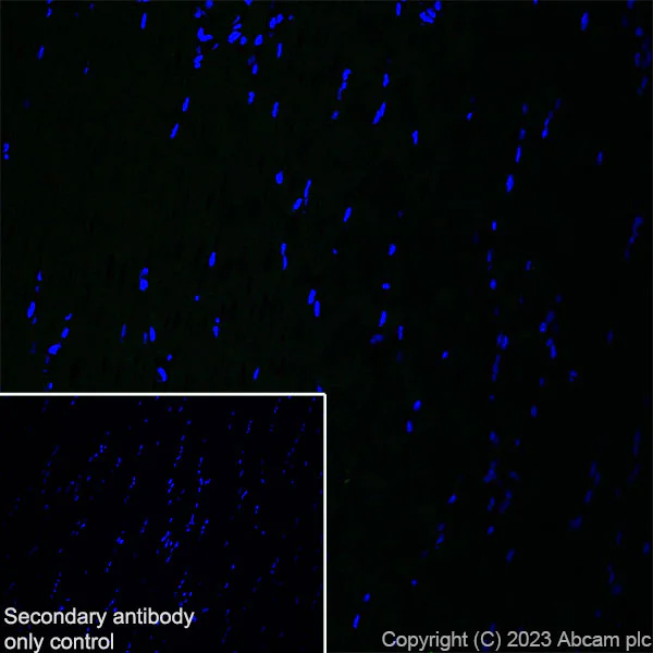
Immunohistochemistry (Frozen sections) - Anti-MAP2 antibody [EPR19691] - Chicken IgY (Chimeric) - BSA and Azide free (AB318994)
This data was developed using ab318993, the same antibody clone in a different buffer formulation.
Immunohistochemical analysis of 4% PFA-fixed, 0.2% Triton X-100 permeabilized frozen Rat skeletal muscle (fresh frozen) tissue labeling MAP2 with ab318993 at 1/2000 (0.486 ug/ml) dilution followed by ab150169 Goat Anti-Chicken IgY H&L (Alexa Fluor® 488) at 1/1000 2 ug/mL dilution (Green).
Negative control : confocal image showing no staining on rat skeletal muscle. The nuclear counterstain was DAPI (Blue). The section was incubated with ab318993 for 60 mins at room temperature. The section was then mounted using Fluoromount®.The immunostaining was performed on a Leica Biosystems BOND® RX instrument. Image was taken with a confocal microscope (Leica-Microsystems, TCS SP8).
Secondary antibody control : Secondary antibody is ab150169 Goat Anti-Chicken IgY H&L (Alexa Fluor® 488)at 1/1000 2 ug/mL dilution.
- Flow Cyt (Intra)
Supplier Data
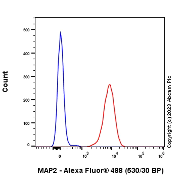
Flow Cytometry (Intracellular) - Anti-MAP2 antibody [EPR19691] - Chicken IgY (Chimeric) - BSA and Azide free (AB318994)
This data was developed using ab318993, the same antibody clone in a different buffer formulation.
Flow cytometric analysis of 4% paraformaldehyde fixed 90% methanol permeabilized PC-12 (rat adrenal gland pheochromocytoma cell) cells labelling MAP2 with ab318993 at 1/1000 dilution (0.1 ug) (Red) compared with a Chicken IgY (Black) isotype control and an unlabelled control (cells without incubation with primary antibody and secondary antibody) (Blue).
Goat anti-Chicken IgY H&L (Alexa Fluor® 488, ab150173) at 1/5000 dilution was used as the secondary antibody.
- IHC-Fr
Supplier Data
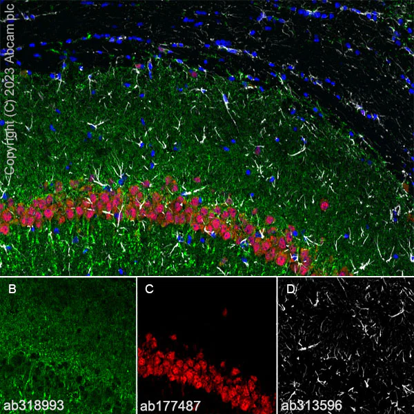
Immunohistochemistry (Frozen sections) - Anti-MAP2 antibody [EPR19691] - Chicken IgY (Chimeric) - BSA and Azide free (AB318994)
This data was developed using ab318993, the same antibody clone in a different buffer formulation.
Immunohistochemical analysis of 4% PFA-fixed, 0.2% Triton X-100 permeabilized frozen Rat hippocampus (fresh frozen) tissue labeling MAP2 with ab318993 at 1/2000 (0.486 ug/ml) dilution followed by ab150169 Goat Anti-Chicken IgY H&L (Alexa Fluor® 488) at 1/1000 2 ug/mL dilution (Green).
Panel A : merged staining of anti-MAP2 (ab318993, green), anti-NeuN (ab177487, red) and anti-GFAP (ab313596, grey) on rat hippocampus.
Panel B : anti-MAP2 stained on rat hippocampus.
Panel C : anti-NeuN stained in neuron of rat hippocampus.
Panel D : anti-GFAP stained in astrocytes of rat hippocampus.
The nuclear counterstain was DAPI (Blue). The section was incubated with ab318993, ab177487 and ab313596 for 60 mins at room temperature. The section was then mounted using Fluoromount®.The immunostaining was performed on a Leica Biosystems BOND® RX instrument. Image was taken with a confocal microscope (Leica-Microsystems, TCS SP8).
Secondary antibody control : Secondary antibody is ab150169 Goat Anti-Chicken IgY H&L (Alexa Fluor® 488)at 1/1000 2 ug/mL dilution.
- IHC-P
Supplier Data
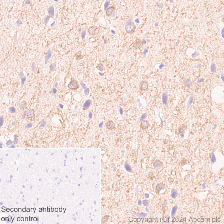
Immunohistochemistry (Formalin/PFA-fixed paraffin-embedded sections) - Anti-MAP2 antibody [EPR19691] - Chicken IgY (Chimeric) - BSA and Azide free (AB318994)
This data was developed using ab318993, the same antibody clone in a different buffer formulation.
Immunohistochemical analysis of paraffin-embedded Rat cerebrum tissue labeling MAP2 with ab318993 at 1/50000 (0.019 ug/ml) dilution, followed by a ready to use LeicaDS9800 (Bond™ Polymer Refine Detection).
Positive staining on rat cerebrum.
The section was incubated with ab318993 for 10 mins at room temperature and followed by anti-Chicken IgY antibody (ab97136) for 8 mins during the LeicaDS9800 kit staining procedure.
The immunostaining was performed on a Leica Biosystems BOND® RX instrument
Counterstained with Hematoxylin.
Secondary antibody only control : Secondary antibody is a ready to use LeicaDS9800 (Bond™ Polymer Refine Detection).
Heat mediated antigen retrieval was performed with Citrate buffer (pH 6.0, Epitope Retrieval Solution 1) for 10 mins
- IHC-P
Supplier Data
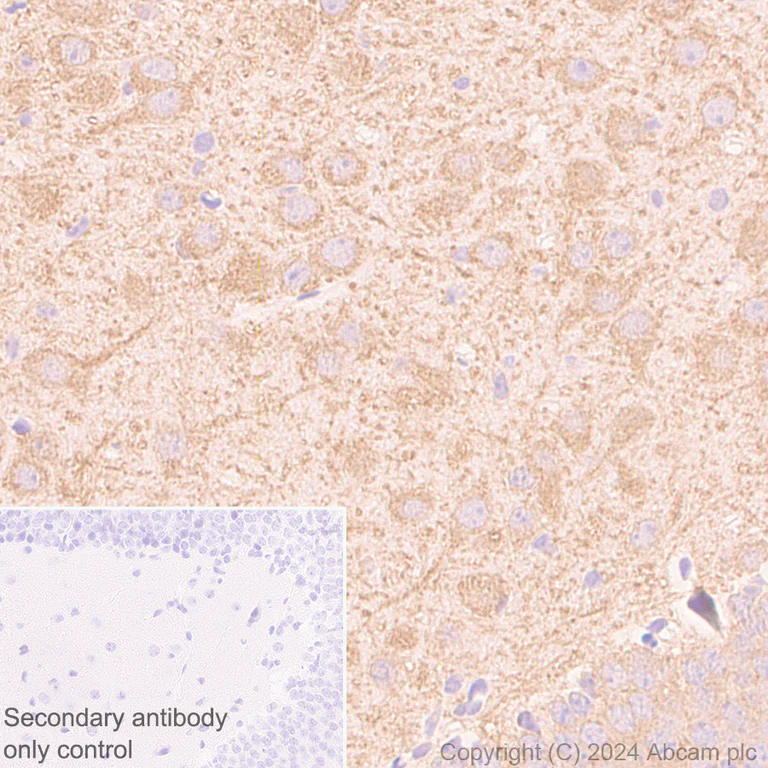
Immunohistochemistry (Formalin/PFA-fixed paraffin-embedded sections) - Anti-MAP2 antibody [EPR19691] - Chicken IgY (Chimeric) - BSA and Azide free (AB318994)
This data was developed using ab318993, the same antibody clone in a different buffer formulation.
Immunohistochemical analysis of paraffin-embedded Mouse cerebrum tissue labeling MAP2 with ab318993 at 1/50000 (0.019 ug/ml) dilution, followed by a ready to use LeicaDS9800 (Bond™ Polymer Refine Detection).
Positive staining on mouse cerebrum.
The section was incubated with ab318993 for 10 mins at room temperature and followed by anti-Chicken IgY antibody (ab97136) for 8 mins during the LeicaDS9800 kit staining procedure.
The immunostaining was performed on a Leica Biosystems BOND® RX instrument
Counterstained with Hematoxylin.
Secondary antibody only control : Secondary antibody is a ready to use LeicaDS9800 (Bond™ Polymer Refine Detection).
Heat mediated antigen retrieval was performed with Citrate buffer (pH 6.0, Epitope Retrieval Solution 1) for 10 mins
- Flow Cyt (Intra)
Supplier Data
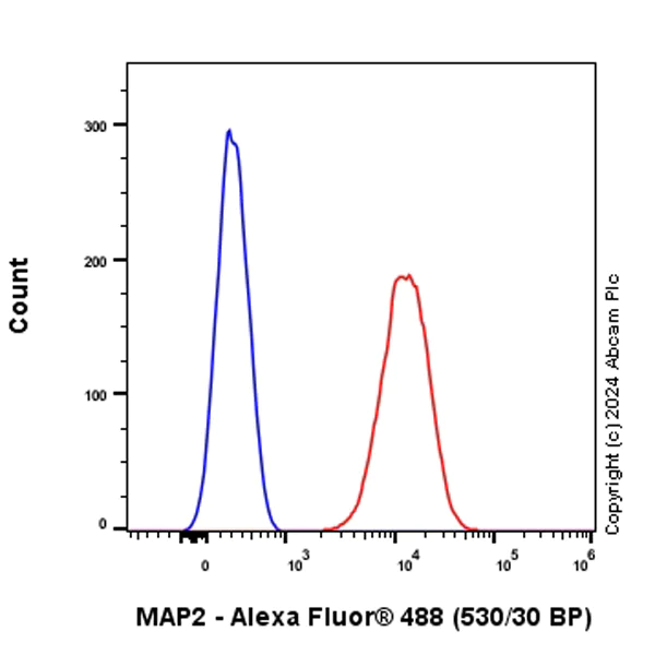
Flow Cytometry (Intracellular) - Anti-MAP2 antibody [EPR19691] - Chicken IgY (Chimeric) - BSA and Azide free (AB318994)
This data was developed using ab318993, the same antibody clone in a different buffer formulation.
Flow cytometric analysis of 4% paraformaldehyde fixed 90% methanol permeabilized Neuro-2a (mouse neuroblastoma neuroblast) cells labelling MAP2 with ab318993 at 1/1000 dilution (0.1 ug) (Red) compared with a Chicken IgY (Black) isotype control and an unlabelled control (cells without incubation with primary antibody and secondary antibody) (Blue).
Goat anti-Chicken IgY H&L (Alexa Fluor® 488, ab150173) at 1/5000 dilution was used as the secondary antibody.
- IHC-Fr
Supplier Data
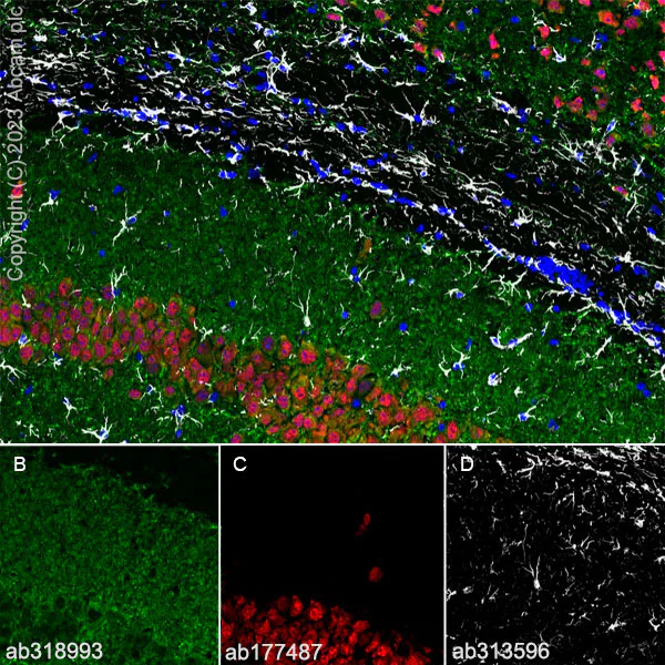
Immunohistochemistry (Frozen sections) - Anti-MAP2 antibody [EPR19691] - Chicken IgY (Chimeric) - BSA and Azide free (AB318994)
This data was developed using ab318993, the same antibody clone in a different buffer formulation.
Immunohistochemical analysis of 4% PFA-fixed, 0.2% Triton X-100 permeabilized frozen Mouse hippocampus (fresh frozen) tissue labeling MAP2 with ab318993 at 1/2000 (0.486 ug/ml) dilution followed by ab150169 Goat Anti-Chicken IgY H&L (Alexa Fluor® 488) at 1/1000 2 ug/mL dilution (Green).
Panel A : merged staining of anti-MAP2 (ab318993, green), anti-NeuN (ab177487, red) and anti-GFAP (ab313596, grey) on mouse hippocampus.
Panel B : anti-MAP2 stained on mouse hippocampus.
Panel C : anti-NeuN stained in neuron of mouse hippocampus.
Panel D : anti-GFAP stained in astrocytes of mouse hippocampus.
The nuclear counterstain was DAPI (Blue). The section was incubated with ab318993, ab177487 and ab313596 for 60 mins at room temperature. The section was then mounted using Fluoromount®.The immunostaining was performed on a Leica Biosystems BOND® RX instrument. Image was taken with a confocal microscope (Leica-Microsystems, TCS SP8).
Secondary antibody control : Secondary antibody is ab150169 Goat Anti-Chicken IgY H&L (Alexa Fluor® 488)at 1/1000 2 ug/mL dilution.
- ICC/IF
Supplier Data
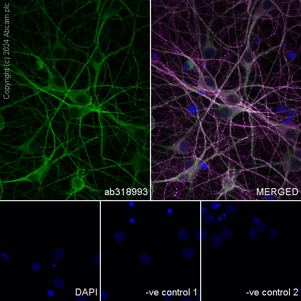
Immunocytochemistry/ Immunofluorescence - Anti-MAP2 antibody [EPR19691] - Chicken IgY (Chimeric) - BSA and Azide free (AB318994)
This data was developed using ab318993, the same antibody clone in a different buffer formulation.
Immunofluorescent analysis of 4% Paraformaldehyde-fixed, 0.1% TritonX-100 permeabilized rat primary neuron cells labelling MAP2 with ab318993 at 1/100 (9.72 ug/ml) dilution, followed by ab150173 Goat Anti-Chicken IgY H&L (Alexa Fluor® 488) preadsorbed antibody at 1/1000 2 ug/ml dilution (Green).
Confocal image showing cytoplasmic staining in rat primary neurons (shown in green). The counterstain was observed in magenta. Nuclear DNA was labeled with DAPI (shown in blue).
Confocal scanning Z step was set as 0.3 μm followed by image processing with maximum Z projection.
Image was taken with a confocal microscope (Leica-Microsystems, TCS SP8).
ab11267 Anti-MAP2 mouse monoclonal antibody was used to counterstain tubulin at 1/500 4ug/ml dilution (Magenta). The Nuclear counterstain was DAPI (Blue).
Secondary antibody only control : Secondary antibody is ab150173 Goat Anti-Chicken IgY H&L (Alexa Fluor® 488) preadsorbed at 1/1000 2 ug/ml dilution.
- IHC-P
Supplier Data
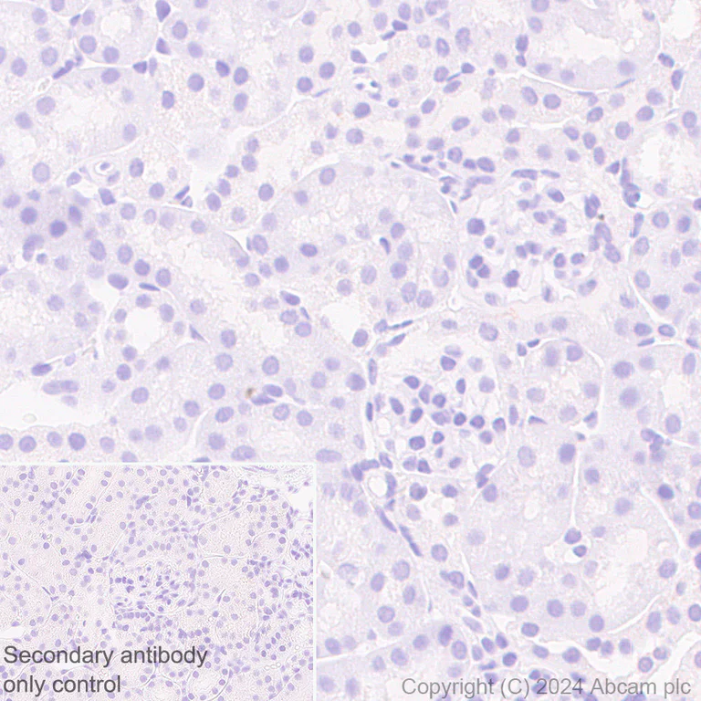
Immunohistochemistry (Formalin/PFA-fixed paraffin-embedded sections) - Anti-MAP2 antibody [EPR19691] - Chicken IgY (Chimeric) - BSA and Azide free (AB318994)
This data was developed using ab318993, the same antibody clone in a different buffer formulation.
Immunohistochemical analysis of paraffin-embedded Mouse kidney tissue labeling MAP2 with ab318993 at 1/50000 (0.019 ug/ml) dilution, followed by a ready to use LeicaDS9800 (Bond™ Polymer Refine Detection).
Negative control : no staining on mouse kidney.
The section was incubated with ab318993 for 10 mins at room temperature and followed by anti-Chicken IgY antibody (ab97136) for 8 mins during the LeicaDS9800 kit staining procedure.
The immunostaining was performed on a Leica Biosystems BOND® RX instrument
Counterstained with Hematoxylin.
Secondary antibody only control : Secondary antibody is a ready to use LeicaDS9800 (Bond™ Polymer Refine Detection).
Heat mediated antigen retrieval was performed with Citrate buffer (pH 6.0, Epitope Retrieval Solution 1) for 10 mins
- ICC/IF
Supplier Data
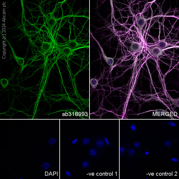
Immunocytochemistry/ Immunofluorescence - Anti-MAP2 antibody [EPR19691] - Chicken IgY (Chimeric) - BSA and Azide free (AB318994)
This data was developed using ab318993, the same antibody clone in a different buffer formulation.
Immunofluorescent analysis of 4% Paraformaldehyde-fixed, 0.1% TritonX-100 permeabilized Mouse primary neuron cells labelling MAP2 with ab318993 at 1/100 (9.72 ug/ml) dilution, followed by ab150173 Goat Anti-Chicken IgY H&L (Alexa Fluor® 488) preadsorbed antibody at 1/1000 2 ug/ml dilution (Green).
Confocal image showing cytoplasmic staining in mouse primary neurons (shown in green). The counterstain was observed in magenta. Nuclear DNA was labeled with DAPI (shown in blue).
Confocal scanning Z step was set as 0.3 μm followed by image processing with maximum Z projection.
Image was taken with a confocal microscope (Leica-Microsystems, TCS SP8).
ab11267 Anti-MAP2 mouse monoclonal antibody was used to counterstain tubulin at 1/500 4ug/ml dilution (Magenta). The Nuclear counterstain was DAPI (Blue).
Secondary antibody only control : Secondary antibody is ab150173 Goat Anti-Chicken IgY H&L (Alexa Fluor® 488) preadsorbed at 1/1000 2 ug/ml dilution.
- IHC-P
Supplier Data
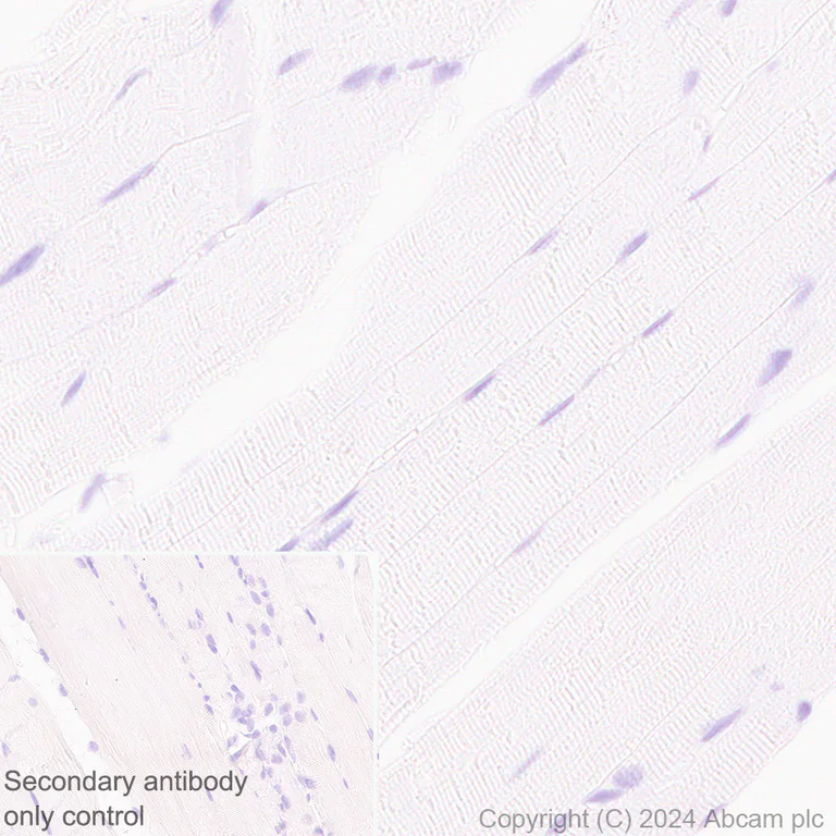
Immunohistochemistry (Formalin/PFA-fixed paraffin-embedded sections) - Anti-MAP2 antibody [EPR19691] - Chicken IgY (Chimeric) - BSA and Azide free (AB318994)
This data was developed using ab318993, the same antibody clone in a different buffer formulation.
Immunohistochemical analysis of paraffin-embedded Mouse skeletal muscle tissue labeling MAP2 with ab318993 at 1/50000 (0.019 ug/ml) dilution, followed by a ready to use LeicaDS9800 (Bond™ Polymer Refine Detection).
Negative control : no staining on mouse skeletal muscle
The section was incubated with ab318993 for 10 mins at room temperature and followed by anti-Chicken IgY antibody (ab97136) for 8 mins during the LeicaDS9800 kit staining procedure.
The immunostaining was performed on a Leica Biosystems BOND® RX instrument
Counterstained with Hematoxylin.
Secondary antibody only control : Secondary antibody is a ready to use LeicaDS9800 (Bond™ Polymer Refine Detection).
Heat mediated antigen retrieval was performed with Citrate buffer (pH 6.0, Epitope Retrieval Solution 1) for 10 mins
- IHC-Fr
Supplier Data
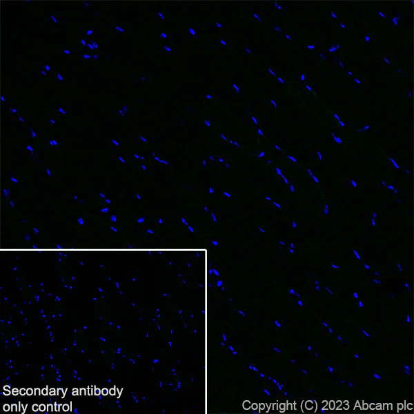
Immunohistochemistry (Frozen sections) - Anti-MAP2 antibody [EPR19691] - Chicken IgY (Chimeric) - BSA and Azide free (AB318994)
This data was developed using ab318993, the same antibody clone in a different buffer formulation.
Immunohistochemical analysis of 4% PFA-fixed, 0.2% Triton X-100 permeabilized frozen Mouse skeletal muscle (fresh frozen) tissue labeling MAP2 with ab318993 at 1/2000 (0.486 ug/ml) dilution followed by ab150169 Goat Anti-Chicken IgY H&L (Alexa Fluor® 488) at 1/1000 2 ug/mL dilution (Green).
Negative control : confocal image showing no staining on mouse skeletal muscle. The nuclear counterstain was DAPI (Blue). The section was incubated with ab318993 for 60 mins at room temperature. The section was then mounted using Fluoromount®.The immunostaining was performed on a Leica Biosystems BOND® RX instrument. Image was taken with a confocal microscope (Leica-Microsystems, TCS SP8).
Secondary antibody control : Secondary antibody is ab150169 Goat Anti-Chicken IgY H&L (Alexa Fluor® 488)at 1/1000 2 ug/mL dilution.
Related conjugates and formulations (6)
-
Anti-MAP2 antibody [HM-2] - Neuronal Marker
-
Anti-MAP2 antibody [EPR19691] - Neuronal Marker - Mouse IgG2a (Chimeric)
-
Anti-MAP2 antibody [EPR19691] - Mouse IgG2a (Chimeric) - BSA and Azide free
-
Anti-MAP2 antibody [EPR19691] - Neuronal Marker - Goat IgG (Chimeric)
-
Anti-MAP2 antibody [EPR19691] - Goat IgG (Chimeric) - BSA and Azide free
-
Anti-MAP2 antibody [EPR19691] - Neuronal Marker - Chicken IgY (Chimeric)
Complementary Products


Primary Antibodies
AB177487
Anti-NeuN antibody [EPR12763] - Neuronal Marker
BOND RX™ Validated|RabMAb|Recombinant|Lab Essentials|20ul selling size|Advanced Validation

- 5
- 76
Primary Antibodies
AB78078
Anti-beta III Tubulin antibody [2G10] - Neuronal Marker
BOND RX™ Validated|KO Validated|Lab Essentials

- 5
- 41
Primary Antibodies
AB178846
Anti-Iba1 antibody [EPR16588] - Microglia marker
20ul selling size|BOND RX™ Validated|RabMAb|Recombinant|Lab Essentials

- 5
- 50
Primary Antibodies
AB278054
Anti-GFAP antibody [RM1003] - Astrocyte Marker
BOND RX™ Validated|20ul selling size|RabMAb|Recombinant

- 0
- 0
Reactivity data
Product details
This chicken monoclonal chimeric antibody (ab318994) is the carrier-free version of ab318993.
This chicken monoclonal chimeric antibody has been engineered from a RabMAb parent antibody (ab183830). By design, some rabbit sequence is retained as part of the variable domain. When multiplexing with other rabbit-derived antibodies, using cross absorbed FC-reactive secondary antibodies are recommended.
What are the advantages of a recombinant monoclonal antibody?
This product is a recombinant monoclonal antibody, which offers several advantages including:
- - High batch-to-batch consistency and reproducibility
- - Improved sensitivity and specificity
- - Long-term security of supply
- - Animal-free batch production
For more information, read more on recombinant antibodies.
Conjugation ready
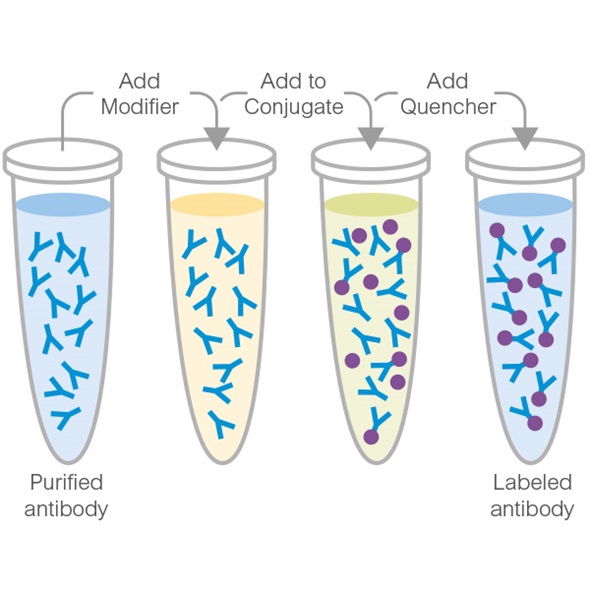
Our carrier-free antibodies are typically supplied in a PBS-only formulation, purified and free of BSA, sodium azide and glycerol. This conjugation-ready format is designed for use with fluorochromes, metal isotopes, oligonucleotides, and enzymes, which makes them ideal for antibody labelling, functional and cell-based assays, flow-based assays (e.g. mass cytometry) and Multiplex Imaging applications.
Use our conjugation kits for antibody conjugates that are ready-to-use in as little as 20 minutes with 1 minute hands-on-time and 100% antibody recovery: available for fluorescent dyes, HRP, biotin and gold.
Compatibility
This product is compatible with the Maxpar® Antibody Labeling Kit from Fluidigm, without the need for antibody preparation. Maxpar® is a trademark of Fluidigm Canada Inc.
Properties and storage information
Form
Purification technique
Storage buffer
Shipped at conditions
Appropriate short-term storage conditions
Appropriate long-term storage conditions
Product protocols
- Visit the General protocols
- Visit the Troubleshooting
Target data
Alternative Names
Product promise
Please note: All products are 'FOR RESEARCH USE ONLY. NOT FOR USE IN DIAGNOSTIC OR THERAPEUTIC PROCEDURES'.
For licensing inquiries, please contact partnerships@abcam.com