- Over 130 publications
- Trusted since 2010

- IHC-P
Supplier Data
Immunohistochemistry (Formalin/PFA-fixed paraffin-embedded sections) - Anti-MMP2 antibody [6E3F8] (AB86607)
Immunohistochemistry of human pancreas carcinoma staining MMP2 with ab86607 at 1μg/ml.

- IHC-P
Supplier Data
Immunohistochemistry (Formalin/PFA-fixed paraffin-embedded sections) - Anti-MMP2 antibody [6E3F8] (AB86607)
IHC image of ab86607 staining in human breast adenocarcinoma formalin fixed paraffin embedded tissue section, performed on a Leica BondTM system using the standard protocol F. The section was pre-treated using heat mediated antigen retrieval with sodium citrate buffer (pH6, epitope retrieval solution 1) for 20 mins. The section was then incubated with ab86607, 1μg/ml, for 15 mins at room temperature and detected using an HRP conjugated compact polymer system. DAB was used as the chromogen. The section was then counterstained with haematoxylin and mounted with DPX.
For other IHC staining systems (automated and non-automated) customers should optimize variable parameters such as antigen retrieval conditions, primary antibody concentration and antibody incubation times.

- IHC-P
PubMed
Immunohistochemistry (Formalin/PFA-fixed paraffin-embedded sections) - Anti-MMP2 antibody [6E3F8] (AB86607)
Immunohistochemical (parraffin-embedded sections) staining for MMP-2 in atheroslerotic lesions of aortic root in from right to left Ad-MIFi, Ad-EGFP, NS control and non-DM mouse control groups using ab86607 at a dilution of 1/200.
Image from Sun H et al., J Cell Mol Med. 2015;19(4):836-49. Fig 6.; doi: 10.1111/jcmm.12521. Reproduced under the Creative Commons license http://creativecommons.org/licenses/by/4.0/.

- WB
Lab
Western blot - Anti-MMP2 antibody [6E3F8] (AB86607)
This blot was produced using a 4-12% Bis-tris gel under the MOPS buffer system. The gel was run at 200V for 50 minutes before being transferred onto a Nitrocellulose membrane at 30V for 70 minutes. The membrane was then blocked for an hour using 2% Bovine Serum Albumin before being incubated with ab86607 overnight at 4°C. Antibody binding was detected using an anti-mouse antibody conjugated to HRP, and visualised using ECL development solution ab133406.
All lanes:
Western blot - Anti-MMP2 antibody [6E3F8] (ab86607) at 1 µg/mL
Lane 1:
Human liver tissue lysate - total protein (<a href='/en-us/products/unavailable/human-liver-tissue-lysate-total-protein-ab29889'>ab29889</a>) at 10 µg
Lane 2:
A375 (Human melanoma cell line) Whole Cell Lysate at 10 µg
Secondary
All lanes:
Western blot - Goat Anti-Mouse IgG H&L (HRP) preadsorbed (<a href='/en-us/products/secondary-antibodies/goat-mouse-igg-h-l-hrp-preadsorbed-ab97040'>ab97040</a>) at 1/50000 dilution
Predicted band size: 73 kDa
Observed band size: 100 kDa,70 kDa,75 kDa
true
Exposure time: 4min

- WB
Unknown
Western blot - Anti-MMP2 antibody [6E3F8] (AB86607)
All lanes:
Western blot - Anti-MMP2 antibody [6E3F8] (ab86607) at 1 µg/mL
Lane 1:
NIH/3T3 (Mouse embryonic fibroblast cell line) Whole Cell Lysate at 10 µg
Lane 2:
Liver (Mouse) Tissue Lysate at 10 µg
Lane 3:
PC-12 (Rat adrenal pheochromocytoma cell line) Whole Cell Lysate at 10 µg
Lane 4:
Spleen (Rat) Tissue Lysate at 10 µg
Secondary
All lanes:
Western blot - Goat Anti-Mouse IgG H&L (HRP) preadsorbed (<a href='/en-us/products/secondary-antibodies/goat-mouse-igg-h-l-hrp-preadsorbed-ab97040'>ab97040</a>) at 1/5000 dilution
Predicted band size: 73 kDa
Observed band size: 17 kDa,75 kDa
true
Exposure time: 20min
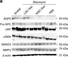
- WB
CiteAb
Western blot - Anti-MMP2 antibody [6E3F8] (AB86607)
Western Blotting using Anti-MMP2 antibody [6E3F8], ab86607. Publication image from Adler, K. B. et al., 2020, Nat Commun, 32111836. Legend direct from paper.
LSC-Sec inhalation treatment promotes alveolar repair.a Representative immunostaining of lung sections for von Willebrand Factor (vWF), pro-surfactant protein C (Pro-SPC) and aquaporin 5 (AQP5). Scale bar = 100 µm. b–d Quantification of percent pixel intensity of vWF+ (b), percent ProSPC+ nuclei (c), and percent pixel intensity of AQP5+ (d); each dot represents data from one animal; n = 12 biological independent animals. e–f Immunoblot analysis of aquaporin 5 (AQP5), pro-surfactant protein C (Pro-SPC), von Willebrand Factor (vWF), alpha smooth muscle actin (αSMA), SMAD3, matrix metalloproteinase 2 (MMP-2), and beta-actin loading control (B-actin) from lung protein lysate (e) with corresponding quantification of protein levels as fold of sham control (f); each dot represents data from one animal; n = 3 biological independent animals. g–h Representative cytokine array with quantification of relative intensity (g) and corresponding quantification of relative intensity (h). Throughout, data are mean ± s.d. P-value as indicated by non-parametric one-way ANOVA.
false
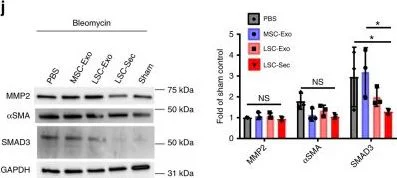
- WB
CiteAb
Western blot - Anti-MMP2 antibody [6E3F8] (AB86607)
Western Blotting using Anti-MMP2 antibody [6E3F8], ab86607. Publication image from Adler, K. B. et al., 2020, Nat Commun, 32111836. Legend direct from paper.
Therapeutic potential of exosome inhalation treatment in rats with pulmonary fibrosis.a Size analysis of fresh, frozen and lyophilized exosome particles by NanoSight. b Representative transmission electron micrograph (TEM) of LSC-Exo. Left scale bar = 0.2 µm. Right scale bar = 50 nm. c Immunoblot analysis of CD63, CD81, TSG101, and CD9 protein in LSC-Exo and MSC-Exo. d The experimental study schematic of the exosome study in SD rats; n = 12 biological independent animals per group. e Representative H&E staining Top : Scale bar = 100 µm Bottom : Scale bar = 50 µm. f Representative Gomori’s trichrome staining Scale bar = 100 µm (bottom); muscle fibers (red), collagen (blue), nuclei (black-purple) and erythrocytes (red). g Representative picrosirius red staining; Collagen types I and III (red); Scale bar = 50 µm. h Quantification of fibrosis by Ashcroft score; Each dot represents data from one animal; n = 12 biological independent animals. Ashcroft score was performed by averaging the score from one blinded and one non-blinded scorer. i Quantification of pulmonary hydroxyproline levels; *P ≤ 0.05; each dot represents data from one animal; n = 4 biological independent animals. j Immunoblot analysis of matrix metalloproteinase 2 (MMP-2), alpha smooth muscle actin (αSMA), SMAD3 and GAPDH loading control. k Representative immunostaining of lung sections for aquaporin 5 (AQP5), alpha-smooth muscle actin (αSMA), and von Willebrand Factor (vWF). Scale bar = 75 µm. l–n Quantification of pixel intensity of AQP5 (l),α-SMA (m), and vWF (n). Each dot represents data from one animal; n = 4 biological independent animals. Throughout, data are mean ± s.d. P-value as indicated by non-parametric one-way ANOVA.
false
Related conjugates and formulations (1)
-
Anti-MMP2 antibody [6E3F8] - BSA and Azide free
Alternative Products


Primary Antibodies
AB181286
Anti-MMP2 antibody [EPR17003-25]
RabMAb|Recombinant|20ul selling size

- 0
- 0
Primary Antibodies
AB215986
Anti-MMP2 antibody [EPR1184] - Low endotoxin, Azide free
RabMAb|Recombinant

- 0
- 0
Complementary Products
Primary Antibodies
AB283575
BOND RX™ Validated|20ul selling size|RabMAb|Recombinant|KO Validated|Trial Size

- 0
- 0
Primary Antibodies
AB115777
Anti-beta Actin antibody [SP124] - Cytoskeleton Marker
20ul selling size|RabMAb|Lab Essentials|Recombinant

- 4
- 1
Primary Antibodies
AB124964
Anti-alpha smooth muscle Actin antibody [EPR5368]
BOND RX™ Validated|RabMAb|Recombinant|KO Validated|20ul selling size

- 5
- 19
Primary Antibodies
AB51074
BOND RX™ Validated|RabMAb|Recombinant|KO Validated|20ul selling size

- 4
- 20
Reactivity data
Product details
What is this antibody validated in?
Anti-MMP2 antibody [6E3F8] (ab86607) is a mouse monoclonal antibody and is validated for use in Western Blot (WB), Immunohistochemistry (IHC-P) in Human, Mouse, Rat samples.
What is the molecular weight of MMP2?
Anti-MMP2 [6E3F8] (ab86607) specifically detects a band for MMP2 (UniProt: P08253) at a molecular weight of 74kDa.
Trusted by the scientific community
Anti-MMP2 [6E3F8] (ab86607) was first used in a scientific publication in 2010 and has been cited over 130 times in peer-reviewed journals.
Reviewed by scientists
Anti-MMP2 [6E3F8] (ab86607) has over 5 independent reviews from customers.
Other related products
We have a range of other formats of antibody clone [6E3F8] also available for your convenience: ab86607, Carrier free - ab264081
Want a custom formulation?
This antibody clone is manufactured by Abcam. If you require a custom buffer formulation or conjugation for your experiments, please contact orders@abcam.com
Properties and storage information
Form
Purity
Storage buffer
Shipped at conditions
Appropriate short-term storage duration
Appropriate short-term storage conditions
Appropriate long-term storage conditions
Aliquoting information
Storage information
Supplementary information
This supplementary information is collated from multiple sources and compiled automatically.
Biological function summary
Matrix metalloproteinase-2 is mainly involved in the degradation of type IV and V collagens gelatin and fibronectin. As part of the metalloproteinase family it works alongside other MMPs to maintain tissue homeostasis and repair. MMP-2 forms part of a complex network that ensures the timely degradation of matrix components balancing synthesis and breakdown. It remains regulated by tissue inhibitors of metalloproteinases (TIMPs) preventing excessive degradation that could lead to tissue damage.
Pathways
MMP-2 plays a significant role within the extracellular matrix (ECM) remodeling and angiogenesis pathways. It interacts with various proteins including integrins and TIMP-2 to modulate cellular behaviors such as migration and invasion. MMP-2 contributes to processes like wound healing and embryonic development through its involvement in ECM degradation and new tissue formation.
Product protocols
- Visit the General protocols
- Visit the Troubleshooting
Target data
Alternative Names
Publications (180)
Recent publications for all applications. Explore the full listand refine your search
International journal of molecular sciences 26: PubMed40565190
2025
Applications
Unspecified application
Species
Unspecified reactive species
European archives of oto-rhino-laryngology : official journal of the European Federation of Oto-Rhino-Laryngological Societies (EUFOS) : affiliated with the German Society for Oto-Rhino-Laryngology - Head and Neck Surgery 282:5645-5653 PubMed40468054
2025
Applications
Unspecified application
Species
Unspecified reactive species
Scientific reports 15:11096 PubMed40169699
2025
Applications
Unspecified application
Species
Unspecified reactive species
iScience 28:111880 PubMed40104069
2025
Applications
Unspecified application
Species
Unspecified reactive species
Veterinary sciences 12: PubMed40005905
2025
Applications
Unspecified application
Species
Unspecified reactive species
Frontiers in immunology 16:1486072 PubMed39958337
2025
Applications
Unspecified application
Species
Unspecified reactive species
Life (Basel, Switzerland) 15: PubMed39860018
2025
Applications
Unspecified application
Species
Unspecified reactive species
Scientific reports 15:1787 PubMed39805936
2025
Applications
Unspecified application
Species
Unspecified reactive species
Cell biology and toxicology 41:21 PubMed39753834
2025
Applications
Unspecified application
Species
Unspecified reactive species
Redox biology 77:103397 PubMed39427444
2024
Applications
Unspecified application
Species
Unspecified reactive species
Product promise
Please note: All products are 'FOR RESEARCH USE ONLY. NOT FOR USE IN DIAGNOSTIC OR THERAPEUTIC PROCEDURES'.
For licensing inquiries, please contact partnerships@abcam.com