Anti-Neurofascin antibody [EPR19004]
- RabMAb
- Recombinant
- What is this?
Be the first to review this product! Submit a review
|
(2 Publications )

- ICC/IF
Unknown
Immunocytochemistry/ Immunofluorescence - Anti-Neurofascin antibody [EPR19004] (AB184378)
Immunofluorescent analysis of 4% Paraformaldehyde-fixed, 0.1% TritonX-100 permeabilized mouse primary neuron cells labelling Neurofascin with ab184378 at 1/50 dilution, followed by ab150077 Goat Anti-Rabbit IgG H&L (Alexa Fluor® 488) antibody at 1/1000 dilution (2 μg/mL) (Green). Confocal image showing positive staining in mouse primary neuron cell. Confocal scanning Z step was set as 0.3 μm followed by image processing with maximum Z projection. ab11267 Anti-MAP2 mouse monoclonal antibody was used to counterstain tubulin at 1/500 dilution (4 μg/mL) followed by ab150120 Goat Anti-Mouse IgG H&L (Alexa Fluor® 594) at 1/1000 dilution (2 μg/mL) (Red). The nuclear counterstain was DAPI (Blue).
- IHC-P
Supplier Data
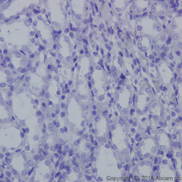
Immunohistochemistry (Formalin/PFA-fixed paraffin-embedded sections) - Anti-Neurofascin antibody [EPR19004] (AB184378)
Immunohistochemical analysis of paraffin-embedded Rat kidney tissue labeling Neurofascin with ab184378 at 1/500 dilution, followed by Goat Anti-Rabbit IgG H&L (HRP) (ab97051) secondary antibody at 1/500 dilution. Negative labeling on the epithelial cells of rat kidney is observed. Counter stained with Hematoxylin.
Perform heat mediated antigen retrieval with Tris/EDTA buffer pH 9.0 before commencing with IHC staining protocol.
- IHC-P
Supplier Data
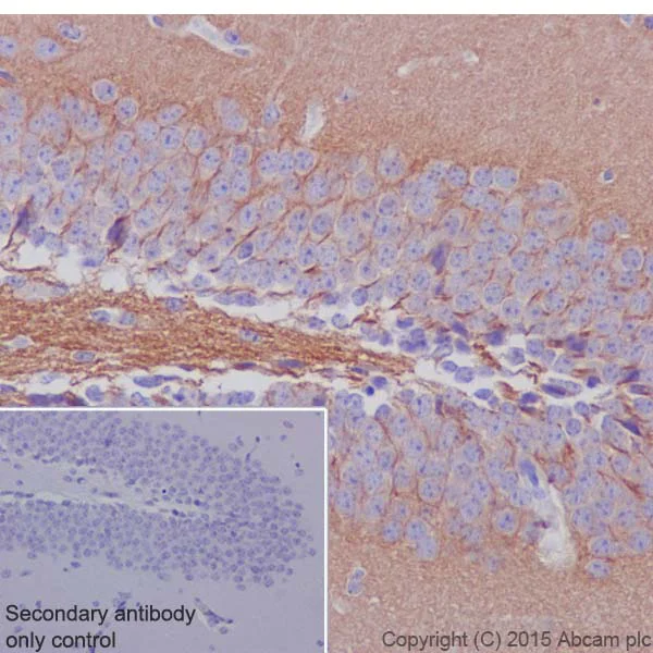
Immunohistochemistry (Formalin/PFA-fixed paraffin-embedded sections) - Anti-Neurofascin antibody [EPR19004] (AB184378)
Immunohistochemical analysis of paraffin-embedded Mouse hippocampus tissue labeling Neurofascin with ab184378 at 1/500 dilution, followed by Goat Anti-Rabbit IgG H&L (HRP) (ab97051) secondary antibody at 1/500 dilution. Membrane and cytoplasm staining on the neuron cells of mouse hippocampus is observed. Counter stained with Hematoxylin.
Secondary antibody only control : Used PBS instead of primary antibody, secondary antibody is Goat Anti-Rabbit IgG H&L (HRP) (ab97051) at 1/500 dilution.
Perform heat mediated antigen retrieval with Tris/EDTA buffer pH 9.0 before commencing with IHC staining protocol.
- IHC-P
Supplier Data
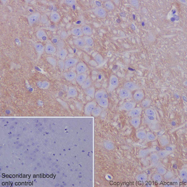
Immunohistochemistry (Formalin/PFA-fixed paraffin-embedded sections) - Anti-Neurofascin antibody [EPR19004] (AB184378)
Immunohistochemical analysis of paraffin-embedded Rat hippocampus tissue labeling Neurofascin with ab184378 at 1/500 dilution, followed by Goat Anti-Rabbit IgG H&L (HRP) (ab97051) secondary antibody at 1/500 dilution. Membrane and cytoplasm staining on the neuron cells of rat hippocampus is observed. Counter stained with Hematoxylin.
Secondary antibody only control : Used PBS instead of primary antibody, secondary antibody is Goat Anti-Rabbit IgG H&L (HRP) (ab97051) at 1/500 dilution.
Perform heat mediated antigen retrieval with Tris/EDTA buffer pH 9.0 before commencing with IHC staining protocol.
- IHC-P
Supplier Data
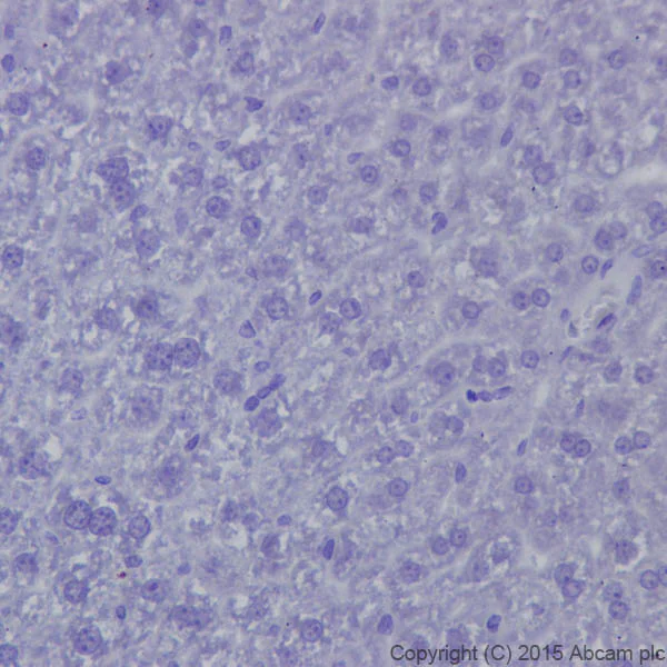
Immunohistochemistry (Formalin/PFA-fixed paraffin-embedded sections) - Anti-Neurofascin antibody [EPR19004] (AB184378)
Immunohistochemical analysis of paraffin-embedded Mouse liver tissue labeling Neurofascin with ab184378 at 1/500 dilution, followed by Goat Anti-Rabbit IgG H&L (HRP) (ab97051) secondary antibody at 1/500 dilution. Negative labeling on the mouse liver is observed. Counter stained with Hematoxylin.
Perform heat mediated antigen retrieval with Tris/EDTA buffer pH 9.0 before commencing with IHC staining protocol.
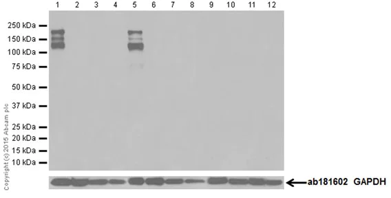
- WB
Supplier Data
Western blot - Anti-Neurofascin antibody [EPR19004] (AB184378)
Blocking/Dilution buffer : 5% NFDM/TBST.
The molecular weight and expression profile observed are consistent with what has been described in the literature (PMID : 23160985).
All lanes:
Western blot - Anti-Neurofascin antibody [EPR19004] (ab184378) at 1/5000 dilution
Lane 1:
Mouse brain lysate at 10 µg
Lane 2:
Mouse heart lysate at 10 µg
Lane 3:
Mouse kidney lysate at 10 µg
Lane 4:
Mouse spleen lysate at 10 µg
Lane 5:
Rat brain lysate at 10 µg
Lane 6:
Rat heart lysate at 10 µg
Lane 7:
Rat kidney lysate at 10 µg
Lane 8:
Rat spleen lysate at 10 µg
Lane 9:
C6 (Rat glial tumor cell line) lysate at 10 µg
Lane 10:
RAW 264.7 (Mouse macrophage cell line transformed with Abelson murine leukemia virus) lysate at 10 µg
Lane 11:
PC-12 (Rat adrenal gland pheochromocytoma cell line) lysate at 10 µg
Lane 12:
NIH/3T3 (Mouse embryonic fibroblast cell line) lysate at 10 µg
Secondary
All lanes:
Western blot - Goat Anti-Rabbit IgG H&L (HRP) (<a href='/en-us/products/secondary-antibodies/goat-rabbit-igg-h-l-hrp-ab97051'>ab97051</a>) at 1/100000 dilution
Predicted band size: 150 kDa
Observed band size: 125 kDa,155 kDa,186 kDa
false
Exposure time: 3min
Related conjugates and formulations (1)
-
Anti-Neurofascin antibody [EPR19004] - BSA and Azide free
Alternative Products


Primary Antibodies
AB271948
Anti-Neurofascin antibody [EPR19003] - BSA and Azide free
RabMAb|Recombinant

- 0
- 0

Complementary Products

Primary Antibodies
AB223343
Anti-68kDa Neurofilament/NF-L antibody [EPR22035-112]
20ul selling size|RabMAb|Recombinant

- 0
- 0


Reactivity data
Product details
Patented technology
Our RabMAb® technology is a patented hybridoma-based technology for making rabbit monoclonal antibodies. For details on our patents, please refer to RabMAb® patents.
What are the advantages of a recombinant monoclonal antibody?
This product is a recombinant monoclonal antibody, which offers several advantages including:
- - High batch-to-batch consistency and reproducibility
- - Improved sensitivity and specificity
- - Long-term security of supply
- - Animal-free batch production
For more information, read more on recombinant antibodies.
Properties and storage information
Form
Purification technique
Storage buffer
Shipped at conditions
Appropriate short-term storage duration
Appropriate short-term storage conditions
Appropriate long-term storage conditions
Aliquoting information
Storage information
Supplementary information
This supplementary information is collated from multiple sources and compiled automatically.
Biological function summary
This neural protein plays an integral role in developing and securing the nervous system's structural organization. Neurofascin helps form the nodes of Ranvier which are critical for saltatory conduction enhancing rapid and efficient nerve impulse transmission. It also interacts with other proteins such as Ankyrin G and Contactin to maintain the stability and function of neuronal domains. Through these interactions Neurofascin facilitates the proper conduction and coordination of electrical signals along neurons.
Pathways
Neurofascin is essential for neural signal transmission and maintenance pathways. It prominently participates in the axon guidance and myelination pathways working alongside proteins like Caspr (Contactin-associated protein) and Ankyrin B. These pathways ensure the growth and maintenance of myelin sheaths supporting nerve insulation and efficient signal relay. Neurofascin's interaction with these proteins reinforces the nodes of Ranvier's organization promoting stable and rapid signal conduction in the peripheral and central nervous systems.
Product protocols
- Visit the General protocols
- Visit the Troubleshooting
Target data
Alternative Names
Publications (2)
Recent publications for all applications. Explore the full listand refine your search
The Journal of clinical investigation 129:2222-2236 PubMed30869655
2019
Applications
Unspecified application
Species
Unspecified reactive species
Stem cells (Dayton, Ohio) 34:984-96 PubMed26676415
2015
Applications
Unspecified application
Species
Unspecified reactive species
Product promise
Please note: All products are 'FOR RESEARCH USE ONLY. NOT FOR USE IN DIAGNOSTIC OR THERAPEUTIC PROCEDURES'.
For licensing inquiries, please contact partnerships@abcam.com