Anti-Presenilin 1/PS-1 antibody [EP2000Y] - BSA and Azide free
- BOND RX™ Validated
- KO Validated
- RabMAb
- Recombinant
- What is this?
Be the first to review this product! Submit a review
|
(9 Publications )
- IHC-P
Unknown
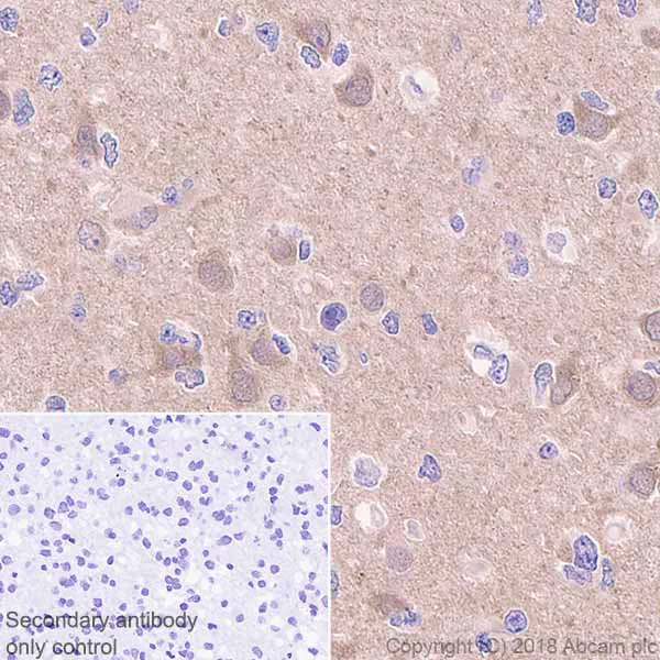
Immunohistochemistry (Formalin/PFA-fixed paraffin-embedded sections) - Anti-Presenilin 1/PS-1 antibody [EP2000Y] - BSA and Azide free (AB227985)
Immunohistochemistry (Formalin/PFA-fixed paraffin-embedded sections) analysis of Human glioma tissue sections labeling Presenilin 1/PS-1 with Purified ab76083 at 1 : 100 dilution (1.31 μg/ml). Heat mediated antigen retrieval was performed using ab93684 (Tris/EDTA buffer, pH 9.0). ImmunoHistoProbe one step HRP Polymer (ready to use) was used as the secondary antibody. Negative control : PBS instead of the primary antibody. Hematoxylin was used as a counterstain.
This data was developed using the same antibody clone in a different buffer formulation containing PBS, BSA, glycerol, and sodium azide (ab76083).
- IHC-P
Lab
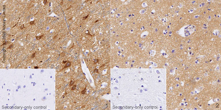
Immunohistochemistry (Formalin/PFA-fixed paraffin-embedded sections) - Anti-Presenilin 1/PS-1 antibody [EP2000Y] - BSA and Azide free (AB227985)
This data was developed using ab76083, the same antibody clone in a different buffer formulation.
Immunohistochemical analysis of paraffin-embedded human cerebral cortex and human Alzheimer's brain tissue* labelling PSEN1 with ab76083 at 1ug/ml followed by a ready to use LeicaDS9800 (Bond™ Polymer Refine Detection).
Positive staining in human cerebral cortex and negative staining in Alzheimer's brain.
The section was incubated with ab76083 for 30 mins at room temperature. The immunostaining was performed on a Leica Biosystems BOND® RX instrument. Counterstained with Hematoxylin.
Secondary antibody only control : Secondary antibody is a ready to use LeicaDS9800 (Bond™ Polymer Refine Detection).
Heat mediated antigen retrieval with sodium citrate buffer (pH 6.0 epitope retrieval solution1) for 20 mins.
*Tissue obtained from the Human Research Tissue Bank, supported by the NIHR Cambridge Biomedical Research Centre.
- WB
Lab
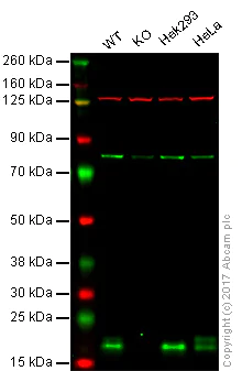
Western blot - Anti-Presenilin 1/PS-1 antibody [EP2000Y] - BSA and Azide free (AB227985)
**Lanes 1 - 4 : ** Merged signal (red and green). Green - ab76083 observed at 18 kDa. Red - loading control, ab18058, observed at 130 kDa. ab76083 was shown to recognize Presenilin 1/PS-1 in wild-type HAP1 cells as signal was lost at the expected MW in Presenilin 1/PS-1 knockout cells. Additional cross-reactive bands were observed in the wild-type and knockout cells. Wild-type and Presenilin 1/PS-1 knockout samples were subjected to SDS-PAGE. ab76083 and ab18058 (Mouse anti-Vinculin loading control) were incubated overnight at 4°C at 1/5000 dilution and 1/20000 dilution respectively. Blots were developed with Goat anti-Rabbit IgG H&L (IRDye® 800CW) preabsorbed ab216773 and Goat anti-Mouse IgG H&L (IRDye® 680RD) preabsorbed ab216776 secondary antibodies at 1/10000 dilution for 1 hour at room temperature before imaging. This data was developed using the same antibody clone in a different buffer formulation containing PBS, BSA, glycerol, and sodium azide (ab76083).
Lane 1:
Western blot - Anti-Presenilin 1/PS-1 antibody [EP2000Y] (<a href='/en-us/products/primary-antibodies/presenilin-1-ps-1-antibody-ep2000y-ab76083'>ab76083</a>)
Lanes 2 - 4:
Western blot - Anti-Presenilin 1/PS-1 antibody [EP2000Y] - BSA and Azide free (ab227985) at 1/5000 dilution
Lane 1:
Wild-type HAP1 whole cell lysate at 20 µg
Lane 2:
Presenilin 1/PS-1 knockout HAP1 whole cell lysate at 20 µg
Lane 3:
HEK293 whole cell lysate at 20 µg
Lane 4:
HeLa whole cell lysate at 20 µg
Predicted band size: 53 kDa
false
- WB
Lab
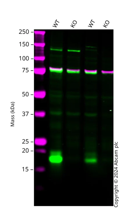
Western blot - Anti-Presenilin 1/PS-1 antibody [EP2000Y] - BSA and Azide free (AB227985)
Western blot : Anti-PSEN1 antibody [EP2000Y] (ab76083) staining at 1/2500 dilution, shown in green; Mouse anti-CANX [CANX/1543] (ab238078) loading control staining at 1/20000 dilution, shown in magenta. In Western blot, ab76083 was shown to bind specifically to PSEN1. A band was observed at 18 kDa in wild-type PC-3 cell lysates with no signal observed at this size in PSEN1 knockout cell line. To generate this image, wild-type and PSEN1 knockout PC-3 cell lysates were analysed. First, samples were run on an SDS-PAGE gel then transferred onto a nitrocellulose membrane. Membranes were blocked in 3 % milk in TBS-0.1 % Tween® 20 (TBS-T) before incubation with primary antibodies overnight at 4 °C. Blots were washed four times in TBS-T, incubated with secondary antibodies for 1 h at room temperature, washed again four times then imaged. Secondary antibodies used were Goat anti-Rabbit IgG H&L 800CW and Goat anti-Mouse IgG H&L 680RD at 1/20000 dilution.
All lanes:
Western blot - Anti-Presenilin 1/PS-1 antibody [EP2000Y] (<a href='/en-us/products/primary-antibodies/presenilin-1-ps-1-antibody-ep2000y-ab76083'>ab76083</a>) at 1/2500 dilution
Lane 1:
Wild-type PC-3 cell lysate at 20 µg
Lane 2:
PSEN1 knockout PC-3 cell lysate at 20 µg
Lane 3:
Wild-type HAP1 cell lysate at 20 µg
Lane 4:
PSEN1 knockout HAP1 cell lysate at 20 µg
Secondary
All lanes:
Goat anti-Rabbit IgG H&L 800CW and Goat anti-Mouse IgG H&L 680RD at 1/20000 dilution
false
Related conjugates and formulations (1)
-
Anti-Presenilin 1/PS-1 antibody [EP2000Y]
Alternative Products


Primary Antibodies
AB76131
Anti-Presenilin 1/PS-1 (phospho S310) antibody [EP2001Y]
20ul selling size|RabMAb

- 4
- 1
Complementary Products


Primary Antibodies
AB183612
Anti-BACE1 antibody [EPR19523]
BOND RX™ Validated|20ul selling size|RabMAb|Recombinant|KO Validated

- 5
- 9
Primary Antibodies
AB32136
Anti-Amyloid Precursor Protein antibody [Y188]
BOND RX™ Validated|20ul selling size|RabMAb|Recombinant|KO Validated

- 4
- 9
Primary Antibodies
AB51249
Anti-Presenilin 2/AD5 antibody [EP1515Y]
BOND RX™ Validated|20ul selling size|RabMAb|Recombinant|KO Validated

- 4
- 7

Reactivity data
Product details
ab227985 is the carrier-free version of ab76083.
Patented technology
Our RabMAb® technology is a patented hybridoma-based technology for making rabbit monoclonal antibodies. For details on our patents, please refer to RabMAb® patents.
What are the advantages of a recombinant monoclonal antibody?
This product is a recombinant monoclonal antibody, which offers several advantages including:
- - High batch-to-batch consistency and reproducibility
- - Improved sensitivity and specificity
- - Long-term security of supply
- - Animal-free batch production
For more information, read more on recombinant antibodies.
Conjugation ready
Our carrier-free antibodies are typically supplied in a PBS-only formulation, purified and free of BSA, sodium azide and glycerol. This conjugation-ready format is designed for use with fluorochromes, metal isotopes, oligonucleotides, and enzymes, which makes them ideal for antibody labelling, functional and cell-based assays, flow-based assays (e.g. mass cytometry) and Multiplex Imaging applications.
Use our conjugation kits for antibody conjugates that are ready-to-use in as little as 20 minutes with 1 minute hands-on-time and 100% antibody recovery: available for fluorescent dyes, HRP, biotin and gold.
Compatibility
This product is compatible with the Maxpar® Antibody Labeling Kit from Fluidigm, without the need for antibody preparation. Maxpar® is a trademark of Fluidigm Canada Inc.
Properties and storage information
Form
Purification technique
Storage buffer
Shipped at conditions
Appropriate short-term storage conditions
Appropriate long-term storage conditions
Storage information
Supplementary information
This supplementary information is collated from multiple sources and compiled automatically.
Biological function summary
Presenilin 1 plays a fundamental role in the cleavage of amyloid precursor protein (APP) within the transmembrane domain. This cleavage results in the formation of amyloid-beta peptides which are of significant interest in neurobiology. Presenilin 1 is an integral component of the gamma-secretase complex that includes nicastrin APH-1 and PEN-2. This complex involvement underlines the enzyme's importance in cell signaling pathways and modulation of synaptic functions. The functionality at the synaptic level is particularly important given its expression pattern and involvement in neural processes.
Pathways
The action of presenilin 1 is closely linked to the Notch signaling pathway and the amyloidogenic pathway. Presenilin 1 facilitates the intramembrane cleavage of the Notch receptor releasing the Notch intracellular domain that translocates to the nucleus and affects gene transcription. The protein's role in processing APP into amyloid-beta peptides links it directly to Alzheimer's disease pathology. The protein Tax also a component of the gamma-secretase complex shows interdependencies in these pathways.
Product protocols
- Visit the General protocols
- Visit the Troubleshooting
Target data
Alternative Names
Publications (9)
Recent publications for all applications. Explore the full list and refine your search
Molecular neurodegeneration 12:15 PubMed28193235
2017
Applications
Unspecified application
Species
Unspecified reactive species
FASEB journal : official publication of the Federation of American Societies for Experimental Biology 31:1382-1397 PubMed28034848
2016
Applications
Unspecified application
Species
Unspecified reactive species
Oncotarget 7:10650-62 PubMed26872378
2016
Applications
IHC-P, WB
Species
Human, Human
PloS one 9:e100373 PubMed24972054
2014
Applications
WB
Species
Human
Journal of neurochemistry 131:94-100 PubMed24865334
2014
Applications
Unspecified application
Species
Unspecified reactive species
Proceedings of the National Academy of Sciences of 111:E1230-9 PubMed24639524
2014
Applications
Unspecified application
Species
Mouse
Carcinogenesis 34:2622-8 PubMed23740836
2013
Applications
WB
Species
Unspecified reactive species
Neurobiology of aging 34:2389-98 PubMed23673309
2013
Applications
WB
Species
Mouse
The Journal of biological chemistry 287:34709-21 PubMed22891244
2012
Applications
WB
Species
Rat
Product promise
Please note: All products are 'FOR RESEARCH USE ONLY. NOT FOR USE IN DIAGNOSTIC OR THERAPEUTIC PROCEDURES'.
For licensing inquiries, please contact partnerships@abcam.com