Anti-Smad2 antibody [EP784Y] - BSA and Azide free
- RabMAb
- Advanced Validation
- Recombinant
- KO Validated
- What is this?
Be the first to review this product! Submit a review
|
(1 Publication)
- ICC/IF
Unknown
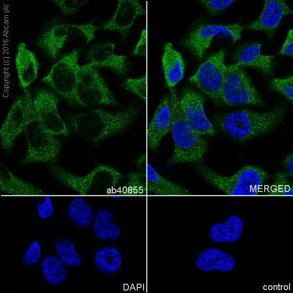
Immunocytochemistry/ Immunofluorescence - Anti-Smad2 antibody [EP784Y] - BSA and Azide free (AB157371)
Immunofluorescence staining of HeLa cells with purified ab40855 at a working dilution of 1/500, counter-stained with DAPI. The secondary antibody was an Alexa Fluor® 488 conjugated goat anti-rabbit (ab150077), used at a dilution of 1/1000. The cells were fixed in 4% PFA and permeabilized using 0.1% Triton X 100. The negative control is shown in bottom right hand panel - for the negative control, PBS was used instead of the primary antibody.
This data was developed using the same antibody clone in a different buffer formulation containing PBS, BSA, glycerol, and sodium azide (ab40855).
- IHC-P
Unknown
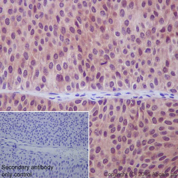
Immunohistochemistry (Formalin/PFA-fixed paraffin-embedded sections) - Anti-Smad2 antibody [EP784Y] - BSA and Azide free (AB157371)
This IHC data was generated using the same anti-Smad2 antibody clone, EP784Y, in a different buffer formulation (cat# ab40855).
ab40855 stainingSmad2 in human bladder carcinoma tissue sections by Immunohistochemistry (IHC-P - paraformaldehyde-fixed, paraffin-embedded sections). Tissue was fixed with paraformaldehyde and antigen retrieval was by heat mediation in a EDTA buffer. Samples were incubated with primary antibody at a dilution of 1/50. A ImmunoHistoProbe one step HRP Polymer was used as a secondary antibody, ready to use.
Negative control 1 : PBS in place of primary antibody.
- ICC/IF
Unknown
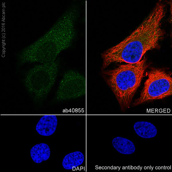
Immunocytochemistry/ Immunofluorescence - Anti-Smad2 antibody [EP784Y] - BSA and Azide free (AB157371)
ab40855 staining Smad2 in HeLa (human cervix adenocarcinoma) cells by ICC/IF (Immunocytochemistry/immunofluorescence). Cells were fixed with 4% Paraformaldehyde and permeabilized with 0.1% Triton X-100. Samples were incubated with primary antibody at a dilution of 1/100. A goat anti rabbit IgG (Alexa Fluor® 488) (ab150077) was used as the secondary antibody at a dilution of 1/1000. ab195889 was used as a counterstain for primary antibody ab40855 at 1/1000. DAPI was used as a nuclear counterstain and PBS as a negative control.
This data was developed using the same antibody clone in a different buffer formulation containing PBS, BSA, glycerol, and sodium azide (ab40855).
- IHC-P
Unknown
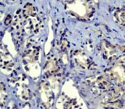
Immunohistochemistry (Formalin/PFA-fixed paraffin-embedded sections) - Anti-Smad2 antibody [EP784Y] - BSA and Azide free (AB157371)
ab40855 at a 1 : 100 dilution staining Smad2 in human prostate carcinoma tissue.This data was developed using the same antibody clone in a different buffer formulation containing PBS, BSA, glycerol, and sodium azide (ab40855).
- Flow Cyt (Intra)
Unknown
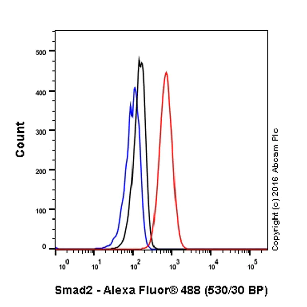
Flow Cytometry (Intracellular) - Anti-Smad2 antibody [EP784Y] - BSA and Azide free (AB157371)
ab40855 staining Smad2 in the human cell line HeLa (Human epithelial cell line from cervix adenocarcinoma) by intracellular flow cytometry. Cells were fixed with 4% paraformaldehyde and the sample was incubated with the primary antibody at a dilution of 1/20. A goat anti rabbit IgG (Alexa Fluor® 488) at a dilution of 1/2000 was used as the secondary antibody.
Isoytype control : Rabbit monoclonal IgG (Black)
Unlabelled control : Cell without incubation with primary antibody and secondary antibody (Blue)
This data was developed using the same antibody clone in a different buffer formulation containing PBS, BSA, glycerol, and sodium azide (ab40855).
- Flow Cyt (Intra)
Unknown
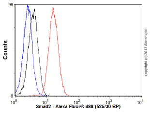
Flow Cytometry (Intracellular) - Anti-Smad2 antibody [EP784Y] - BSA and Azide free (AB157371)
Overlay histogram showing PC3 cells stained with ab40855 (red line). The cells were fixed with 80% methanol (5 min) and then permeabilized with 0.1% PBS-Tween for 20 min. The cells were then incubated in 1x PBS / 10% normal goat serum / 0.3M glycine to block non-specific protein-protein interactions followed by the antibody (ab40855, 1/100 dilution) for 30 min at 22°C. The secondary antibody used was Alexa Fluor® 488 goat anti-rabbit IgG (H&L) (ab150077) at 1/2000 dilution for 30 min at 22°C. Isotype control antibody (black line) was rabbit IgG (monoclonal) (1μg/1x106 cells) used under the same conditions. Unlabelled sample (blue line) was also used as a control. Acquisition of >5,000 events were collected using a 20mW Argon ion laser (488nm) and 525/30 bandpass filter. This data was developed using the same antibody clone in a different buffer formulation containing PBS, BSA, glycerol, and sodium azide (ab40855).

- IP
Unknown
Immunoprecipitation - Anti-Smad2 antibody [EP784Y] - BSA and Azide free (AB157371)
ab40855 (purified) at 1/20 immunoprecipitating EGFR in HeLa (Human epithelial cell line from cervix adenocarcinoma) whole cell lysate.
Lane 1 (input) : HeLa whole cell lysate (10μg)
Lane 2 (+) : ab40855 + HeLa whole cell lysate.
Lane 3 (-) : Rabbit monoclonal IgG (ab172730) instead of ab40855 in HeLa whole cell lysate.
For western blotting, VeriBlot for IP Detection Reagent (HRP) (ab131366), was used for detection at 1/1000 dilution.
Blocking/Diluting buffer and concentration : 5% NFDM/TBST.
This data was developed using the same antibody clone in a different buffer formulation containing PBS, BSA, glycerol, and sodium azide (ab40855).
All lanes:
Immunoprecipitation - Anti-Smad2 antibody [EP784Y] (<a href='/en-us/products/primary-antibodies/smad2-antibody-ep784y-ab40855'>ab40855</a>)
Predicted band size: 52 kDa
false
- WB
Lab
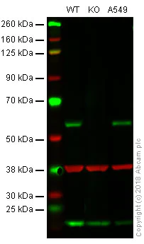
Western blot - Anti-Smad2 antibody [EP784Y] - BSA and Azide free (AB157371)
Lanes 1 - 3 : Merged signal (red and green). Green - ab40855 observed at 58 kDa. Red - loading control, ab9484, observed at 37 kDa.
ab40855 was shown to specifically react with Smad2 in wild-type HeLa cells as signal was lost in Smad2 knockout cells. Wild-type and SMAD2 knockout samples were subjected to SDS-PAGE. ab40855 and ab9484 (Mouse anti-GAPDH loading control) were incubated overnight at 4°C at 1/10000 dilution and 1/20000 dilution respectively. Blots were developed with Goat anti-Rabbit IgG H&L (IRDye® 800CW) preabsorbed ab216773 and Goat anti-Mouse IgG H&L (IRDye® 680RD) preabsorbed ab216776 secondary antibodies at 1/20000 dilution for 1 hour at room temperature before imaging.
This data was developed using the same antibody clone in a different buffer formulation containing PBS, BSA, glycerol, and sodium azide (ab40855).
All lanes:
Western blot - Anti-Smad2 antibody [EP784Y] (<a href='/en-us/products/primary-antibodies/smad2-antibody-ep784y-ab40855'>ab40855</a>) at 1/1000 dilution
Lane 1:
Wild-type HeLa whole cell lysate at 20 µg
Lane 2:
Smad2 knockout HeLa whole cell lysate at 20 µg
Lane 3:
A549 whole cell lysate at 20 µg
Predicted band size: 52 kDa
false
- WB
Supplier Data
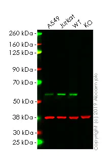
Western blot - Anti-Smad2 antibody [EP784Y] - BSA and Azide free (AB157371)
This data was developed using the same antibody clone in a different buffer formulation (ab40855).
Lanes 1 - 4 : Merged signal (red and green). Green - ab40855 observed at 58 kDa. Red - loading control, ab8245 observed at 37 kDa.
ab40855 was shown to react with Smad2 in wild-type HeLa. Loss of signal was observed when knockout cell line ab255430 (knockout cell lysate ab263833) was used. Wild-type and Smad2 knockout samples were subjected to SDS-PAGE. ab40855 and Anti-GAPDH antibody [6C5] - Loading Control (ab8245) were incubated overnight at 4°C at 1 in 2000 dilution and 1 in 20000 dilution respectively. Blots were developed with Goat anti-Rabbit IgG H&L (IRDye® 800CW) preadsorbed (ab216773) and Goat anti-Mouse IgG H&L (IRDye® 680RD) preadsorbed (ab216776) secondary antibodies at 1 in 20000 dilution for 1 hour at room temperature before imaging.
All lanes:
Western blot - Anti-Smad2 antibody [EP784Y] (<a href='/en-us/products/primary-antibodies/smad2-antibody-ep784y-ab40855'>ab40855</a>) at 1/2000 dilution
Lane 1:
A549 cell lysate at 20 µg
Lane 2:
Jurkat cell lysate at 20 µg
Lane 2:
Western blot - Human SMAD2 knockout HeLa cell line (<a href='/en-us/products/cell-lines/human-smad2-knockout-hela-cell-line-ab255430'>ab255430</a>)
Lane 3:
Wild-type HeLa cell lysate at 20 µg
Lane 4:
SMAD2 knockout HeLa cell lysate at 20 µg
Secondary
All lanes:
Western blot - Goat anti-Rabbit IgG H&L (IRDye® 800CW) preadsorbed (<a href='/en-us/products/secondary-antibodies/goat-rabbit-igg-h-l-irdye-800cw-preadsorbed-ab216773'>ab216773</a>) at 1/20000 dilution
Predicted band size: 52 kDa
Observed band size: 37 kDa,55 kDa
false
- ChIC/CUT&RUN-seq
Lab
ChIC/CUT&RUN sequencing - Anti-Smad2 antibody [EP784Y] - BSA and Azide free (AB157371)
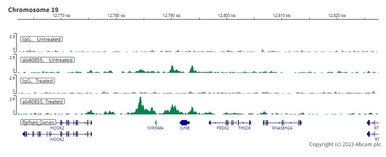
This data was developed using the same antibody clone in a different buffer formulation (ab40855). ChIC/CUT&RUN was performed using a pAG-MNAse at a final concentration of 700 ng/mL, 2.5 x 10^5 HaCaT (Human keratinocyte cell line) cells (treated with 7ng/ml TGF-β for 1h) and 5 µg of ab40855 [EP784Y]. The resulting DNA was sequenced on the Illumina NovaSeq 6000 to a depth of 10 million reads. The negative IgG control ab172730 is also shown. Additional screenshots of mapped reads can be found in the Protocol booklet in the Product Protocol section. The University of Geneva owns patents relevant to ChIC (Chromatin Immuno-Cleavage) methods.
- ChIC/CUT&RUN-seq
Lab
ChIC/CUT&RUN sequencing - Anti-Smad2 antibody [EP784Y] - BSA and Azide free (AB157371)
This data was developed using ab40855, the same antibody clone in a different buffer formulation.
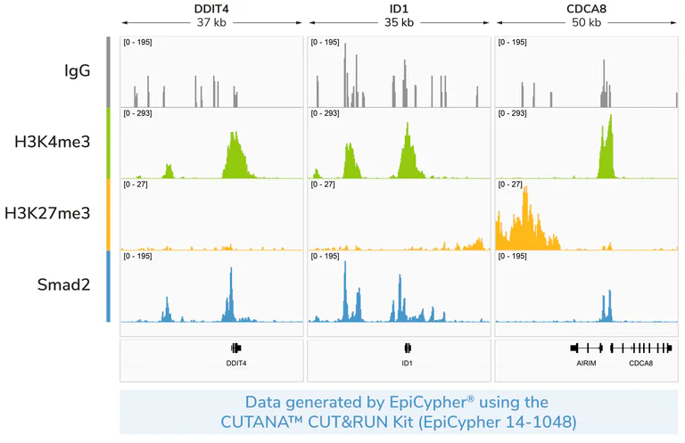
CUT&RUN profiling with Smad2 antibody reveals the expected genomic enrichment pattern in cells. Representative genome browser tracks show CUT&RUN data generated using the CUTANA™ CUT&RUN Kit (EpiCypher 14-1048) with Smad2 antibody (Abcam ab40855, 0.5 µg). 500,000 HeLa cells were used per reaction. IgG, H3K4me3, and H3K27me3 antibodies were included as controls to assess non-specific background, active promoters, and repressed chromatin, respectively. Libraries were prepared using the CUTANA™ CUT&RUN Library Prep Kit (EpiCypher 14-1001). Sequencing was performed with paired-end 50 bp reads, and data were processed on CUTANA™ Cloud (cloud.epicypher.com) by alignment to the hg38 genome. Images were generated using Integrative Genomics Viewer (IGV, Broad Institute).
- ChIC/CUT&RUN-seq
Lab
ChIC/CUT&RUN sequencing - Anti-Smad2 antibody [EP784Y] - BSA and Azide free (AB157371)
This data was developed using ab40855, the same antibody clone in a different buffer formulation.
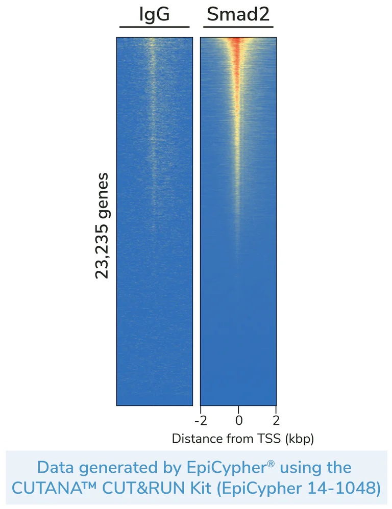
CUT&RUN profiling with Smad2 antibody demonstrates robust genome-wide enrichment in cells. Heatmaps of genome-wide signal flanking annotated transcription start sites (TSSs, +/- 2 kbp) display CUT&RUN data generated using the CUTANA™ CUT&RUN Kit (EpiCypher 14-1048) with Smad2 antibody (Abcam ab40855, 0.5 µg). 500,000 HeLa cells were used per reaction. IgG antibody was included as a negative control to assess non-specific background. Libraries were prepared using the CUTANA™ CUT&RUN Library Prep Kit (EpiCypher 14-1001). Sequencing was performed with paired-end 50 bp reads, and data were processed on CUTANA™ Cloud (cloud.epicypher.com) by alignment to the hg38 genome. Heatmaps were generated using deepTools (Ramírez et al., Nucleic Acids Res. 2014; PMID : 24799436). Row-linked data are ranked by intensity relative to Smad2, with red indicating high localized enrichment and blue denoting background.
Related conjugates and formulations (3)
-
Anti-Smad2 antibody [EP784Y]
-
519 Alexa Fluor® 488
Alexa Fluor® 488 Anti-Smad2 antibody [EP784Y]
-
578 PE
PE Anti-Smad2 antibody [EP784Y]
Alternative Products


Primary Antibodies
AB184557
Anti-Smad2 (phospho S250) antibody [EPR2855(2)]
BOND RX™ Validated|20ul selling size|RabMAb|Recombinant|KO Validated

- 0
- 0
Primary Antibodies
AB33875
20ul selling size|RabMAb|Advanced Validation|Recombinant|KO Validated

- 4
- 2
Primary Antibodies
AB219598
Anti-Smad2 (phospho S255) antibody [EPR2856(N)] - BSA and Azide free
Recombinant|Advanced Validation|RabMAb

- 0
- 0
Primary Antibodies
AB188334
Anti-Smad2 (phospho S255) antibody [EPR2856(N)]
20ul selling size|Recombinant|Advanced Validation|RabMAb

- 5
- 2

Primary Antibodies
AB216454
Anti-Smad2 antibody [EP567Y] - BSA and Azide free
RabMAb|Advanced Validation|Recombinant|KO Validated

- 0
- 0
Complementary Products

Sample Prep & Detection Kits
AB270705
5’ Feature Barcode Antibody Conjugation Kit - Lightning-Link® - Oligos 11-20

- 0
- 0
Primary Antibodies
AB40854
20ul selling size|Recombinant|KO Validated|Advanced Validation|RabMAb

- 5
- 10
Primary Antibodies
AB280888
Anti-Smad2 (phospho S467) antibody [EPR23681-40]
BOND RX™ Validated|20ul selling size|RabMAb|Recombinant|KO Validated

- 5
- 2
Primary Antibodies
AB215715
Anti-TGF beta 1 antibody [EPR21143]
20ul selling size|BOND RX™ Validated|RabMAb|Recombinant|KO Validated

- 5
- 7
Primary Antibodies
AB52903
Anti-SMAD3 (pS423/425) + SMAD5 (pS463/465) + SMAD1 (pS463/465) + SMAD2 (pS465/467) antibody [EP823Y]
20ul selling size|Recombinant|Advanced Validation|RabMAb

- 4
- 21
Reactivity data
Product details
ab157371 is the carrier-free version of ab40855.
Patented technology
Our RabMAb® technology is a patented hybridoma-based technology for making rabbit monoclonal antibodies. For details on our patents, please refer to RabMAb® patents.
Conjugation ready
Our carrier-free antibodies are typically supplied in a PBS-only formulation, purified and free of BSA, sodium azide and glycerol. This conjugation-ready format is designed for use with fluorochromes, metal isotopes, oligonucleotides, and enzymes, which makes them ideal for antibody labelling, functional and cell-based assays, flow-based assays (e.g. mass cytometry) and Multiplex Imaging applications.
Use our conjugation kits for antibody conjugates that are ready-to-use in as little as 20 minutes with 1 minute hands-on-time and 100% antibody recovery: available for fluorescent dyes, HRP, biotin and gold.
Compatibility
This product is compatible with the Maxpar® Antibody Labeling Kit from Fluidigm, without the need for antibody preparation. Maxpar® is a trademark of Fluidigm Canada Inc.
Properties and storage information
Form
Purification technique
Purification notes
Storage buffer
Shipped at conditions
Appropriate short-term storage conditions
Appropriate long-term storage conditions
Storage information
Supplementary information
This supplementary information is collated from multiple sources and compiled automatically.
Biological function summary
Smad2 acts as an intracellular mediator for TGF-β signaling a pathway important for regulating cell proliferation differentiation and apoptosis. Smad2 typically functions as part of a heteromeric complex with Smad4 another key player in TGF-β signaling. Upon activation phosphorylated Smad2 combines with Smad4 to form a complex that translocates into the nucleus. This complex then binds to specific DNA sequences to regulate the transcription of target genes involved in processes such as cell growth inhibition and extracellular matrix production.
Pathways
Smad2 is integral to the TGF-β and activin receptor signaling pathways. These pathways are essential in controlling cell growth and immune responses. Smad2 interacts with other proteins such as Smad3 in addition to Smad4 to modulate gene expression effectively. The interaction between Smad2 and these proteins ensures precise control of cellular responses to external stimuli emphasizing its pivotal role in maintaining cellular homeostasis.
Product protocols
- Visit the General protocols
- Visit the Troubleshooting
- Download chicCutRunSequencingBooklet|en
Target data
Alternative Names
Publications (1)
Recent publications for all applications. Explore the full list and refine your search
Biochemistry and biophysics reports 35:101497 PubMed37534324
2023
Applications
Unspecified application
Species
Unspecified reactive species
Product promise
Please note: All products are 'FOR RESEARCH USE ONLY. NOT FOR USE IN DIAGNOSTIC OR THERAPEUTIC PROCEDURES'.
For licensing inquiries, please contact partnerships@abcam.com