Anti-Sodium/Hydrogen Exchanger 3/NHE-3 antibody [EPR26951-3]
- BOND RX™ Validated
- 20ul selling size
- RabMAb
- Recombinant
- Advanced Validation
- What is this?
Be the first to review this product! Submit a review
|
(1 Publication)
- mIHC
Lab
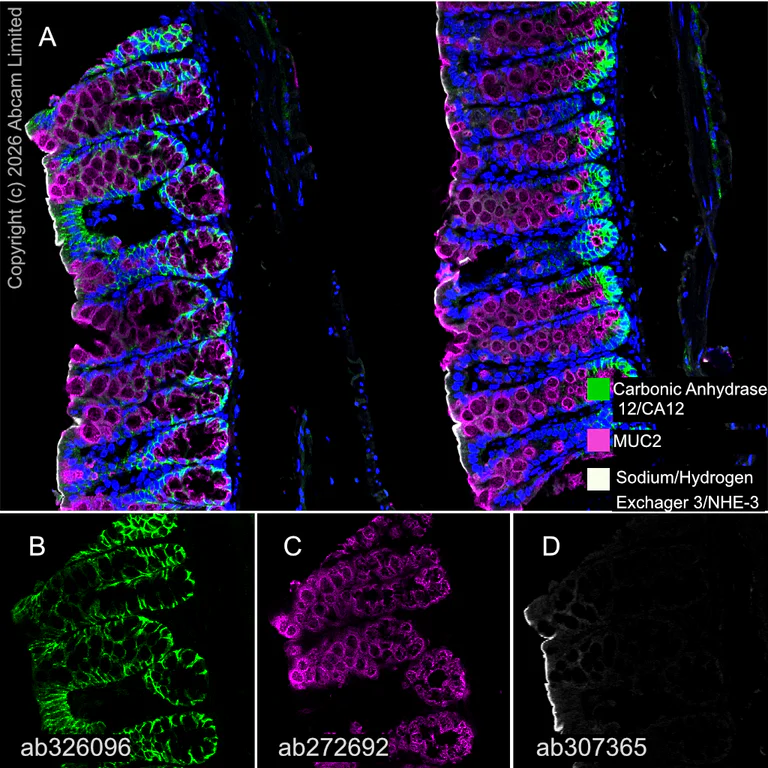
Multiplex immunohistochemistry - Anti-Sodium/Hydrogen Exchanger 3/NHE-3 antibody [EPR26951-3] (AB307365)
Multiplex immunohistochemistry analysis of formalin/PFA-fixed paraffin-embedded mouse colon tissue staining Carbonic Anhydrase 12/CA12 with ab326096 at a 1/1000 dilution (0.527 μg/ml), ab272692 anti-MUC2 at a 1/4000 dilution (0.253 μg/ml) and ab307365 anti-Sodium/Hydrogen Exchanger 3/NHE-3 at a 1/5000 dilution (0.126 μg/ml).
Panel A : merged staining of anti-Carbonic Anhydrase 12/CA12 (green; Opal™520), anti-MUC2 (magenta; Opal™690) and anti-Sodium/Hydrogen Exchanger 3/NHE-3 (gray; Opal™570) on mouse colon.
Panel B : anti-Carbonic Anhydrase 12/CA12 staining epithelium in mouse colon.
Panel C : anti-MUC2 staining goblet cells in mouse colon.
Panel D : anti-Sodium/Hydrogen Exchanger 3/NHE-3 staining apical and luminal facing edges of surface cells in mouse colon.
Nuclear DNA was labeled with DAPI (shown in blue).
The section was incubated in three rounds of staining : in the order of ab326096, ab272692 and ab307365 for 30 mins at room temperature. Each round was followed by a separate fluorescent tyramide signal amplification system.
The immunostaining was performed on a Leica Biosystems BOND® RX instrument with an Opal™ 4-color kit using Opal Polymer HRP Ms + Rb secondary reagent. Image acquisition was performed with a Leica STELLARIS confocal microscope.
Heat mediated antigen retrieval was performed with Tris-EDTA buffer (pH 9.0, Epitope Retrieval Solution2) for 20 mins.
- IHC-P
Supplier Data
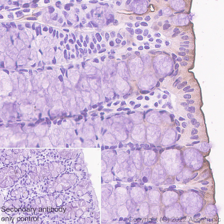
Immunohistochemistry (Formalin/PFA-fixed paraffin-embedded sections) - Anti-Sodium/Hydrogen Exchanger 3/NHE-3 antibody [EPR26951-3] (AB307365)
Immunohistochemical analysis of paraffin-embedded Mouse colon tissue lABeling Sodium/Hydrogen Exchanger 3/NHE-3 with ab307365 at 1/4000 (0.126 ug/ml) followed by a ready to use Goat Anti-RABbit IgG H&L (HRP polymer). Apical staining in mouse colon (PMID : 33904662).The section was incubated with ab307365 at 4ЎгC overnight. Counterstained with Hematoxylin. Secondary antibody only control : Secondary antibody is a ready to use Goat Anti-RABbit IgG H&L (HRP polymer). Heat mediated antigen retrieval was performed using ab93684 (Tris/EDTA buffer, pH 9.0)
- IHC-Fr
Supplier Data
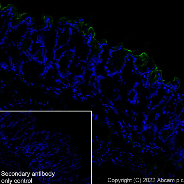
Immunohistochemistry (Frozen sections) - Anti-Sodium/Hydrogen Exchanger 3/NHE-3 antibody [EPR26951-3] (AB307365)
Immunohistochemical analysis of 4% PFA-fixed, 0.2% Triton X-100 permeabilized frozen Mouse colon (fresh) tissue labeling Sodium/Hydrogen Exchanger 3/NHE-3 with ab307365 at 1/50 (10.06 ug/ml) dilution followed by ab150081 Goat Anti-RABbit IgG H&L (Alexa Fluor® 488) preadsorbed at 1/1000 2 ug/mL dilution (Green). Confocal image showing positive staining along the apical margin of surface enterocytes on mouse colon. The nuclear counterstain was DAPI (Blue). The section was incubated with ab307365 for 60 mins at room temperature. The section was then mounted using Fluoromount®. The immunostaining was performed on a Leica Biosystems BOND® RX instrument. Image was taken with a confocal microscope (Leica-Microsystems, TCS SP8). The nuclear counterstain was DAPI (Blue). Secondary antibody control : Secondary antibody is ab150081 Goat Anti-Rabbit IgG H&L (Alexa Fluor® 488) preadsorbed at 1/1000 2 ug/mL dilution.
- IHC-Fr
Supplier Data
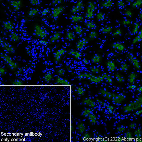
Immunohistochemistry (Frozen sections) - Anti-Sodium/Hydrogen Exchanger 3/NHE-3 antibody [EPR26951-3] (AB307365)
Immunohistochemical analysis of 4% PFA-fixed, 0.2% Triton X-100 permeabilized frozen Mouse kidney (fresh) tissue labeling Sodium/Hydrogen Exchanger 3/NHE-3 with ab307365 at 1/50 (10.06 ug/ml) dilution followed by ab150081 Goat Anti-Rabbit IgG H&L (Alexa Fluor® 488) preadsorbed at 1/1000 2 ug/mL dilution (Green). Confocal image showing positive staining in the brush border membrane of proximal tubular cells on mouse kidney. The nuclear counterstain was DAPI (Blue). The section was incubated with ab307365 for 60 mins at room temperature. The section was then mounted using Fluoromount®. The immunostaining was performed on a Leica Biosystems BOND® RX instrument. Image was taken with a confocal microscope (Leica-Microsystems, TCS SP8). The nuclear counterstain was DAPI (Blue). Secondary antibody control : Secondary antibody is ab150081 Goat Anti-Rabbit IgG H&L (Alexa Fluor® 488) preadsorbed at 1/1000 2 ug/mL dilution.
- IHC-Fr
Supplier Data
Immunohistochemistry (Frozen sections) - Anti-Sodium/Hydrogen Exchanger 3/NHE-3 antibody [EPR26951-3] (AB307365)
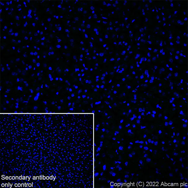
Immunohistochemical analysis of 4% PFA-fixed, 0.2% Triton X-100 permeabilized frozen Mouse liver (fresh) tissue labeling Sodium/Hydrogen Exchanger 3/NHE-3 with ab307365 at 1/50 (10.06 ug/ml) dilution followed by ab150081 Goat Anti-Rabbit IgG H&L (Alexa Fluor® 488) preadsorbed at 1/1000 2 ug/mL dilution (Green). Negative control : confocal image showing no staining on mouse liver. The nuclear counterstain was DAPI (Blue). The section was incubated with ab307365 for 60 mins at room temperature. The section was then mounted using Fluoromount®. The immunostaining was performed on a Leica Biosystems BOND® RX instrument. Image was taken with a confocal microscope (Leica-Microsystems, TCS SP8). The nuclear counterstain was DAPI (Blue). Secondary antibody control : Secondary antibody is ab150081 Goat Anti-Rabbit IgG H&L (Alexa Fluor® 488) preadsorbedat 1/1000 2 ug/mL dilution.
- mIHC
Lab
Multiplex immunohistochemistry - Anti-Sodium/Hydrogen Exchanger 3/NHE-3 antibody [EPR26951-3] (AB307365)
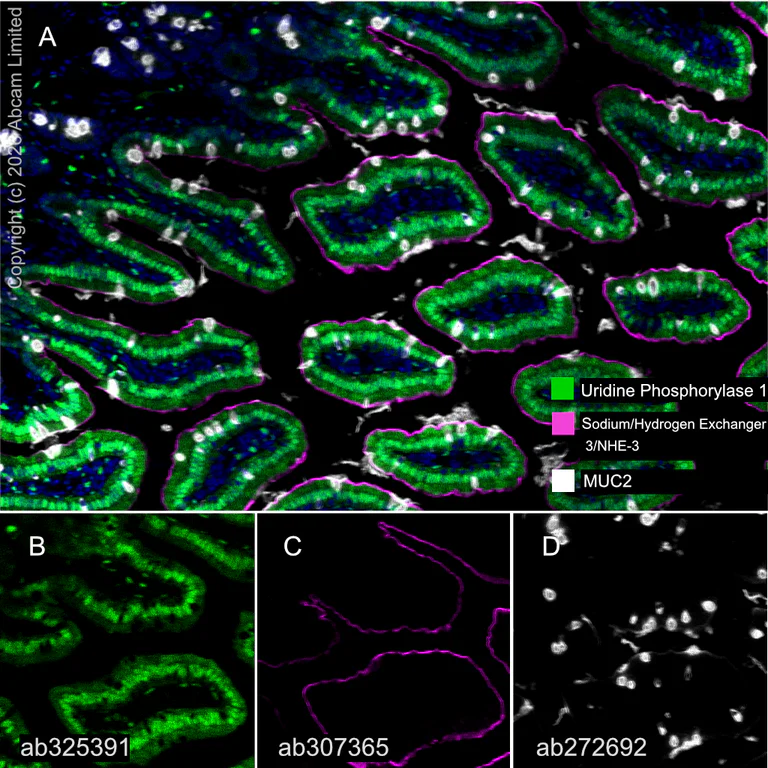
Multiplex immunohistochemistry analysis of formalin/PFA-fixed paraffin-embedded mouse small intestine tissue staining Uridine Phosphorylase 1 with ab325391 at a 1/2000 (0.258 μg/ml) dilution, ab307365 anti-Sodium/Hydrogen Exchanger 3/NHE-3 used at a 1/5000 (0.126 μg/ml) dilution and ab272692 anti-MUC2 used at a 1/2000 (0.26 μg/ml) dilution.
Panel A : anti-Uridine Phosphorylase 1 (green; Opal™690), anti-Sodium/Hydrogen Exchanger 3/NHE-3 (magenta; Opal™520), anti-MUC2 (gray; Opal™570) on mouse small intestine.
Panel B : anti-Uridine Phosphorylase 1 staining cytoplasm and nucleus of epithelium in mouse small intestine.
Panel C : anti-Sodium/Hydrogen Exchanger 3/NHE-3 staining apical and luminal facing edges of surface cells in mouse small intestine.
Panel D : anti-MUC2 staining on goblet cells in mouse small intestine.
Nuclear DNA was labeled with DAPI (shown in blue).
The section was incubated in three rounds of staining : in the order of ab325391, ab307365 and ab272692 for 30 mins at room temperature. Each round was followed by a separate fluorescent tyramide signal amplification system.
The immunostaining was performed on a Leica Biosystems BOND® RX instrument with an Opal™ 4-color kit. Image acquisition was performed with Leica SP8 confocal microscope.
Heat mediated antigen retrieval was performed with Tris-EDTA buffer (pH 9.0, Epitope Retrieval Solution2) for 20 mins.
- IHC-P
Supplier Data
Immunohistochemistry (Formalin/PFA-fixed paraffin-embedded sections) - Anti-Sodium/Hydrogen Exchanger 3/NHE-3 antibody [EPR26951-3] (AB307365)
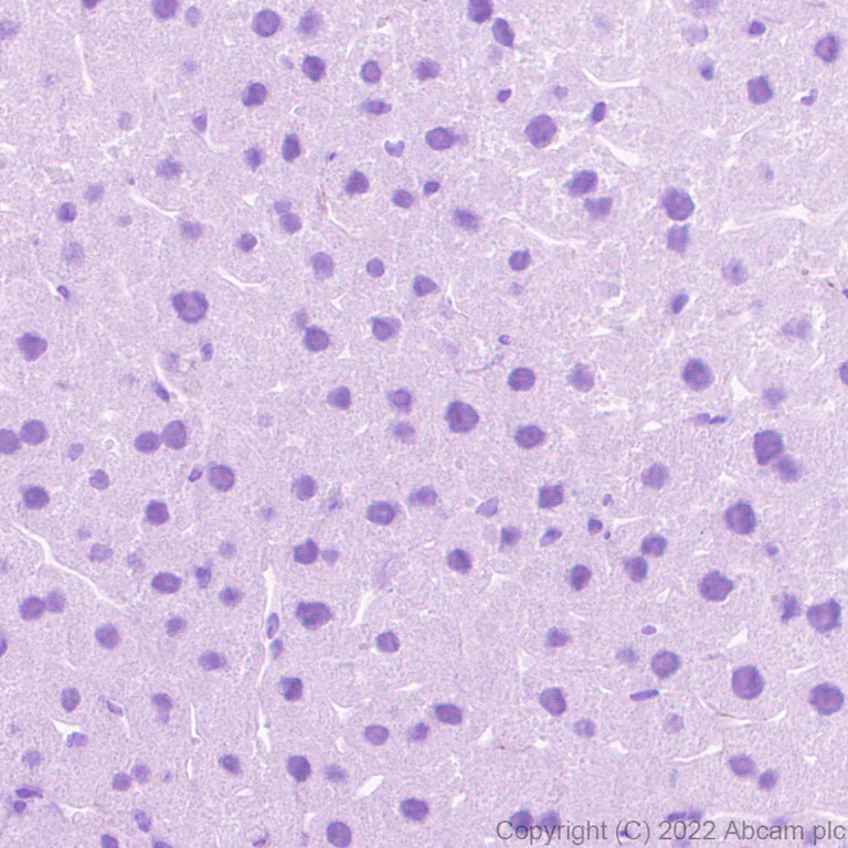
Immunohistochemical analysis of paraffin-embedded Mouse liver tissue lABeling Sodium/Hydrogen Exchanger 3/NHE-3 with ab307365 at 1/4000 (0.126 ug/ml) followed by a ready to use Goat Anti-RABbit IgG H&L (HRP polymer). Negative control : no staining on mouse liver.The section was incubated with ab307365 at 4ЎгC overnight. Counterstained with Hematoxylin. Secondary antibody only control : Secondary antibody is a ready to use Goat Anti-RABbit IgG H&L (HRP polymer). Heat mediated antigen retrieval was performed using ab93684 (Tris/EDTA buffer, pH 9.0)
- IHC-P
Supplier Data
Immunohistochemistry (Formalin/PFA-fixed paraffin-embedded sections) - Anti-Sodium/Hydrogen Exchanger 3/NHE-3 antibody [EPR26951-3] (AB307365)
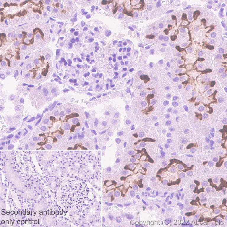
Immunohistochemical analysis of paraffin-embedded Rat kidney tissue lABeling Sodium/Hydrogen Exchanger 3/NHE-3 with ab307365 at 1/4000 (0.126 ug/ml) followed by a ready to use Goat Anti-RABbit IgG H&L (HRP polymer). Positive staining on the brush border in rat kidney.The section was incubated with ab307365 at 4ЎгC overnight. Counterstained with Hematoxylin. Secondary antibody only control : Secondary antibody is a ready to use Goat Anti-RABbit IgG H&L (HRP polymer). Heat mediated antigen retrieval was performed using ab93684 (Tris/EDTA buffer, pH 9.0)
- IHC-P
Supplier Data
Immunohistochemistry (Formalin/PFA-fixed paraffin-embedded sections) - Anti-Sodium/Hydrogen Exchanger 3/NHE-3 antibody [EPR26951-3] (AB307365)
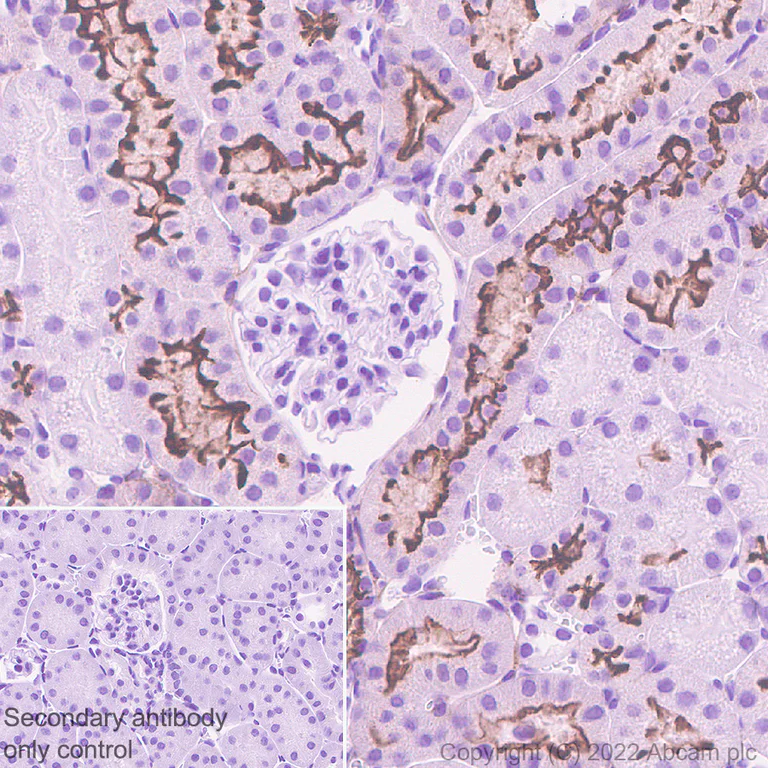
Immunohistochemical analysis of paraffin-embedded Mouse kidney tissue lABeling Sodium/Hydrogen Exchanger 3/NHE-3 with ab307365 at 1/4000 (0.126 ug/ml) followed by a ready to use Goat Anti-RABbit IgG H&L (HRP polymer). Positive staining on the brush border in mouse kidney (PMID : 8238556).The section was incubated with ab307365 at 4ЎгC overnight. Counterstained with Hematoxylin. Secondary antibody only control : Secondary antibody is a ready to use Goat Anti-RABbit IgG H&L (HRP polymer). Heat mediated antigen retrieval was performed using ab93684 (Tris/EDTA buffer, pH 9.0)
- IHC-P
Supplier Data
Immunohistochemistry (Formalin/PFA-fixed paraffin-embedded sections) - Anti-Sodium/Hydrogen Exchanger 3/NHE-3 antibody [EPR26951-3] (AB307365)
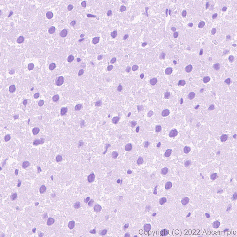
Immunohistochemical analysis of paraffin-embedded Rat liver tissue lABeling Sodium/Hydrogen Exchanger 3/NHE-3 with ab307365 at 1/4000 (0.126 ug/ml) followed by a ready to use Goat Anti-RABbit IgG H&L (HRP polymer). Negative control : no staining on rat liver.The section was incubated with ab307365 at 4ЎгC overnight. Counterstained with Hematoxylin. Secondary antibody only control : Secondary antibody is a ready to use Goat Anti-RABbit IgG H&L (HRP polymer). Heat mediated antigen retrieval was performed using ab93684 (Tris/EDTA buffer, pH 9.0)
- IHC-P
Supplier Data
Immunohistochemistry (Formalin/PFA-fixed paraffin-embedded sections) - Anti-Sodium/Hydrogen Exchanger 3/NHE-3 antibody [EPR26951-3] (AB307365)
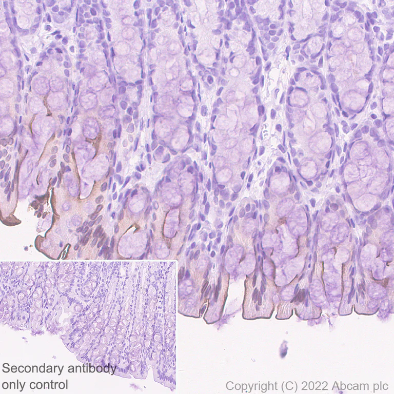
Immunohistochemical analysis of paraffin-embedded Rat colon tissue lABeling Sodium/Hydrogen Exchanger 3/NHE-3 with ab307365 at 1/4000 (0.126 ug/ml) followed by a ready to use Goat Anti-RABbit IgG H&L (HRP polymer). Apical staining in rat colon.The section was incubated with ab307365 at 4ЎгC overnight. Counterstained with Hematoxylin. Secondary antibody only control : Secondary antibody is a ready to use Goat Anti-RABbit IgG H&L (HRP polymer). Heat mediated antigen retrieval was performed using ab93684 (Tris/EDTA buffer, pH 9.0)
- mIHC
Lab
Multiplex immunohistochemistry - Anti-Sodium/Hydrogen Exchanger 3/NHE-3 antibody [EPR26951-3] (AB307365)
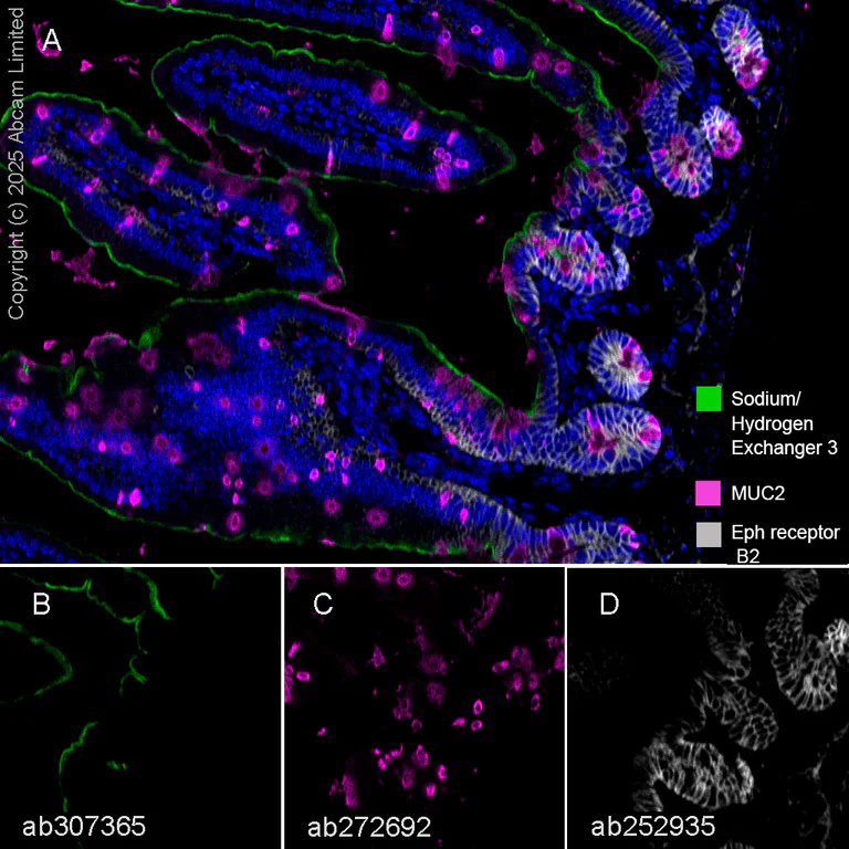
Immunohistochemistry analysis of Formalin/PFA-fixed paraffin-embedded sections mouse small intestine tissue labelling Sodium/Hydrogen Exchanger 3 with ab307365 at 1 : 5000 dilution (B), MUC2 with ab272692 at 1 : 2000 dilution (C) Eph receptor B2 with ab252935 at 1 : 500 dilution (D). Opal Polymer HRP Ms + Rb was used as a secondary antibody, and DAPI was used for a nuclear counter stain. Heat mediated antigen retrieval with Tris-EDTA buffer (pH 9.0, epitope retrieval solution2) for 20 mins.
Panel A : merged staining of anti-Sodium/Hydrogen Exchanger 3 (green; Opal™520), anti-MUC2 (magenta; Opal™690) and anti-Eph receptor B2 (gray; Opal™570) on mouse small intestine.
Panel B : anti-Sodium/Hydrogen Exchanger 3 staining apical and luminal facing edges of surface cells in mouse small intestine.
Panel C : anti-MUC2 staining goblet cells in mouse small intestine.
Panel D : anti-Eph receptor B2 staining membrane of stem cells in mouse small intestine.
Nuclear DNA was labeled with DAPI (shown in blue).
The section was incubated in three rounds of staining : in the order of ab307365, ab272692 and ab252935 for 30 mins at room temperature. Each round was followed by a separate fluorescent tyramide signal amplification system.
- mIHC
Lab
Multiplex immunohistochemistry - Anti-Sodium/Hydrogen Exchanger 3/NHE-3 antibody [EPR26951-3] (AB307365)
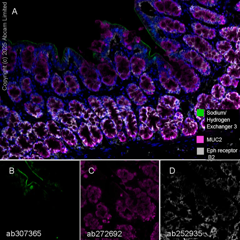
Immunohistochemistry analysis of Formalin/PFA-fixed paraffin-embedded sections rat colon tissue labelling Sodium/Hydrogen Exchanger 3 with ab307365 at 1 : 5000 dilution (B), MUC2 with ab272692 at 1 : 2000 dilution (C) Eph receptor B2 with ab252935 at 1 : 500 dilution (D). Opal Polymer HRP Ms + Rb was used as a secondary antibody, and DAPI was used for a nuclear counter stain. Heat mediated antigen retrieval with Tris-EDTA buffer (pH 9.0, epitope retrieval solution2) for 20 mins.
Panel A : merged staining of anti-Sodium/Hydrogen Exchanger 3 (green; Opal™520), anti-MUC2 (magenta; Opal™690) and anti-Eph receptor B2 (gray; Opal™570) on rat colon.
Panel B : anti-Sodium/Hydrogen Exchanger 3 staining apical and luminal facing edges of surface cells in rat colon.
Panel C : anti-MUC2 staining goblet cells in rat colon.
Panel D : anti-Eph receptor B2 staining membrane of stem cells in rat colon.
Nuclear DNA was labeled with DAPI (shown in blue).
The section was incubated in three rounds of staining : in the order of ab307365, ab272692 and ab252935 for 30 mins at room temperature. Each round was followed by a separate fluorescent tyramide signal amplification system.
- mIHC
Lab
Multiplex immunohistochemistry - Anti-Sodium/Hydrogen Exchanger 3/NHE-3 antibody [EPR26951-3] (AB307365)
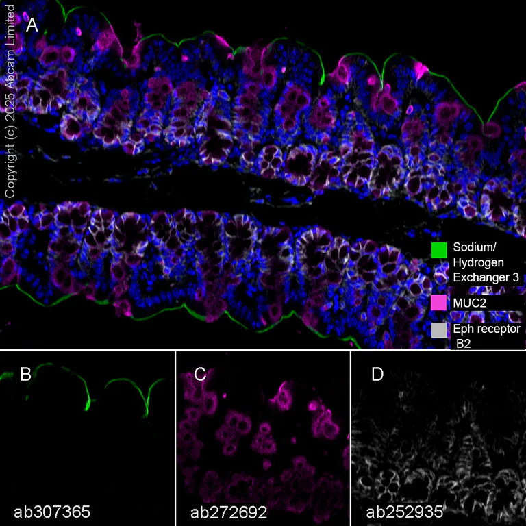
Immunohistochemistry analysis of Formalin/PFA-fixed paraffin-embedded sections mouse colon tissue labelling Sodium/Hydrogen Exchanger 3 with ab307365 at 1 : 5000 dilution (B), MUC2 with ab272692 at 1 : 2000 dilution (C) Eph receptor B2 with ab252935 at 1 : 500 dilution (D). Opal Polymer HRP Ms + Rb was used as a secondary antibody, and DAPI was used for a nuclear counter stain. Heat mediated antigen retrieval with Tris-EDTA buffer (pH 9.0, epitope retrieval solution2) for 20 mins.
Panel A : merged staining of anti-Sodium/Hydrogen Exchanger 3 (green; Opal™520), anti-MUC2 (magenta; Opal™690) and anti-Eph receptor B2 (gray; Opal™570) on mouse colon.
Panel B : anti-Sodium/Hydrogen Exchanger 3 staining apical and luminal facing edges of surface cells in mouse colon.
Panel C : anti-MUC2 staining goblet cells in mouse colon.
Panel D : anti-Eph receptor B2 staining membrane of stem cells in mouse colon.
Nuclear DNA was labeled with DAPI (shown in blue).
The section was incubated in three rounds of staining : in the order of ab307365, ab272692 and ab252935 for 30 mins at room temperature. Each round was followed by a separate fluorescent tyramide signal amplification system.
- mIHC
Lab
Multiplex immunohistochemistry - Anti-Sodium/Hydrogen Exchanger 3/NHE-3 antibody [EPR26951-3] (AB307365)
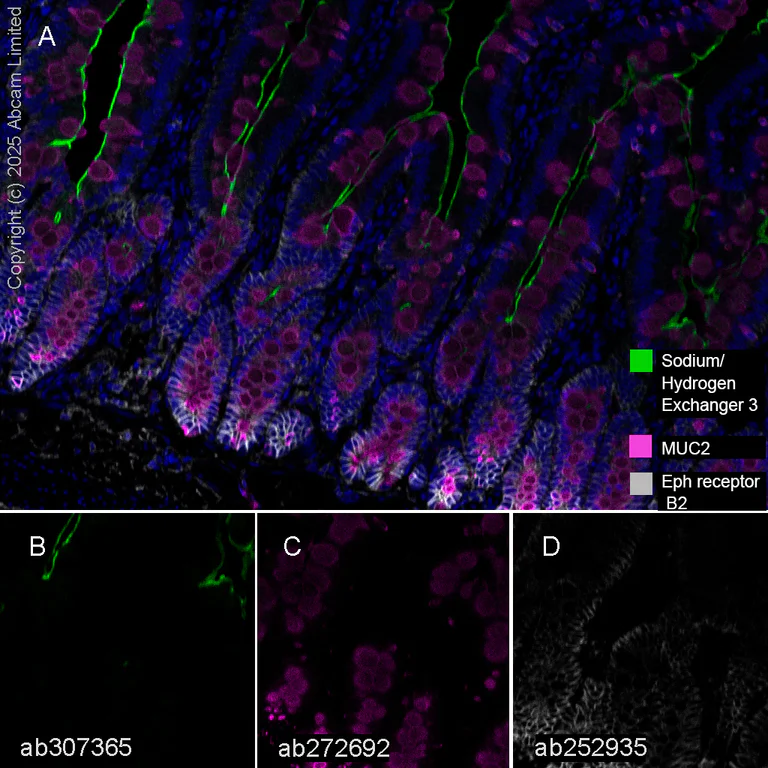
Immunohistochemistry analysis of Formalin/PFA-fixed paraffin-embedded sections rat small intestine tissue labelling Sodium/Hydrogen Exchanger 3 with ab307365 at 1 : 5000 dilution (B), MUC2 with ab272692 at 1 : 2000 dilution (C) Eph receptor B2 with ab252935 at 1 : 500 dilution (D). Opal Polymer HRP Ms + Rb was used as a secondary antibody, and DAPI was used for a nuclear counter stain. Heat mediated antigen retrieval with Tris-EDTA buffer (pH 9.0, epitope retrieval solution2) for 20 mins.
Panel A : merged staining of anti-Sodium/Hydrogen Exchanger 3 (green; Opal™520), anti-MUC2 (magenta; Opal™690) and anti-Eph receptor B2 (gray; Opal™570) on rat small intestine.
Panel B : anti-Sodium/Hydrogen Exchanger 3 staining apical and luminal facing edges of surface cells in rat small intestine.
Panel C : anti-MUC2 staining goblet cells in rat small intestine.
Panel D : anti-Eph receptor B2 staining membrane of stem cells in rat small intestine.
Nuclear DNA was labeled with DAPI (shown in blue).
The section was incubated in three rounds of staining : in the order of ab307365, ab272692 and ab252935 for 30 mins at room temperature. Each round was followed by a separate fluorescent tyramide signal amplification system.
Related conjugates and formulations (1)
-
Anti-Sodium/Hydrogen Exchanger 3/NHE-3 antibody [EPR26951-3] - BSA and Azide free
Complementary Products

Primary Antibodies
AB181602
Anti-GAPDH antibody [EPR16891] - Loading Control
KD Validated|RabMAb|Recombinant|Lab Essentials|20ul selling size

- 5
- 23
Primary Antibodies
AB313640
Anti-SLC12A1/NKCC2 antibody [EPR28176-90]
BOND RX™ Validated|20ul selling size|RabMAb|Recombinant

- 0
- 0
Primary Antibodies
AB316191
Anti-SLC12A3 antibody [EPR27106-48]
BOND RX™ Validated|20ul selling size|Recombinant|RabMAb|Advanced Validation

- 0
- 0
Reactivity data
Product details
Patented technology
Our RabMAb® technology is a patented hybridoma-based technology for making rabbit monoclonal antibodies. For details on our patents, please refer to RabMAb® patents.
What are the advantages of a recombinant monoclonal antibody?
This product is a recombinant monoclonal antibody, which offers several advantages including:
- - High batch-to-batch consistency and reproducibility
- - Improved sensitivity and specificity
- - Long-term security of supply
- - Animal-free batch production
For more information, read more on recombinant antibodies.
Properties and storage information
Form
Purification technique
Storage buffer
Shipped at conditions
Appropriate short-term storage duration
Appropriate short-term storage conditions
Appropriate long-term storage conditions
Aliquoting information
Storage information
Supplementary information
This supplementary information is collated from multiple sources and compiled automatically.
Biological function summary
The NHE-3 transporter facilitates the reabsorption of sodium ions contributing to cellular pH regulation and volume control. It operates as part of a larger multiprotein complex involving various regulatory proteins that fine-tune its activity. The exchanger activity is critical for maintaining homeostasis and proper cellular function influencing processes like nutrient absorption and acid-base balance in different tissues.
Pathways
The NHE-3 transporter plays significant roles in fluid reabsorption and bicarbonate reabsorption pathways. It functions in concert with other transporters and channels such as the Na-K ATPase pump which is key to maintaining sodium and potassium gradients. These pathways highlight the exchanger's contributions to renal and digestive systems emphasizing its interaction with proteins such as carbonic anhydrase within these processes.
Product protocols
- Visit the General protocols
- Visit the Troubleshooting
Target data
Alternative Names
Publications (1)
Recent publications for all applications. Explore the full listand refine your search
Frontiers in physiology 16:1544274 PubMed40177358
2025
Applications
Unspecified application
Species
Unspecified reactive species
Product promise
Please note: All products are 'FOR RESEARCH USE ONLY. NOT FOR USE IN DIAGNOSTIC OR THERAPEUTIC PROCEDURES'.
For licensing inquiries, please contact partnerships@abcam.com