Anti-Spinophilin/Neurabin 2 antibody [EPR26265-105]
- BOND RX™ Validated
- 20ul selling size
- RabMAb
- Recombinant
- What is this?
Be the first to review this product! Submit a review
|
(0 Publication)
Rabbit Recombinant Monoclonal Spinophilin/Neurabin 2 antibody. Suitable for IHC-P, IHC-Fr and reacts with Human, Mouse, Rat samples.
View Alternative Names
PPP1R6, PPP1R9B, Neurabin-2, Neurabin-II, Protein phosphatase 1 regulatory subunit 9B, Spinophilin
- IHC-P
Supplier Data
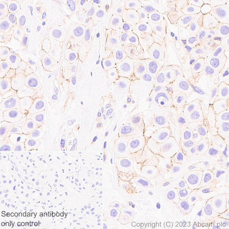
Immunohistochemistry (Formalin/PFA-fixed paraffin-embedded sections) - Anti-Spinophilin/Neurabin 2 antibody [EPR26265-105] (AB309135)
Immunohistochemical analysis of paraffin-embedded Human breast cancer tissue labeling Spinophilin/Neurabin 2 with ab309135 at 1/5000 (0.098 ug/ml) followed by a ready to use LeicaDS9800 (Bond™ Polymer Refine Detection). Positive staining on human breast cancer (PMID : 25857299). The section was incubated with ab309135 for 30 mins at room temperature. The immunostaining was performed on a Leica Biosystems BOND® RX instrument. Incubate slides with 3% Hydrogen Peroxide for 10 mins at room temperature after secondary antibody incubation to reduce the background. Counterstained with Hematoxylin. Secondary antibody only control : Secondary antibody is a ready to use LeicaDS9800 (Bond™ Polymer Refine Detection). Heat mediated antigen retrieval was performed with Tris-EDTA buffer (pH 9.0, Epitope Retrieval Solution2) for 20 mins
- IHC-P
Supplier Data
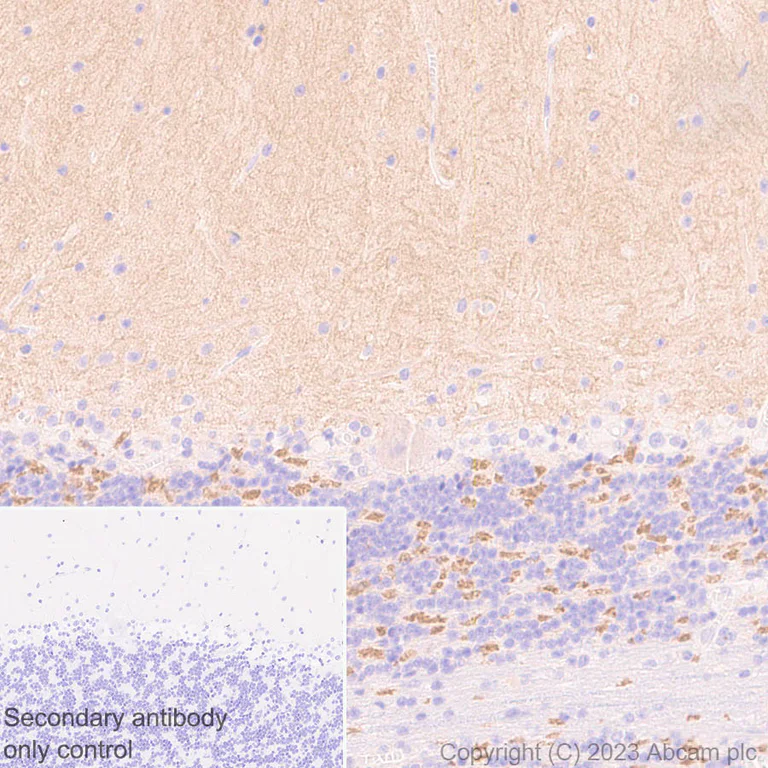
Immunohistochemistry (Formalin/PFA-fixed paraffin-embedded sections) - Anti-Spinophilin/Neurabin 2 antibody [EPR26265-105] (AB309135)
Immunohistochemical analysis of paraffin-embedded Human cerebellum tissue labeling Spinophilin/Neurabin 2 with ab309135 at 1/5000 (0.098 ug/ml) followed by a ready to use LeicaDS9800 (Bond™ Polymer Refine Detection). Positive staining on human cerebellum. The section was incubated with ab309135 for 30 mins at room temperature.The immunostaining was performed on a Leica Biosystems BOND® RX instrument. Incubate slides with 3% Hydrogen Peroxide for 10 mins at room temperature after secondary antibody incubation to reduce the background. Counterstained with Hematoxylin. Secondary antibody only control : Secondary antibody is a ready to use LeicaDS9800 (Bond™ Polymer Refine Detection). Heat mediated antigen retrieval was performed with Tris-EDTA buffer (pH 9.0, Epitope Retrieval Solution2) for 20 mins
- IHC-P
Supplier Data
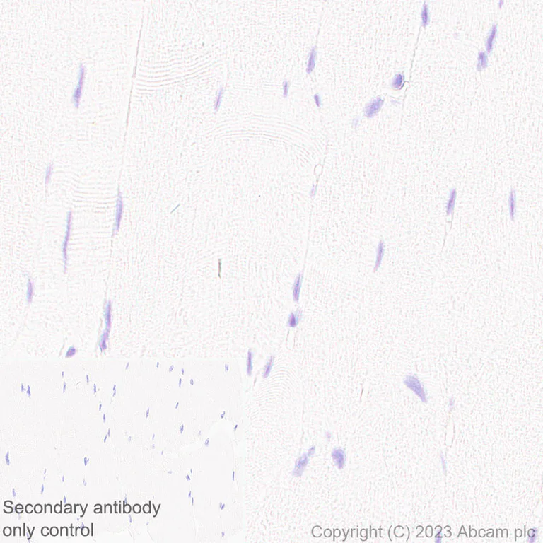
Immunohistochemistry (Formalin/PFA-fixed paraffin-embedded sections) - Anti-Spinophilin/Neurabin 2 antibody [EPR26265-105] (AB309135)
Immunohistochemical analysis of paraffin-embedded Human skeletal muscl tissue labeling Spinophilin/Neurabin 2 with ab309135 at 1/5000 (0.098 ug/ml) followed by a ready to use LeicaDS9800 (Bond™ Polymer Refine Detection). Low expression : no staining on human skeletal muscle. The section was incubated with ab309135 for 30 mins at room temperature. The immunostaining was performed on a Leica Biosystems BOND® RX instrument. Incubate slides with 3% Hydrogen Peroxide for 10 mins at room temperature after secondary antibody incubation to reduce the background. Counterstained with Hematoxylin. Secondary antibody only control : Secondary antibody is a ready to use LeicaDS9800 (Bond™ Polymer Refine Detection). Heat mediated antigen retrieval was performed with Tris-EDTA buffer (pH 9.0, Epitope Retrieval Solution2) for 20 mins
- IHC-P
Supplier Data
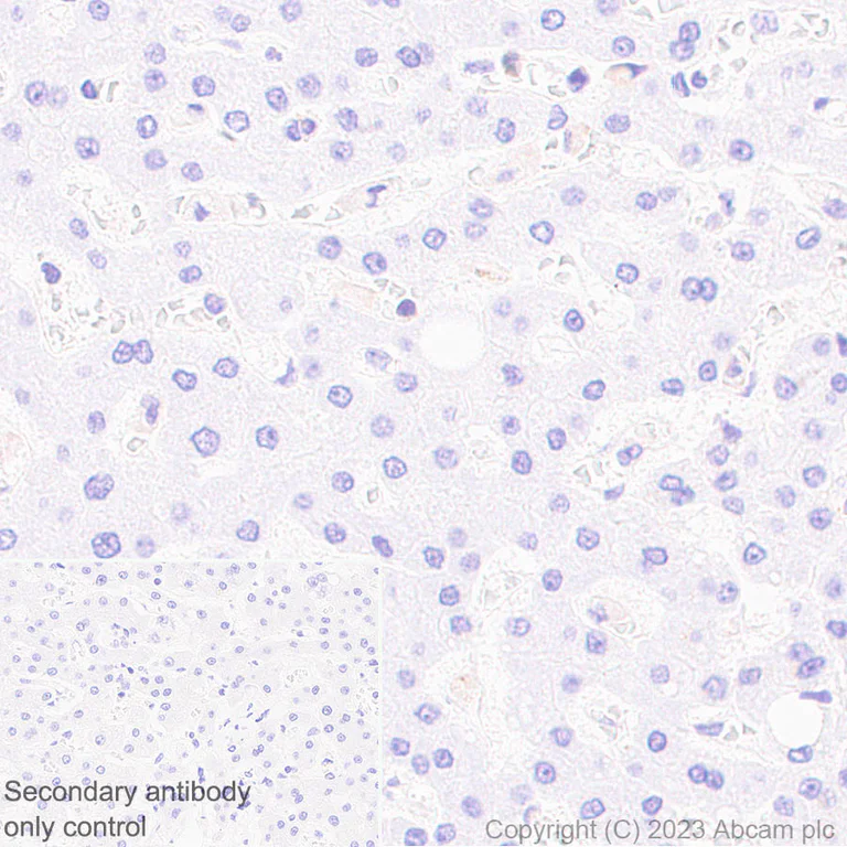
Immunohistochemistry (Formalin/PFA-fixed paraffin-embedded sections) - Anti-Spinophilin/Neurabin 2 antibody [EPR26265-105] (AB309135)
Immunohistochemical analysis of paraffin-embedded Human liver tissue labeling Spinophilin/Neurabin 2 with ab309135 at 1/5000 (0.098 ug/ml) followed by a ready to use LeicaDS9800 (Bond™ Polymer Refine Detection). Low expression : almost no staining on human liver. The section was incubated with ab309135 for 30 mins at room temperature. The immunostaining was performed on a Leica Biosystems BOND® RX instrument. Incubate slides with 3% Hydrogen Peroxide for 10 mins at room temperature after secondary antibody incubation to reduce the background. Counterstained with Hematoxylin. Secondary antibody only control : Secondary antibody is a ready to use LeicaDS9800 (Bond™ Polymer Refine Detection). Heat mediated antigen retrieval was performed with Tris-EDTA buffer (pH 9.0, Epitope Retrieval Solution2) for 20 mins
- IHC-P
Supplier Data
Immunohistochemistry (Formalin/PFA-fixed paraffin-embedded sections) - Anti-Spinophilin/Neurabin 2 antibody [EPR26265-105] (AB309135)
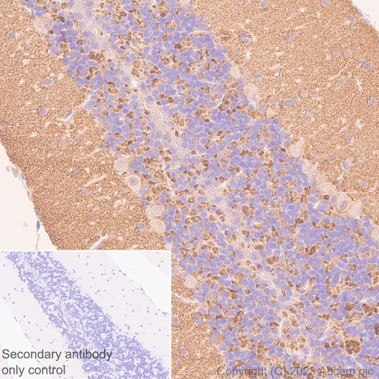
Immunohistochemical analysis of paraffin-embedded Mouse cerebellum tissue labeling Spinophilin/Neurabin 2 with ab309135 at 1/5000 (0.098 ug/ml) followed by a ready to use LeicaDS9800 (Bond™ Polymer Refine Detection) was used. Positive staining on mouse cerebellum (PMID : 9275233). The section was incubated with ab309135 for 30 mins at room temperature. The immunostaining was performed on a Leica Biosystems BOND® RX instrument. Incubate slides with 3% Hydrogen Peroxide for 10 mins at room temperature after secondary antibody incubation to reduce the background. Counterstained with Hematoxylin. Secondary antibody only control : Secondary antibody is a ready to use LeicaDS9800 (Bond™ Polymer Refine Detection) was used. Heat mediated antigen retrieval was performed with Tris-EDTA buffer (pH 9.0, Epitope Retrieval Solution2) for 20 mins
- IHC-P
Supplier Data
Immunohistochemistry (Formalin/PFA-fixed paraffin-embedded sections) - Anti-Spinophilin/Neurabin 2 antibody [EPR26265-105] (AB309135)
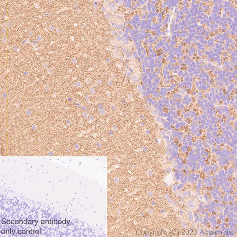
Immunohistochemical analysis of paraffin-embedded Rat cerebellum tissue labeling Spinophilin/Neurabin 2 with ab309135 at 1/5000 (0.098 ug/ml) followed by a ready to use LeicaDS9800 (Bond™ Polymer Refine Detection) was used. Positive staining on rat cerebellum (PMID : 9275233). The section was incubated with ab309135 for 30 mins at room temperature. The immunostaining was performed on a Leica Biosystems BOND® RX instrument. Incubate slides with 3% Hydrogen Peroxide for 10 mins at room temperature after secondary antibody incubation to reduce the background. Counterstained with Hematoxylin. Secondary antibody only control : Secondary antibody is a ready to use LeicaDS9800 (Bond™ Polymer Refine Detection) was used. Heat mediated antigen retrieval was performed with Tris-EDTA buffer (pH 9.0, Epitope Retrieval Solution2) for 20 mins
- IHC-Fr
Supplier Data
Immunohistochemistry (Frozen sections) - Anti-Spinophilin/Neurabin 2 antibody [EPR26265-105] (AB309135)
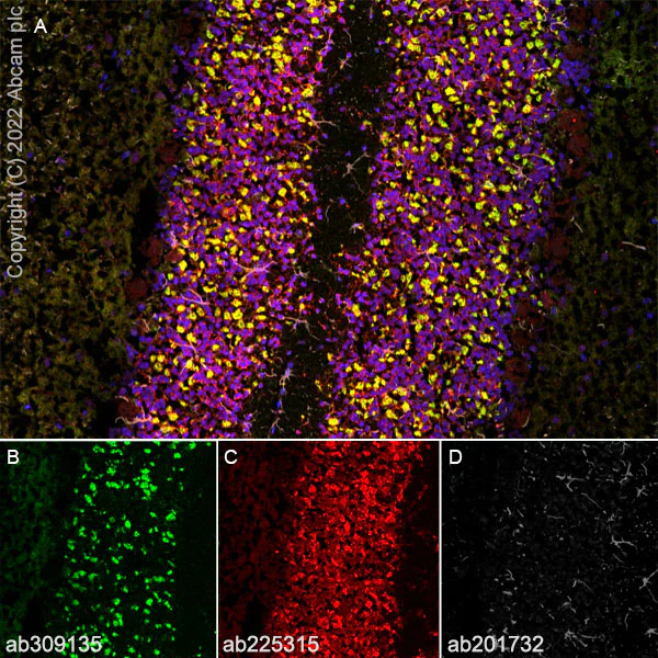
Immunohistochemical analysis of 4% PFA-fixed, 0.2% Triton X-100 permeabilized frozen Rat cerebellum (fresh) tissue labeling Spinophilin/Neurabin 2 with ab309135 at 1/2000 (0.246 ug/ml) dilution followed by ab150081 Goat Anti-Rabbit IgG H&L (Alexa Fluor® 488) preadsorbed at 1/1000 2 ug/mL dilution (Green). Panel A : merged staining of anti-Neurabin-2 (ab309135, green), anti-Map2 (ab225315, red) and anti-GFAP (ab201732, grey) on rat cerebellum. Panel B : anti-Neurabin-2 stained on rat cerebellum. Panel C : anti-Map2 stained in neurons of rat cerebellum. Panel D : anti-GFAP stained in astrocytes of rat cerebellum. The section was incubated in two rounds of staining : in the order of ab309135 and ab225315, ab201732 for 1 hr at room temperature. The nuclear counterstain was DAPI (Blue). The section was then mounted using Fluoromount®. The immunostaining was performed on a Leica Biosystems BOND® RX instrument. The image was taken with a confocal microscope (Leica-Microsystems, TCS SP8). is observed. The nuclear counterstain was DAPI (Blue). Secondary antibody control : Secondary antibody is ab150081 Goat Anti-Rabbit IgG H&L (Alexa Fluor® 488) preadsorbed at 1/1000 2 ug/mL dilution.
- IHC-Fr
Supplier Data
Immunohistochemistry (Frozen sections) - Anti-Spinophilin/Neurabin 2 antibody [EPR26265-105] (AB309135)
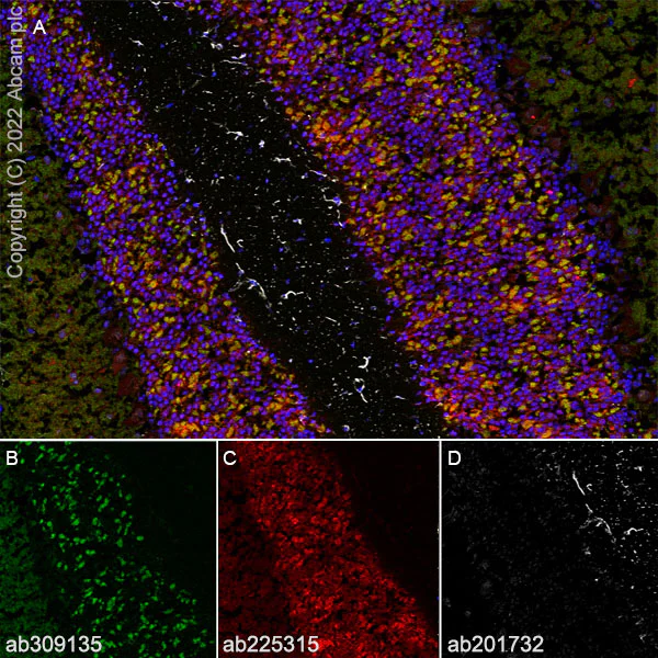
Immunohistochemical analysis of 4% PFA-fixed, 0.2% Triton X-100 permeabilized frozen Mouse cerebellum (fresh) tissue labeling Spinophilin/Neurabin 2 with ab309135 at 1/2000 (0.246 ug/ml) dilution followed by ab150081 Goat Anti-Rabbit IgG H&L (Alexa Fluor® 488) preadsorbed at 1/1000 2 ug/mL dilution (Green). Panel A : merged staining of anti-Neurabin-2 (ab309135, green), anti-Map2 (ab225315, red) and anti-GFAP (ab201732, grey) on mouse cerebellum.Panel B : anti-Neurabin-2 stained on mouse cerebellum.Panel C : anti-Map2 stained in neurons of mouse cerebellum.Panel D : anti-GFAP stained in astrocytes of mouse cerebellum. The section was incubated in two rounds of staining : in the order of ab309135 and ab225315, ab201732 for 1 hr at room temperature. The nuclear counterstain was DAPI (Blue). The section was then mounted using Fluoromount®. The immunostaining was performed on a Leica Biosystems BOND® RX instrument. The image was taken with a confocal microscope (Leica-Microsystems, TCS SP8). is observed. The nuclear counterstain was DAPI (Blue). Secondary antibody control : Secondary antibody is ab150081 Goat Anti-Rabbit IgG H&L (Alexa Fluor® 488) preadsorbed at 1/1000 2 ug/mL dilution.
- IHC-P
Supplier Data
Immunohistochemistry (Formalin/PFA-fixed paraffin-embedded sections) - Anti-Spinophilin/Neurabin 2 antibody [EPR26265-105] (AB309135)
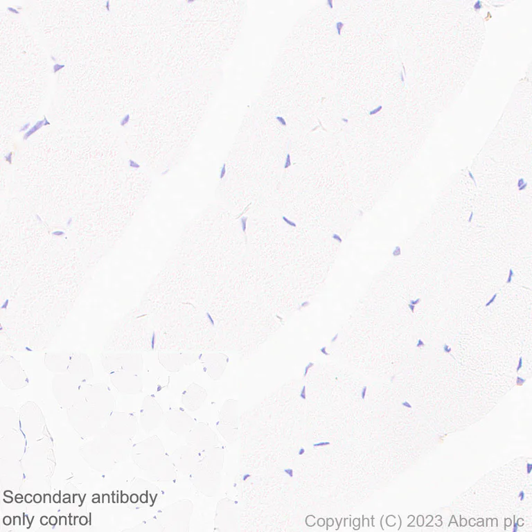
Immunohistochemical analysis of paraffin-embedded Mouse skeletal muscle tissue labeling Spinophilin/Neurabin 2 with ab309135 at 1/5000 (0.098 ug/ml) followed by a ready to use LeicaDS9800 (Bond™ Polymer Refine Detection). Low expression : no staining on mouse skeletal muscle (PMID : 9452470). The section was incubated with ab309135 for 30 mins at room temperature. The immunostaining was performed on a Leica Biosystems BOND® RX instrument. Incubate slides with 3% Hydrogen Peroxide for 10 mins at room temperature after secondary antibody incubation to reduce the background. Counterstained with Hematoxylin. Secondary antibody only control : Secondary antibody is a ready to use LeicaDS9800 (Bond™ Polymer Refine Detection). Heat mediated antigen retrieval was performed with Tris-EDTA buffer (pH 9.0, Epitope Retrieval Solution2) for 20 mins
- IHC-Fr
Supplier Data
Immunohistochemistry (Frozen sections) - Anti-Spinophilin/Neurabin 2 antibody [EPR26265-105] (AB309135)
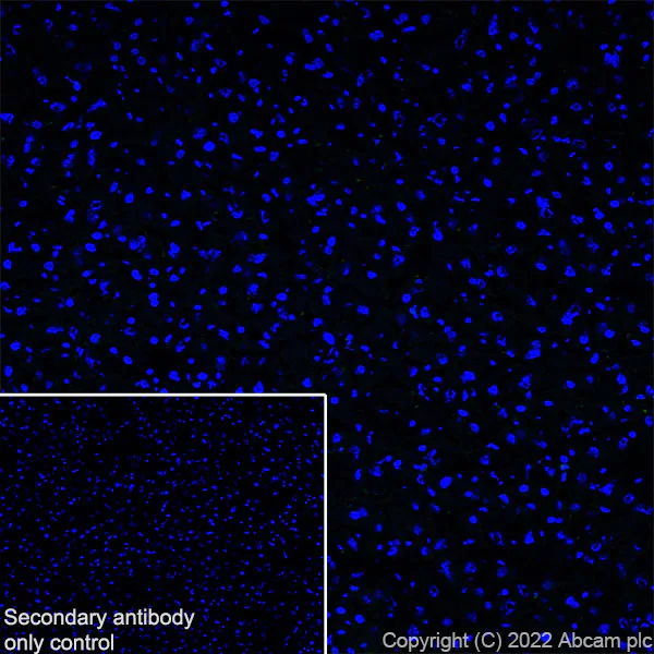
Immunohistochemical analysis of 4% PFA-fixed, 0.2% Triton X-100 permeabilized frozen Mouse liver (fresh) tissue labeling Spinophilin/Neurabin 2 with ab309135 at 1/2000 (0.246 ug/ml) dilution followed by ab150081 Goat Anti-Rabbit IgG H&L (Alexa Fluor® 488) preadsorbed at 1/1000 2 ug/mL dilution (Green). Low expression : confocal image showing no staining on mouse liver (PMID : 9452470). The nuclear counterstain was DAPI (Blue). The section was incubated with ab309135 for 60 mins at room temperature. The section was then mounted using Fluoromount®. The immunostaining was performed on a Leica Biosystems BOND® RX instrument. The image was taken with a confocal microscope (Leica-Microsystems, TCS SP8). is observed. The nuclear counterstain was DAPI (Blue). Secondary antibody control : Secondary antibody is ab150081 Goat Anti-Rabbit IgG H&L (Alexa Fluor® 488) preadsorbed at 1/1000 2 ug/mL dilution.
- IHC-Fr
Supplier Data
Immunohistochemistry (Frozen sections) - Anti-Spinophilin/Neurabin 2 antibody [EPR26265-105] (AB309135)
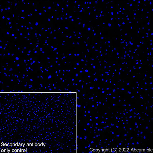
Immunohistochemical analysis of 4% PFA-fixed, 0.2% Triton X-100 permeabilized frozen Rat liver (fresh) tissue labeling Spinophilin/Neurabin 2 with ab309135 at 1/2000 (0.246 ug/ml) dilution followed by ab150081 Goat Anti-Rabbit IgG H&L (Alexa Fluor® 488) preadsorbed at 1/1000 2 ug/mL dilution (Green). Low expression : confocal image showing no staining on rat liver (PMID : 9452470). The nuclear counterstain was DAPI (Blue). The section was incubated with ab309135 for 60 mins at room temperature. The section was then mounted using Fluoromount®. The immunostaining was performed on a Leica Biosystems BOND® RX instrument. The image was taken with a confocal microscope (Leica-Microsystems, TCS SP8). is observed. The nuclear counterstain was DAPI (Blue). Secondary antibody control : Secondary antibody is ab150081 Goat Anti-Rabbit IgG H&L (Alexa Fluor® 488) preadsorbed at 1/1000 2 ug/mL dilution.
Related conjugates and formulations (1)
-
Anti-Spinophilin/Neurabin 2 antibody [EPR26265-105] - BSA and Azide free
Complementary Products
Primary Antibodies
AB179471
Anti-Integrin beta 1 antibody [EPR16895]
20ul selling size|RabMAb|Recombinant|KO Validated

- 5
- 5

Primary Antibodies
AB238135
Anti-PSD95 antibody [EPR23124-118] - Synaptic Marker
BOND RX™ Validated|KO Validated|RabMAb|Recombinant|Lab Essentials|20ul selling size

- 5
- 5

Reactivity data
Product details
Patented technology
Our RabMAb® technology is a patented hybridoma-based technology for making rabbit monoclonal antibodies. For details on our patents, please refer to RabMAb® patents.
What are the advantages of a recombinant monoclonal antibody?
This product is a recombinant monoclonal antibody, which offers several advantages including:
- - High batch-to-batch consistency and reproducibility
- - Improved sensitivity and specificity
- - Long-term security of supply
- - Animal-free batch production
For more information, read more on recombinant antibodies.
Properties and storage information
Form
Purification technique
Storage buffer
Shipped at conditions
Appropriate short-term storage duration
Appropriate short-term storage conditions
Appropriate long-term storage conditions
Aliquoting information
Storage information
Supplementary information
This supplementary information is collated from multiple sources and compiled automatically.
Biological function summary
Spinophilin functions in the regulation of synaptic signaling and structural plasticity contributing to neural communication. It often forms part of signaling complexes binding to protein phosphatase 1 (PP1) to modulate dephosphorylation activities. Spinophilin influences the organization and dynamics of actin filaments affecting the stability and morphology of dendritic spines which are critical sites for synaptic transmission and plasticity. Through these interactions Spinophilin plays an important role in learning and memory processes.
Pathways
Spinophilin interacts with systems involved in synaptic signaling and cytoskeletal organization. It impacts the RhoA/ROCK pathway which is essential for actin filament dynamics by modulating PP1 activity. Additionally Spinophilin indirectly affects the mTOR pathway which is involved in protein synthesis and cell growth. It relates to proteins such as RhoA and mTOR through its role in maintaining the balance of phosphorylation states within the cell.
Product protocols
- Visit the General protocols
- Visit the Troubleshooting
Target data
Product promise
Please note: All products are 'FOR RESEARCH USE ONLY. NOT FOR USE IN DIAGNOSTIC OR THERAPEUTIC PROCEDURES'.
For licensing inquiries, please contact partnerships@abcam.com