Anti-SQSTM1 / p62 antibody [EPR4844] - BSA and Azide free
- RabMAb
- Recombinant
- KO Validated
- What is this?
Be the first to review this product! Submit a review
|
(7 Publications )

- ICC/IF
Lab
Immunocytochemistry/ Immunofluorescence - Anti-SQSTM1 / p62 antibody [EPR4844] - BSA and Azide free (AB219581)
Clone EPR4844 (ab219581) has been successfully conjugated by Abcam. This image was generated using Anti-SQSTM1 / p62 antibody [EPR4844] - Autophagosome Marker (Alexa Fluor® 488). Please refer to ab185015 for protocol details.
ab185015 staining SQSTM1 in wild-type HAP1 cells, untreated and chloroquine-treated (50μM, 24 hours) and chloroquine-treated SQSTM1 knockout HAP1 cells. The cells were fixed with 4% formaldehyde (10min), permeabilized with 0.1% Triton X-100 for 5 minutes and then blocked with 1% BSA/10% normal goat serum/0.3M glycine in 0.1% PBS-Tween for 1h. The cells were then incubated overnight at +4°C with ab185015 at 1/500 dilution (shown in green) and ab195889, Mouse monoclonal to alpha Tubulin (Alexa Fluor® 594), at 1/250 dilution (shown in pseudocolor red). Nuclear DNA was labelled with DAPI (shown in blue).
Image was taken with a confocal microscope (Leica-Microsystems, TCS SP8).
- ICC/IF
Lab
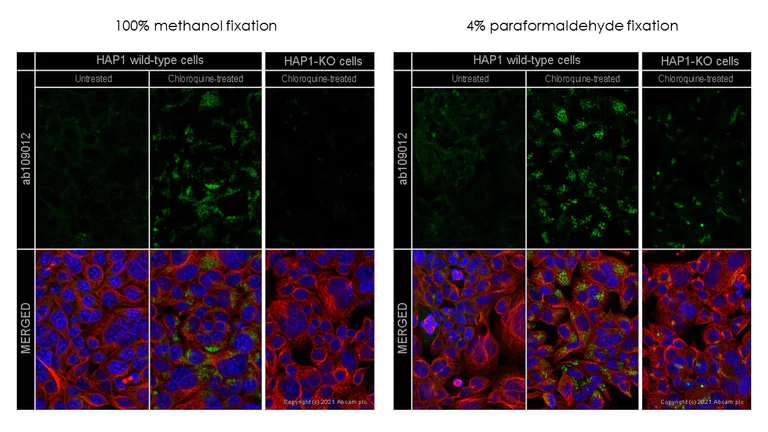
Immunocytochemistry/ Immunofluorescence - Anti-SQSTM1 / p62 antibody [EPR4844] - BSA and Azide free (AB219581)
This data was developed using the same antibody clone in a different buffer formulation (ab109012). ab109012 staining SQSTM1 in wild-type Hap1 cells and SQSTM1 knockout Hap1 cells treated with chloroquine (ab142116, 50μM for 24 hrs). The cells were fixed with 100% methanol (5 min) or with 4% paraformaldehyde (10 min) then permeabilized with 0.1% Triton X-100 for 5 minutes and then blocked with 1% BSA/10% normal goat serum/0.3M glycine in 0.1% PBS-Tween for 1h. The cells were then incubated with ab109012 at 0.1μg/ml concentration and ab7291 (Mouse monoclonal to alpha Tubulin) at 1/1000 dilution overnight at 4°C followed by a further incubation at room temperature for 1h with a goat secondary antibody to rabbit IgG (Alexa Fluor® 488) (ab150081) at 2 μg/ml (shown in green) and a goat secondary antibody to mouse IgG (Alexa Fluor® 594) (ab150120) at 2 μg/ml (shown in red). Nuclear DNA was labelled in blue with DAPI.
Image was taken with a confocal microscope (Leica-Microsystems TCS SP8).
Residual signal is observed in KO cells when paraformaldehyde is used for fixation, therefore we recommend using methanol fixation with this antibody. Alternatively, please use ab240635 or ab207305 which have been KO validated in both paraformaldehyde and methanol fixed cells.
- Flow Cyt (Intra)
Unknown
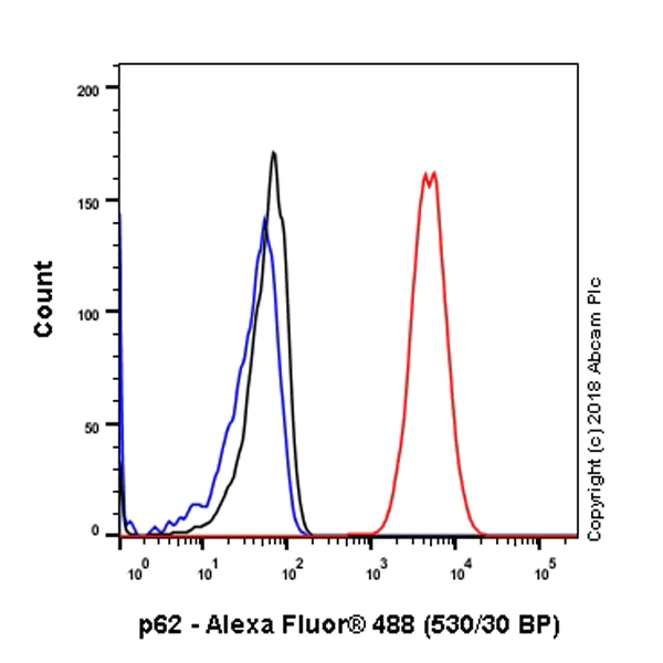
Flow Cytometry (Intracellular) - Anti-SQSTM1 / p62 antibody [EPR4844] - BSA and Azide free (AB219581)
Intracellular Flow Cytometry analysis of HeLa (Human cervix adenocarcinoma epithelial cell) cells labeling SQSTM1 / p62 with purified ab109012 at 1/50 dilution (10μg/ml) (Red). Cells were fixed with 80% Methanol and permeabilised with 0.1% Tween-20. A Goat anti rabbit IgG (Alexa Fluor® 488, ab150077) secondary antibody was used at 1/2000. Isotype control - Rabbit monoclonal IgG (Black). Unlabeled control - Cell without incubation with primary antibody and secondary antibody (Blue).
This data was developed using the same antibody clone in a different buffer formulation containing PBS, BSA, glycerol, and sodium azide (ab109012).
- ICC/IF
Lab
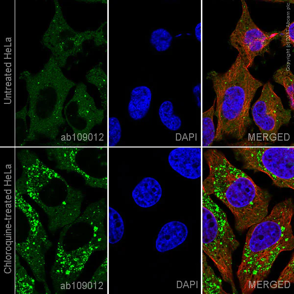
Immunocytochemistry/ Immunofluorescence - Anti-SQSTM1 / p62 antibody [EPR4844] - BSA and Azide free (AB219581)
Purified ab109012 staining SQSTM1/p62 in HeLa cells +/- Chloroquine (50μM, 24 hours). The cells were fixed with 100% methanol (5 min), permeabilized with 0.1% Triton X-100 for 5 minutes and then blocked with 1% BSA/10% normal goat serum/0.3M glycine in 0.1% PBS-Tween for 1h. The cells were then incubated overnight at +4°C with ab109012 at 1μg/ml and ab195889, Mouse monoclonal to alpha Tubulin (Alexa Fluor® 594), at 1/250 dilution (shown in pseudocolor red) followed by a further incubation at room temperature for 1h with a goat secondary antibody to Rabbit IgG (Alexa Fluor® 488) (ab150081) at 2 μg/ml (shown in green). Nuclear DNA was labelled with DAPI (shown in blue).
Image was taken with a confocal microscope (Leica-Microsystems, TCS SP8).
This data was developed using the same antibody clone in a different buffer formulation containing PBS, BSA, glycerol, and sodium azide (ab109012).
- ICC/IF
Lab
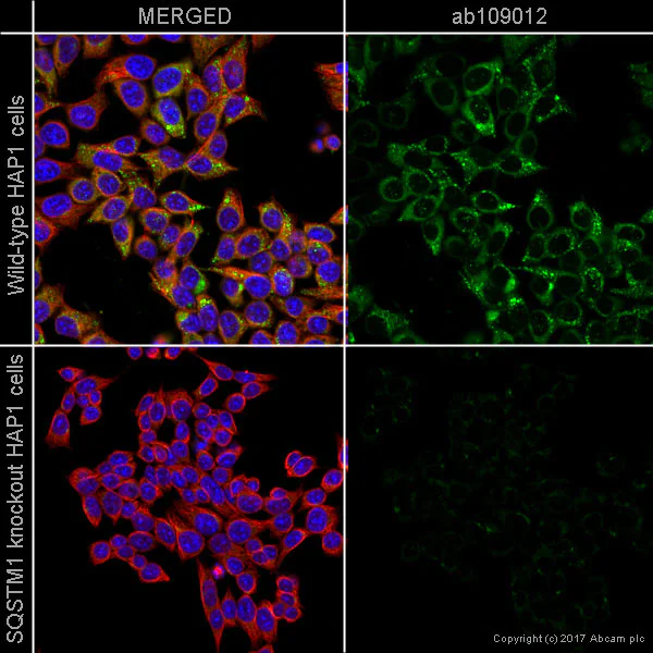
Immunocytochemistry/ Immunofluorescence - Anti-SQSTM1 / p62 antibody [EPR4844] - BSA and Azide free (AB219581)
Purified ab109012 staining SQSTM1 in wild-type HAP1 cells (top panel) and SQSTM1 knockout HAP1 cells (bottom panel). The cells were fixed with 100% methanol (5min), permeabilized with 0.1% Triton X-100 for 5 minutes and then blocked with 1% BSA/10% normal goat serum/0.3M glycine in 0.1% PBS-Tween for 1h. The cells were then incubated with ab109012 at 1μg/ml and ab195889 at 1/250 dilution (shown in pseudocolour red) overnight at +4°C, followed by a further incubation at room temperature for 1h with a goat secondary antibody to Rabbit IgG (Alexa Fluor® 488) (ab150081) at 2 μg/ml (shown in green). Nuclear DNA was labelled in blue with DAPI.
Image was taken with a confocal microscope (Leica-Microsystems, TCS SP8).
This data was developed using the same antibody clone in a different buffer formulation containing PBS, BSA, glycerol, and sodium azide (ab109012).
- ICC/IF
Lab
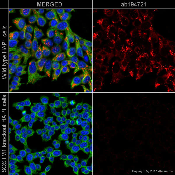
Immunocytochemistry/ Immunofluorescence - Anti-SQSTM1 / p62 antibody [EPR4844] - BSA and Azide free (AB219581)
Clone EPR4844 (ab219581) has been successfully conjugated by Abcam. This image was generated using Anti-SQSTM1 / p62 antibody [EPR4844] (Alexa Fluor® 647). Please refer to ab194721 for protocol details.
ab194721 staining SQSTM1 in wild-type HAP1 cells (top panel) and SQSTM1 knockout HAP1 cells (bottom panel). The cells were fixed with 100% methanol (5min), permeabilized with 0.1% Triton X-100 for 5 minutes and then blocked with 1% BSA/10% normal goat serum/0.3M glycine in 0.1% PBS-Tween for 1h. The cells were then incubated with ab194721 at 1/500 (shown in red) and ab195887 at 1/250 dilution (shown in green). Nuclear DNA was labelled in blue with DAPI.
Image was taken with a confocal microscope (Leica-Microsystems, TCS SP8).
- Flow Cyt (Intra)
Unknown
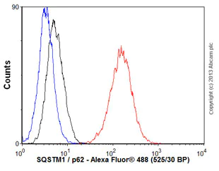
Flow Cytometry (Intracellular) - Anti-SQSTM1 / p62 antibody [EPR4844] - BSA and Azide free (AB219581)
Overlay histogram showing HeLa cells stained with unpurified ab109012 (red line). The cells were fixed with 80% methanol (5 min) and then permeabilized with 0.1% PBS-Tween for 20 min. The cells were then incubated in 1x PBS / 10% normal goat serum / 0.3M glycine to block non-specific protein-protein interactions followed by the antibody (ab109012, 1/100 dilution) for 30 min at 22°C. The secondary antibody used was Alexa Fluor® 488 goat anti-rabbit IgG (H+L) (ab150077) at 1/2000 dilution for 30 min at 22°C. Isotype control antibody (black line) was rabbit IgG (monoclonal) (1μg/1x106 cells) used under the same conditions. Unlabelled sample (blue line) was also used as a control. Acquisition of >5,000 events were collected using a 20mW Argon ion laser (488nm) and 525/30 bandpass filter.
This data was developed using the same antibody clone in a different buffer formulation containing PBS, BSA, glycerol, and sodium azide (ab109012).
- Flow Cyt (Intra)
Unknown
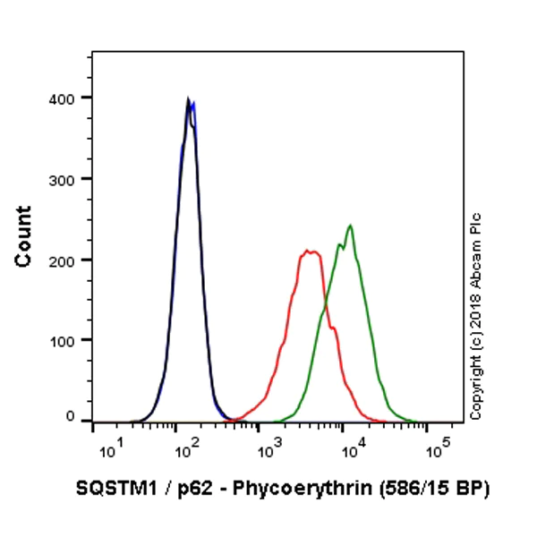
Flow Cytometry (Intracellular) - Anti-SQSTM1 / p62 antibody [EPR4844] - BSA and Azide free (AB219581)
Clone EPR4844 (ab219581) has been successfully conjugated by Abcam. This image was generated using Anti-SQSTM1 / p62 antibody [EPR4844] - Autophagosome Marker (PE). Please refer to ab225094 for protocol details.
Overlay histogram showing HeLa cells untreated (red line) and HeLa cells treated with Chloroquine, 50μM, 24 hours, (green line) stained with ab225094. The cells were fixed with 80% methanol (5 min) and then permeabilized with 0.1% PBS-Triton X-100 for 15 min. The cells were then incubated in 1x PBS / 10% normal goat serum to block non-specific protein-protein interactions followed by the antibody (ab225094, 1/10000 dilution) for 30 min at 22°C.
Isotype control antibody (black line) was Rabbit IgG (monoclonal) Phycoerythrin (ab209478) used at the same concentration and conditions as the primary antibody. Unlabelled sample (blue line) was also used as a control.
Acquisition of >5,000 events were collected using a 50 mW Yellow/Green laser (561nm) and 586/15 bandpass filter.
- WB
Collaborator
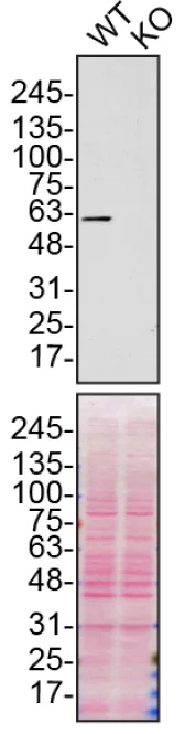
Western blot - Anti-SQSTM1 / p62 antibody [EPR4844] - BSA and Azide free (AB219581)
This data was developed using the same antibody in a different buffer formulation (ab109012).
ab109012 was shown to react with SQSTM1 in wild-type U-2 OS cells in Western blot with loss of signal observed in a SQSTM1 knockout cell line. Wild-type U-2 OS and SQSTM1 knockout cell lysates were subjected to SDS-PAGE. Membranes were blocked in 5% milk in TBST for 1 hr before incubation with ab109012 overnight at 4 °C at a 1/10000 dilution. Blots were incubated with goat anti-rabbit HRP secondary antibodies at 0.2ug/mL before imaging. These data were provided by YCharOS Inc., an open science company with the mission of characterizing commercially available antibody reagents for all human proteins. Abcam and YCharOS are working together to help address the reproducibility crisis by enabling the life science community to better evaluate commercially available antibodies.
All lanes:
Western blot - Anti-SQSTM1 / p62 antibody [EPR4844] - Autophagosome Marker (<a href='/en-us/products/primary-antibodies/sqstm1-p62-antibody-epr4844-autophagosome-marker-ab109012'>ab109012</a>) at 1/10000 dilution
Lane 1:
Wild-type U-2 OS cell lysate at 20 µg
Lane 2:
SQSTM1 knockout U-2 OS cell lysate at 20 µg
Predicted band size: 47 kDa
false
- WB
Lab
Western blot - Anti-SQSTM1 / p62 antibody [EPR4844] - BSA and Azide free (AB219581)
This data was developed using the same antibody clone in a different buffer formulation (ab109012).
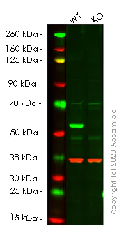
Lanes 1- 2 : Merged signal (red and green). Green - ab109012 observed at 64 kDa. Red - Anti-GAPDH antibody [6C5] - Loading Control (ab8245) observed at 37 kDa.
ab109012 was shown to react with SQSTM1/p62 in wild-type HEK293T cells in western blot. Loss of signal was observed when knockout cell line ab255343 (knockout cell lysate ab263770) was used. Wild-type HEK293T and SQSTM1 knockout HEK293T cell lysates were subjected to SDS-PAGE. Membrane was blocked for 1 hour at room temperature in 0.1% TBST with 3% non-fat dried milk. ab109012 and Anti-GAPDH antibody [6C5] - Loading Control (ab8245) overnight at 4°C at a 1 in 10000 dilution and a 1 in 20000 dilution respectively. Blots were developed with Goat anti-Rabbit IgG H&L (IRDye®800CW) preadsorbed (ab216773) and Goat anti-Mouse IgG H&L (IRDye®680RD) preadsorbed (ab216776) secondary antibodies at 1 in 20000 dilution for 1 hour at room temperature before imaging.
All lanes:
Western blot - Anti-SQSTM1 / p62 antibody [EPR4844] - Autophagosome Marker (<a href='/en-us/products/primary-antibodies/sqstm1-p62-antibody-epr4844-autophagosome-marker-ab109012'>ab109012</a>) at 1/10000 dilution
Lane 1:
Wild-type HEK293T cell lysate at 20 µg
Lane 2:
SQSTM1 knockout HEK293T cell lysate at 20 µg
Lane 2:
Western blot - Human SQSTM1 (p62) knockout HEK-293T cell line (<a href='/en-us/products/cell-lines/human-sqstm1-p62-knockout-hek-293t-cell-line-ab255343'>ab255343</a>)
Predicted band size: 47 kDa
Observed band size: 64 kDa
false
- WB
Lab
Western blot - Anti-SQSTM1 / p62 antibody [EPR4844] - BSA and Azide free (AB219581)
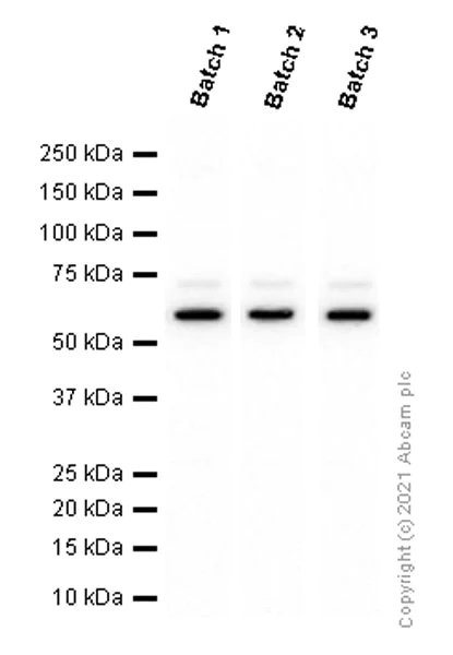
This data was developed using ab109012, the same antibody clone in a different buffer formulation. Different batches of ab109012 were tested on MCF7 (Human breast adenocarcinoma epithelial cell) lysate at 0.4 µg/ml. 15 µg of lysate was loaded in each lane. Bands observed at 62 kDa.
All lanes:
Western blot - Anti-SQSTM1 / p62 antibody [EPR4844] - Autophagosome Marker (<a href='/en-us/products/primary-antibodies/sqstm1-p62-antibody-epr4844-autophagosome-marker-ab109012'>ab109012</a>)
Predicted band size: 47 kDa
false
- WB
Lab
Western blot - Anti-SQSTM1 / p62 antibody [EPR4844] - BSA and Azide free (AB219581)
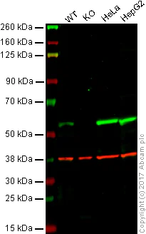
Lanes 1 - 4 : Merged signal (red and green). Green - unpurifed ab109012 observed at 55 kDa. Red - loading control, ab8245, observed at 37 kDa.
ab109012 was shown to specifically react with SQSTM1 in wild-type HAP1 cells. No band was observed when SQSTM1 knockout samples were used. Wild-type and SQSTM1 knockout samples were subjected to SDS-PAGE, ab109012 and ab8245 (Mouse anti GAPDH loading control) were incubated overnight at 4°C at 1/10,000 dilution and 1/20,000 dilution respectively. Blots were developed with Goat anti-Rabbit IgG H&L (IRDye® 800CW) preabsorbed ab216773 and Goat anti-Mouse IgG H&L (IRDye® 680RD) preabsorbed ab216776 secondary antibodies at 1/20,000 dilution for 1 hour at room temperature before imaging.
This data was developed using the same antibody clone in a different buffer formulation containing PBS, BSA, glycerol, and sodium azide (ab109012).
All lanes:
Western blot - Anti-SQSTM1 / p62 antibody [EPR4844] - Autophagosome Marker (<a href='/en-us/products/primary-antibodies/sqstm1-p62-antibody-epr4844-autophagosome-marker-ab109012'>ab109012</a>) at 1/10000 dilution
Lane 1:
Wild-type HAP1 whole cell lysate at 20 µg
Lane 2:
SQSTM1 knockout HAP1 whole cell lysate at 20 µg
Lane 3:
HeLa whole cell lysate at 20 µg
Lane 4:
HepG2 whole cell lysate at 20 µg
Predicted band size: 47 kDa
false
- WB
Lab
Western blot - Anti-SQSTM1 / p62 antibody [EPR4844] - BSA and Azide free (AB219581)
This data was developed using ab109012, the same antibody clone in a different buffer formulation.
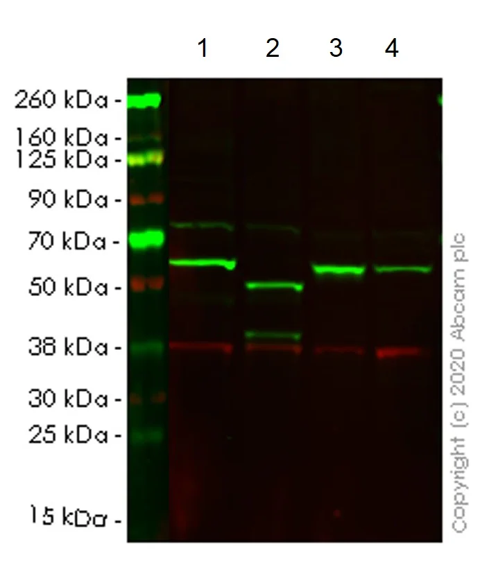
Lanes 1-4 : Merged signal (red and green). Green - ab109012 observed at 55 kDa. Red - loading control ab8245 observed at 36 kDa.
ab109012 Anti-SQSTM1 / p62 antibody [EPR4844] - Autophagosome Marker was shown to specifically react with SQSTM1 / p62 in wild-type HCT116 cells. The band observed in CRISPR/Cas9 edited cell line ab266871 (CRISPR/Cas9 edited cell lysate ab257052) lane below 55 kDa may represent truncated forms and cleaved fragments. This has not been investigated further. Wild-type and SQSTM1 / p62 CRISPR/Cas9 edited samples were subjected to SDS-PAGE. ab109012 and Anti-GAPDH antibody [6C5] - Loading Control (ab8245) were and 1 in 20000 dilution respectively. Blots were developed with Goat anti-Rabbit IgG H&L (IRDye® 800CW) preadsorbed (ab216773) and Goat anti-Mouse IgG H&L (IRDye® 680RD) preadsorbed (ab216776) secondary antibodies at 1 in 20000 dilution for 1 hour at room temperature before imaging.
All lanes:
Western blot - Anti-SQSTM1 / p62 antibody [EPR4844] - Autophagosome Marker (<a href='/en-us/products/primary-antibodies/sqstm1-p62-antibody-epr4844-autophagosome-marker-ab109012'>ab109012</a>)
Lane 1:
Wild-type HCT116 cell lysate at 20 µg
Lane 2:
SQSTM1 CRISPR/Cas9 edited HCT116 cell lysate at 20 µg
Lane 3:
HepG2 cell lysate at 20 µg
Lane 4:
HeLa cell lysate at 20 µg
Secondary
All lanes:
Western blot - Goat anti-Rabbit IgG H&L (IRDye® 800CW) preadsorbed (<a href='/en-us/products/secondary-antibodies/goat-rabbit-igg-h-l-irdye-800cw-preadsorbed-ab216773'>ab216773</a>) at 1/10000 dilution
Predicted band size: 47 kDa
Observed band size: 55 kDa
false
Related conjugates and formulations (9)
-
Anti-SQSTM1 / p62 antibody [EPR4844] - Autophagosome Marker
-
Anti-SQSTM1 / p62 antibody [EPR4844] - Autophagosome Marker – Goat IgG (Chimeric)
-
Anti-SQSTM1 / p62 antibody [EPR4844] - Goat IgG (Chimeric) – BSA and Azide Free
-
660 APC
APC Anti-SQSTM1 / p62 antibody [EPR4844] - Autophagosome Marker
-
519 Alexa Fluor® 488
Alexa Fluor® 488 Anti-SQSTM1 / p62 antibody [EPR4844] - Autophagosome Marker
-
565 Alexa Fluor® 555
Alexa Fluor® 555 Anti-SQSTM1 / p62 antibody [EPR4844] - Autophagosome Marker
-
617 Alexa Fluor® 594
Alexa Fluor® 594 Anti-SQSTM1 / p62 antibody [EPR4844] - Autophagosome Marker
-
665 Alexa Fluor® 647
Alexa Fluor® 647 Anti-SQSTM1 / p62 antibody [EPR4844] - Autophagosome Marker
-
578 PE
PE Anti-SQSTM1 / p62 antibody [EPR4844] - Autophagosome Marker
Alternative Products


Primary Antibodies
AB225453
Alexa Fluor® 647 Anti-SQSTM1 / p62 antibody [EPR18351] - Autophagosome Marker
RabMAb|Recombinant|KO Validated

- 0
- 0
Primary Antibodies
AB225454
PE Anti-SQSTM1 / p62 antibody [EPR18351] - Autophagosome Marker
RabMAb|Recombinant

- 0
- 0
Primary Antibodies
AB237323
Alexa Fluor® 647 Anti-SQSTM1 / p62 (phospho S349) antibody [EPR20451]
RabMAb|Recombinant

- 0
- 0
Primary Antibodies
AB240635
Anti-SQSTM1 / p62 antibody [EPR23101-103] - Autophagosome Marker
20ul selling size|KO Validated|RabMAb|Recombinant|Lab Essentials

- 0
- 0
Primary Antibodies
AB207305
Anti-SQSTM1 / p62 antibody [EPR18351] - Autophagosome Marker
20ul selling size|KO Validated|RabMAb|Recombinant|Lab Essentials|Trial Size

- 5
- 3
Primary Antibodies
AB211324
Anti-SQSTM1 / p62 (phospho S349) antibody [EPR20451]
20ul selling size|KO Validated|RabMAb|Recombinant|Lab Essentials

- 5
- 3
Primary Antibodies
AB91526
Anti-SQSTM1 / p62 antibody - Autophagosome Marker
Lab Essentials|KO Validated

- 4
- 7
Primary Antibodies
AB238445
Anti-SQSTM1 / p62 (phospho S349) antibody [EPR20451] - BSA and Azide free
RabMAb|Recombinant|KO Validated

- 4
- 2
Primary Antibodies
AB227992
Anti-SQSTM1 / p62 antibody [EPR18351] - BSA and Azide free
RabMAb|Recombinant|KO Validated

- 3
- 1
Primary Antibodies
AB237322
Alexa Fluor® 488 Anti-SQSTM1 / p62 (phospho S349) antibody [EPR20451]
RabMAb|Recombinant

- 0
- 0
Complementary Products


Primary Antibodies
AB192890
Anti-LC3B antibody [EPR18709] - Autophagosome Marker
BOND RX™ Validated|RabMAb|Recombinant|KO Validated|Lab Essentials|20ul selling size

- 5
- 19
Primary Antibodies
AB207612
Anti-Beclin 1 antibody [EPR19662]
20ul selling size|KO Validated|RabMAb|Recombinant|Trial Size

- 5
- 10
Primary Antibodies
AB52768
Anti-MAP1LC3A antibody [EP1983Y] - Autophagosome Marker
RabMAb|Recombinant|Lab Essentials|20ul selling size

- 0
- 0
Primary Antibodies
AB32503
BOND RX™ Validated|20ul selling size|RabMAb|Recombinant|KO Validated

- 4
- 12
Reactivity data
Product details
ab219581 is the carrier-free version of ab109012.
Patented technology
Our RabMAb® technology is a patented hybridoma-based technology for making rabbit monoclonal antibodies. For details on our patents, please refer to RabMAb® patents.
What are the advantages of a recombinant monoclonal antibody?
This product is a recombinant monoclonal antibody, which offers several advantages including:
- - High batch-to-batch consistency and reproducibility
- - Improved sensitivity and specificity
- - Long-term security of supply
- - Animal-free batch production
For more information, read more on recombinant antibodies.
Conjugation ready
Our carrier-free antibodies are typically supplied in a PBS-only formulation, purified and free of BSA, sodium azide and glycerol. This conjugation-ready format is designed for use with fluorochromes, metal isotopes, oligonucleotides, and enzymes, which makes them ideal for antibody labelling, functional and cell-based assays, flow-based assays (e.g. mass cytometry) and Multiplex Imaging applications.
Use our conjugation kits for antibody conjugates that are ready-to-use in as little as 20 minutes with 1 minute hands-on-time and 100% antibody recovery: available for fluorescent dyes, HRP, biotin and gold.
Compatibility
This product is compatible with the Maxpar® Antibody Labeling Kit from Fluidigm, without the need for antibody preparation. Maxpar® is a trademark of Fluidigm Canada Inc.
Properties and storage information
Form
Purification technique
Storage buffer
Shipped at conditions
Appropriate short-term storage conditions
Appropriate long-term storage conditions
Storage information
Product protocols
- Visit the General protocols
- Visit the Troubleshooting
Target data
Alternative Names
Publications (7)
Recent publications for all applications. Explore the full list and refine your search
iScience 25:104547 PubMed35754738
2022
Applications
Unspecified application
Species
Unspecified reactive species
iScience 25:104268 PubMed35521515
2022
Applications
Unspecified application
Species
Unspecified reactive species
Medicine and science in sports and exercise 48:1448-58 PubMed27031745
2016
Applications
WB
Species
Unspecified reactive species
The Journal of neuroscience : the official journal of the Society for Neuroscience 36:1316-23 PubMed26818518
2016
Applications
Unspecified application
Species
Unspecified reactive species
Autophagy 11:503-15 PubMed25714412
2015
Applications
Unspecified application
Species
Unspecified reactive species
Autophagy 10:2223-38 PubMed25484082
2014
Applications
Unspecified application
Species
Unspecified reactive species
Journal of virology 88:5152-64 PubMed24574393
2014
Applications
Unspecified application
Species
Unspecified reactive species
Product promise
Please note: All products are 'FOR RESEARCH USE ONLY. NOT FOR USE IN DIAGNOSTIC OR THERAPEUTIC PROCEDURES'.
For licensing inquiries, please contact partnerships@abcam.com