Anti-TSG101 antibody (ab30871) is a rabbit polyclonal antibody detecting TSG101 in Western Blot, IHC-P, ICC/IF. Suitable for Human, Mouse, Rat.
- Over 190 publications
- Trusted since 2006
View Alternative Names
Tumor susceptibility gene 101 protein, ESCRT-I complex subunit TSG101, TSG101
- WB
Project
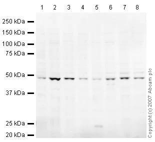
Western blot - Anti-TSG101 antibody (AB30871)
All lanes:
Western blot - Anti-TSG101 antibody (ab30871) at 1 µg/mL
Lane 1:
HeLa (Human epithelial carcinoma cell line) Whole Cell Lysate at 10 µg
Lane 2:
A431 (Human epithelial carcinoma cell line) Whole Cell Lysate at 10 µg
Lane 3:
Jurkat (Human T cell lymphoblast-like cell line) Whole Cell Lysate at 10 µg
Lane 4:
HEK293 Human embryonic kidney cell line Whole Cell Lysate at 10 µg
Lane 5:
HepG2 (Human hepatocellular liver carcinoma cell line) Whole Cell Lysate at 10 µg
Lane 6:
MCF7 (Human breast adenocarcinoma cell line) Whole Cell Lysate at 10 µg
Lane 7:
SHSY-5Y (Human neuroblastoma cell line) Whole Cell Lysate at 10 µg
Lane 8:
U2OS (Human osteosarcoma cell line) Whole Cell Lysate at 10 µg
Secondary
All lanes:
IRDye 680 Conjugated Goat Anti-Rabbit IgG (H+L) at 1/10000 dilution
Predicted band size: 44 kDa
Observed band size: 49 kDa
false
- ICC/IF
Lab
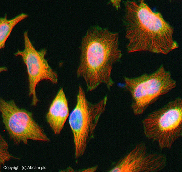
Immunocytochemistry/ Immunofluorescence - Anti-TSG101 antibody (AB30871)
ab30871 stained in HeLa cells. Cells were fixed with 4% paraformaldehyde (10min) at room temperature and incubated with PBS containing 10% goat serum, 0.3 M glycine, 1% BSA and 0.1% Triton for 1h at room temperature to permeabilise the cells and block non-specific protein-protein interactions. The cells were then incubated with the antibody ab30871 at 5μg/ml and ab7291 (Mouse monoclonal to alpha Tubulin - Loading Control) used at a 1/1000 dilution overnight at +4°C. The secondary antibodies were ab150081, Goat Anti-Rabbit IgG H&L (Alexa Fluor® 488) preadsorbed, (pseudo-colored green) and ab150120, Goat polyclonal Secondary Antibody to Mouse IgG - H&L (Alexa Fluor® 594) preadsorbed, (colored red), both used at a 1/1000 dilution for 1 hour at room temperature. DAPI was used to stain the cell nuclei (colored blue) at a concentration of 1.43 μM for 1hour at room temperature.
- IHC-P
Unknown
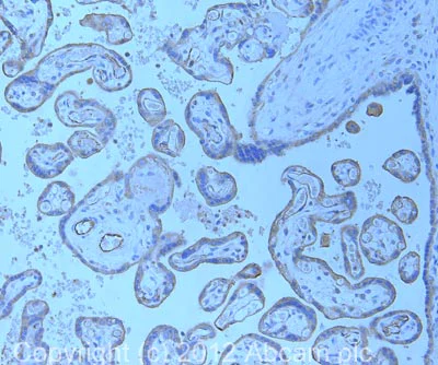
Immunohistochemistry (Formalin/PFA-fixed paraffin-embedded sections) - Anti-TSG101 antibody (AB30871)
IHC image of ab30871 staining in human placenta formalin fixed paraffin embedded tissue section, performed on a Leica BondTM system using the standard protocol F. The section was pre-treated using heat mediated antigen retrieval with sodium citrate buffer (pH6, epitope retrieval solution 1) for 20 mins. The section was then incubated with ab30871, 5µg/ml, for 15 mins at room temperature and detected using an HRP conjugated compact polymer system. DAB was used as the chromogen. The section was then counterstained with haematoxylin and mounted with DPX.
For other IHC staining systems (automated and non-automated) customers should optimize variable parameters such as antigen retrieval conditions, primary antibody concentration and antibody incubation times.

- WB
Lab
Western blot - Anti-TSG101 antibody (AB30871)
This blot was produced using a 4-12% Bis-tris gel under the MOPS buffer system. The gel was run at 200V for 50 minutes before being transferred onto a Nitrocellulose membrane at 30V for 70 minutes. The membrane was then blocked for an hour using 5% Bovine Serum Albumin before being incubated with ab30871 overnight at 4°C. Antibody binding was detected using an anti-rabbit antibody conjugated to HRP, and visualised using ECL development solution.
The doublet seen in Western blot has been described in PMID : 11427703 and may be due to internal initiation at Met10.
All lanes:
Western blot - Anti-TSG101 antibody (ab30871) at 1 µg/mL
Lane 1:
NIH 3T3 (Mouse embryonic fibroblast cell line) Whole Cell Lysate at 10 µg
Lane 2:
PC12 (Rat adrenal pheochromocytoma cell line) Whole Cell Lysate at 10 µg
Secondary
All lanes:
Western blot - Goat Anti-Rabbit IgG H&L (HRP) (<a href='/en-us/products/secondary-antibodies/goat-rabbit-igg-h-l-hrp-ab97051'>ab97051</a>) at 1/10000 dilution
Predicted band size: 44 kDa
Observed band size: 29 kDa,48 kDa,49 kDa
true
Exposure time: 90s

- WB
CiteAb
Western blot - Anti-TSG101 antibody (AB30871)
Western Blotting using Anti-TSG101 antibody, ab30871. Publication image from van Rheenen, J. et al., 2020, Nat Commun, 32111843. Legend direct from paper.
Intercellular sgRNA exchange is EV-mediated.a Schematic of EV isolation workflow and analysis. Conditioned medium is isolated from sgRNA+ MDA-MB-231 donor cells, after which cell debris is pelleted through centrifugation. Conditioned medium is then concentrated by tangential flow filtration, followed by size exclusion chromatography-mediated EV isolation. EVs are then characterized, and used for EV addition experiments in Stoplight+spCas9+ reporter cells. b Nanosight Nanoparticle Tracking Analysis (NTA) showing the size distribution of the isolated EVs (mode = 81 ± 3 nm). c Western blot analysis of EVs and cell lysates for common EV markers (CD9, CD63, ALIX, Flot-1, and TSG101), and EV-negative markers Calnexin and H2B. Representative images as observed in three independent experiments. d Electron microscopy analysis of isolated EVs. Scale bar represents 100 nm. Representative image as observed in 12 separate fields. e An EV RNase protection assay, followed by RT-PCR sgRNAs, shows that sgRNAs are only susceptible to RNase-mediated degradation in the presence of the membrane-disrupting anionic surfactant SDS. Representative data as observed in three independent experiments. f Confocal microscopy images of HEK293T reporter cells (red) that have taken up MDA-MB-231-derived PKH67-labeled EVs (green). Representative image as observed in seven randomly selected fields. g, h EV-mediated activation of the CROSS-FIRE platform using EVs isolated from sgRNA+ MDA-MB-231 donor cells. EVs were added every 72 h for 12 additions with an average dose of 1.1e11 ± 4.9e10 EVs per addition. Cells were analyzed by fluorescence microscopy (g) and flow cytometry (h). Scale bar represents 200 µm. Means + SD, n = 3 biological replicates, Tukey’s multiple comparison test. i EVs from sgRNA+ MDA-MB-231 donor cells activate Stoplight+ spCas9+ HEK293T reporter cells in a dose-dependent manner. EVs were added every 72 h for nine additions with an average concentration of 2.2e10 ± 1e10 (20 µl) or 2.2e11 ± 1e11 (200 µl). Means + SD, n = 3 biological replicates, Tukey’s multiple comparison test. *p < 0.05.
false
- WB
CiteAb
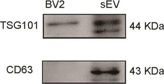
Western blot - Anti-TSG101 antibody (AB30871)
Western Blotting using Anti-TSG101 antibody, ab30871. Publication image from Yang, G. Y. et al., 2021, Theranostics, 33391532. Legend direct from paper.
Characterization of M2 BV2 cells induced by IL-4 and identification of sEVs derived from M2 BV2 cells. (A, B) Representative images of BV2 cells immunostained for Iba-1 (green), CD206 (red), and arginase (red). Cultured systems were treated with 0 or 20 ng/µL IL-4. Cell nuclei were counterstained with DAPI. Scale bar = 50 µm. (C) Western blotting analysis of CD206 and arginase expression in BV2 cells after 0 or 20 ng/µL IL-4 treatment. (D) Representative electron microscopy images showing the phenotype of M2-sEVs. Left image scale bar = 100 nm, right image scale bar = 50 nm. (F) NTA of M2-sEVs isolated by ultracentrifugation from M2 BV2 cells. Data represent the average size distribution profile of three samples and each purification normalized to the total nanoparticle concentrations. Data for each sample was derived from three different videos and analyses. (G) Western blotting analysis of TSG101 and CD63 levels in M2 BV2 cells and M2-sEVs.
false
Alternative Products
Primary Antibodies
AB125011
Anti-TSG101 antibody [EPR7130(B)]
20ul selling size|BOND RX™ Validated|RabMAb|Recombinant

- 4
- 10
Primary Antibodies
AB133586
Anti-TSG101 antibody [EPR7131(B)]
RabMAb|Recombinant|20ul selling size

- 0
- 0
Primary Antibodies
AB216447
Anti-TSG101 antibody [EPR7130(B)] - Low endotoxin, Azide free
BOND RX™ Validated|RabMAb|Recombinant

- 0
- 0
Primary Antibodies
AB214404
Alexa Fluor® 555 Anti-TSG101 antibody [EPR7130(B)]
RabMAb|Recombinant

- 0
- 0
Primary Antibodies
AB207664
Alexa Fluor® 647 Anti-TSG101 antibody [EPR7130(B)]
RabMAb|Recombinant

- 0
- 0
Primary Antibodies
AB207663
Alexa Fluor® 488 Anti-TSG101 antibody [EPR7130(B)]
RabMAb|Recombinant

- 0
- 0

Complementary Products
Primary Antibodies
AB134045
Anti-CD63 antibody [EPR5702] - Late Endosome Marker
20ul selling size|RabMAb|Recombinant|KO Validated|Lab Essentials

- 3
- 6

Primary Antibodies
AB307085
Anti-CD9 antibody [EPR27551-92]
BOND RX™ Validated|20ul selling size|RabMAb|Recombinant|KO Validated

- 0
- 0
Primary Antibodies
AB275377
Anti-ALIX antibody [EPR23653-32]
20ul selling size|RabMAb|Recombinant|KO Validated

- 5
- 2
Reactivity data
Product details
Anti-TSG101 antibody (ab30871) is a rabbit polyclonal antibody and is validated for use in Western Blot (WB), Immunohistochemistry (IHC-P), Immunocytochemistry/immunofluorescence (ICC/IF) in Human, Mouse, Rat samples.
What is the molecular weight of TSG101?
Anti-TSG101 (ab30871) specifically detects a band for TSG101 (UniProt: Q99816) at a molecular weight of 44kDa.
Trusted by the scientific community
Anti-TSG101 (ab30871) was first used in a scientific publication in 2006 and has been cited over 190 times in peer-reviewed journals.
Properties and storage information
Form
Purification technique
Storage buffer
Shipped at conditions
Appropriate short-term storage duration
Appropriate short-term storage conditions
Appropriate long-term storage conditions
Aliquoting information
Storage information
Supplementary information
This supplementary information is collated from multiple sources and compiled automatically.
Biological function summary
TSG101 plays a critical role in the sorting and trafficking of proteins destined for lysosomal degradation or recycling back to the plasma membrane. It is an essential part of the ESCRT machinery which also includes other components like VPS37 and VPS28. TSG101 interacts with specific domains and motifs on target proteins facilitating the recognition and recruitment of these proteins into vesicles. Its involvement in membrane budding and scission makes it important for maintaining cellular homeostasis and protein quality control.
Pathways
TSG101 is integrally involved in the ESCRT pathway and is associated with processes like viral budding and cytokinesis. Within these pathways it collaborates with proteins such as ALIX and the ESCRT-II component VPS25 to mediate vesicle formation and facilitate the trafficking of ubiquitinated cargo. The ESCRT pathway is vital in cellular responses such as membrane repair and is important for the life cycle of certain enveloped viruses which utilize host cell machinery for budding.
Product protocols
- Visit the General protocols
- Visit the Troubleshooting
Target data
Publications (261)
Recent publications for all applications. Explore the full list and refine your search
Clinical proteomics 22:36 PubMed41062929
2025
Applications
Unspecified application
Species
Unspecified reactive species
Journal of extracellular biology 4:e70087 PubMed40994722
2025
Applications
Unspecified application
Species
Unspecified reactive species
Molecular therapy. Nucleic acids 36:102644 PubMed40896585
2025
Applications
Unspecified application
Species
Unspecified reactive species
Stem cell research & therapy 16:450 PubMed40846969
2025
Applications
Unspecified application
Species
Unspecified reactive species
Journal of extracellular biology 4:e70080 PubMed40843441
2025
Applications
Unspecified application
Species
Unspecified reactive species
Cell biology and toxicology 41:78 PubMed40304798
2025
Applications
Unspecified application
Species
Unspecified reactive species
Molecular neurobiology 62:11099-11111 PubMed40261603
2025
Applications
Unspecified application
Species
Unspecified reactive species
Stem cells translational medicine 14: PubMed40257841
2025
Applications
Unspecified application
Species
Unspecified reactive species
The Journal of biological chemistry 301:108305 PubMed39947471
2025
Applications
Unspecified application
Species
Unspecified reactive species
Communications biology 8:228 PubMed39948151
2025
Applications
Unspecified application
Species
Unspecified reactive species
Product promise
Please note: All products are 'FOR RESEARCH USE ONLY. NOT FOR USE IN DIAGNOSTIC OR THERAPEUTIC PROCEDURES'.
For licensing inquiries, please contact partnerships@abcam.com