Anti-VCAM1 antibody [EPR5038(2)] - BSA and Azide free
- RabMAb
- Recombinant
- KO Validated
- What is this?
Be the first to review this product! Submit a review
|
(1 Publication)
- IP
Supplier Data
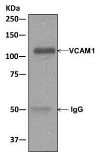
Immunoprecipitation - Anti-VCAM1 antibody [EPR5038(2)] - BSA and Azide free (AB249790)
This data was developed using ab174279, the same antibody clone in a different buffer formulation.
Immunoprecipitation. ab174279 at 1/1000 labeling VCAM1 immunoprecipitated from NIH3T3 cell lysate using ab174279 at 1/10.
All lanes:
Immunoprecipitation - Anti-VCAM1 antibody [EPR5038(2)] (<a href='/en-us/products/primary-antibodies/vcam1-antibody-epr50382-ab174279'>ab174279</a>)
Predicted band size: 81 kDa
Observed band size: 100 kDa
false
- WB
Lab
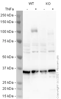
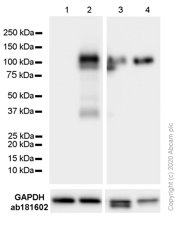
Western blot - Anti-VCAM1 antibody [EPR5038(2)] - BSA and Azide free (AB249790)
ab174279 was shown to react with VCAM1 in wild-type A549 cells in western blot. Loss of signal was observed when VCAM1 knockout sample was used. Membranes were blocked in 3 % milk in TBS-T (0.1 % Tween®) before incubation with ab174279 overnight at 4 °C at a 1 in 1000 dilution. Blots were incubated with HRP conjugated Goat anti-Rabbit (H+L) secondary antibody at 1 in 5000 for 1 hour at room temperature before development with Optiblot ECL reagent (ab133456) and imaging.
All lanes:
Western blot - Anti-VCAM1 antibody [EPR5038(2)] (<a href='/en-us/products/primary-antibodies/vcam1-antibody-epr50382-ab174279'>ab174279</a>) at 1/1000 dilution
Lane 1:
Wild-type A549 cell lysate at 30 µg
Lane 2:
Wild-type A549 TNF-a treated (10 ng/mL, 16h) cell lysate at 30 µg
Lane 3:
VCAM1 knockout A549 cell lysate at 30 µg
Lanes 3 - 4:
Western blot - Human VCAM1 knockout A549 cell line (<a href='/en-us/products/cell-lines/human-vcam1-knockout-a549-cell-line-ab273758'>ab273758</a>)
Lane 4:
VCAM1 knockout A549 TNF-a treated (10 ng/mL, 16h) cell lysate at 30 µg
Predicted band size: 81 kDa
Observed band size: 105 kDa
false
Exposure time: 20s
- WB
Supplier Data
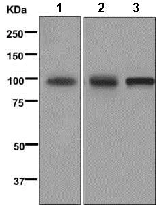
Western blot - Anti-VCAM1 antibody [EPR5038(2)] - BSA and Azide free (AB249790)
This data was developed using ab174279, the same antibody clone in a different buffer formulation.
All lanes:
Western blot - Anti-VCAM1 antibody [EPR5038(2)] (<a href='/en-us/products/primary-antibodies/vcam1-antibody-epr50382-ab174279'>ab174279</a>) at 1/1000 dilution
Lane 1:
Human fetal kidney lysate at 10 µg
Lane 2:
HuT-78 lysate at 10 µg
Lane 3:
NIH3T3 lysate at 10 µg
Predicted band size: 81 kDa
false
- WB
Unknown
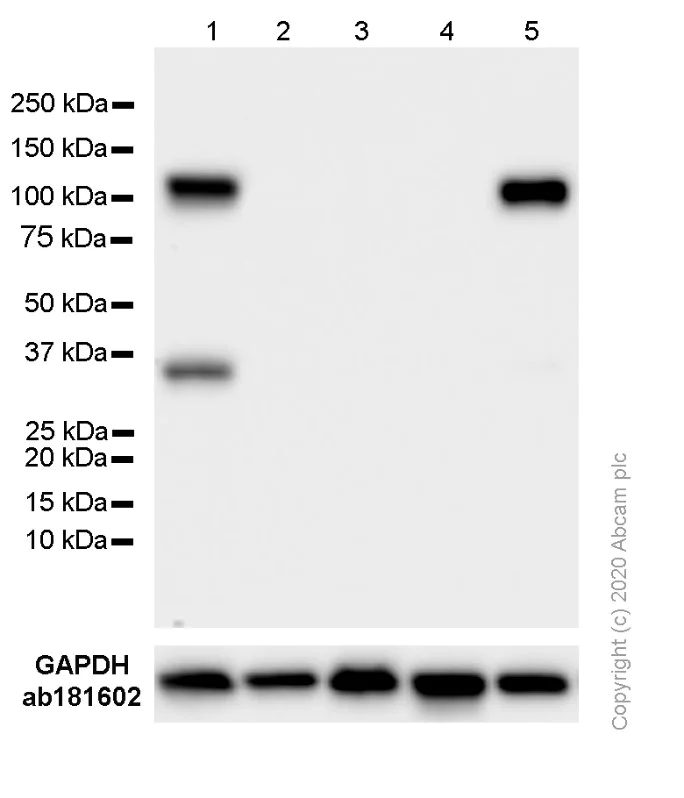
Western blot - Anti-VCAM1 antibody [EPR5038(2)] - BSA and Azide free (AB249790)
This data was developed using ab174279, the same antibody clone in a different buffer formulation.
Blocking and dilution buffer : 5% NFDM/TBST.
All lanes:
Western blot - Anti-VCAM1 antibody [EPR5038(2)] (<a href='/en-us/products/primary-antibodies/vcam1-antibody-epr50382-ab174279'>ab174279</a>) at 1/1000 dilution
Lane 1:
Hut-78 (Human Sezary syndrome cutaneous T lymphocyte) whole cell lysate at 20 µg
Lane 2:
SK-OV-3 (Human ovarian cancer epithelial cell) whole cell lysate at 20 µg
Lane 3:
RAW 264.7 (Mouse Abelson murine leukemia virus-induced tumor macrophage) whole cell lysate at 20 µg
Lane 4:
J774A.1 (Mouse reticulum cell sarcoma monocyte macrophage) whole cell lysate at 20 µg
Lane 5:
LADMAC (Mouse bone marrow monocyte macrophage) whole cell lysate at 20 µg
Secondary
All lanes:
Western blot - Goat Anti-Rabbit IgG H&L (HRP) (<a href='/en-us/products/secondary-antibodies/goat-rabbit-igg-h-l-hrp-ab97051'>ab97051</a>) at 1/20000 dilution
Predicted band size: 81 kDa
Observed band size: 100 kDa
false
Exposure time: 15s
- WB
Unknown
Western blot - Anti-VCAM1 antibody [EPR5038(2)] - BSA and Azide free (AB249790)
This data was developed using ab174279, the same antibody clone in a different buffer formulation.
Blocking and dilution buffer : 5% NFDM/TBST.
All lanes:
Western blot - Anti-VCAM1 antibody [EPR5038(2)] (<a href='/en-us/products/primary-antibodies/vcam1-antibody-epr50382-ab174279'>ab174279</a>) at 1/1000 dilution
Lane 1:
HUVEC (Human umbilical vein endothelial cell) whole cell lysate at 20 µg
Lane 2:
HUVEC treated with 10 ng/ml TNF-α for 16 h, whole cell lysate at 20 µg
Lane 3:
bEND.3 (Mouse brain endothelioma) whole cell lysate at 20 µg
Lane 4:
bEND.3 treated with 10 μg/ml LPS for 24 h, whole cell lysate at 20 µg
Secondary
All lanes:
Western blot - Goat Anti-Rabbit IgG H&L (HRP) (<a href='/en-us/products/secondary-antibodies/goat-rabbit-igg-h-l-hrp-ab97051'>ab97051</a>) at 1/20000 dilution
Predicted band size: 81 kDa
Observed band size: 100 kDa
false
Related conjugates and formulations (1)
-
Anti-VCAM1 antibody [EPR5038(2)]
Complementary Products

Primary Antibodies
AB124964
Anti-alpha smooth muscle Actin antibody [EPR5368]
BOND RX™ Validated|RabMAb|Recombinant|KO Validated|20ul selling size

- 5
- 19
Primary Antibodies
AB115777
Anti-beta Actin antibody [SP124] - Cytoskeleton Marker
20ul selling size|RabMAb|Lab Essentials|Recombinant

- 4
- 1
Primary Antibodies
AB192890
Anti-LC3B antibody [EPR18709] - Autophagosome Marker
BOND RX™ Validated|RabMAb|Recombinant|KO Validated|Lab Essentials|20ul selling size

- 5
- 19
Reactivity data
Product details
ab249790 is the carrier-free version of ab174279.
Patented technology
Our RabMAb® technology is a patented hybridoma-based technology for making rabbit monoclonal antibodies. For details on our patents, please refer to RabMAb® patents.
What are the advantages of a recombinant monoclonal antibody?
This product is a recombinant monoclonal antibody, which offers several advantages including:
- - High batch-to-batch consistency and reproducibility
- - Improved sensitivity and specificity
- - Long-term security of supply
- - Animal-free batch production
For more information, read more on recombinant antibodies.
Conjugation ready
Our carrier-free antibodies are typically supplied in a PBS-only formulation, purified and free of BSA, sodium azide and glycerol. This conjugation-ready format is designed for use with fluorochromes, metal isotopes, oligonucleotides, and enzymes, which makes them ideal for antibody labelling, functional and cell-based assays, flow-based assays (e.g. mass cytometry) and Multiplex Imaging applications.
Use our conjugation kits for antibody conjugates that are ready-to-use in as little as 20 minutes with 1 minute hands-on-time and 100% antibody recovery: available for fluorescent dyes, HRP, biotin and gold.
Compatibility
This product is compatible with the Maxpar® Antibody Labeling Kit from Fluidigm, without the need for antibody preparation. Maxpar® is a trademark of Fluidigm Canada Inc.
Properties and storage information
Form
Purification technique
Storage buffer
Shipped at conditions
Appropriate short-term storage conditions
Appropriate long-term storage conditions
Storage information
Supplementary information
This supplementary information is collated from multiple sources and compiled automatically.
Biological function summary
Adhesion molecules like VCAM1 participate in immune response by recruiting leukocytes to sites of inflammation. VCAM1 helps guide immune cells to directed locations where they perform defense activities. Although VCAM1 is not part of any known large protein complex its interaction with integrins facilitates the migration of immune cells across the endothelium and into tissue.
Pathways
VCAM1 contributes significantly to the leukocyte extravasation process in inflammatory pathways. It plays a role in both the cytokine-cytokine receptor interaction pathway and the NF-kB signaling pathway which are essential for immune response and cell signaling. Through these pathways it interacts with proteins like ICAM1 and various chemokines helping to coordinate the immune cell movement through vascular tissue barriers.
Product protocols
- Visit the General protocols
- Visit the Troubleshooting
Target data
Alternative Names
Publications (1)
Recent publications for all applications. Explore the full listand refine your search
Frontiers in cell and developmental biology 9:775642 PubMed35004677
2021
Applications
Unspecified application
Species
Unspecified reactive species
Product promise
Please note: All products are 'FOR RESEARCH USE ONLY. NOT FOR USE IN DIAGNOSTIC OR THERAPEUTIC PROCEDURES'.
For licensing inquiries, please contact partnerships@abcam.com