Anti-Villin antibody [EPR3491(3)]
- RabMAb
- Recombinant
- What is this?
Be the first to review this product! Submit a review
|
(1 Publication)
- IHC-P
Unknown
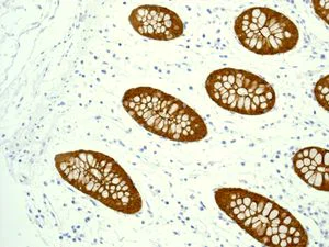
Immunohistochemistry (Formalin/PFA-fixed paraffin-embedded sections) - Anti-Villin antibody [EPR3491(3)] (AB133510)
Immunohistochemical analysis of paraffin-embedded Human colon tissue labelling Villin with ab133510 at 1/100 dilution.
Perform heat mediated antigen retrieval with citrate buffer pH 6 before commencing with IHC staining protocol.

- IHC-P
Unknown
Immunohistochemistry (Formalin/PFA-fixed paraffin-embedded sections) - Anti-Villin antibody [EPR3491(3)] (AB133510)
Immunohistochemical analysis of paraffin-embedded Human colon adenocarcinoma tissue labelling Villin with ab133510 at 1/100 dilution.
Perform heat mediated antigen retrieval with citrate buffer pH 6 before commencing with IHC staining protocol.
- IHC-P
Unknown
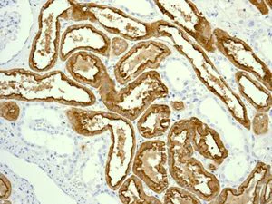
Immunohistochemistry (Formalin/PFA-fixed paraffin-embedded sections) - Anti-Villin antibody [EPR3491(3)] (AB133510)
Immunohistochemical analysis of paraffin-embedded Human kidney tissue labelling Villin with ab133510 at 1/100 dilution.
Perform heat mediated antigen retrieval with citrate buffer pH 6 before commencing with IHC staining protocol.
- WB
Lab
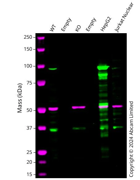
Western blot - Anti-Villin antibody [EPR3491(3)] (AB133510)
Western blot : Anti-VIL1 antibody [EPR3491(3)] (ab133510) staining at 1/5000 dilution, shown in green; Mouse anti-Alpha Tubulin [DM1A] (ab7291) loading control staining at 1/20000 dilution, shown in magenta. In Western blot, ab133510 was shown to bind specifically to VIL1. A band was observed at 93 kDa in wild-type A549 cell lysates with no signal observed at this size in VIL1 knockout cell line. To generate this image, wild-type and VIL1 knockout A549 cell lysates were analysed. First, samples were run on an SDS-PAGE gel then transferred onto a nitrocellulose membrane. Membranes were blocked in 3 % milk in TBS-0.1 % Tween® 20 (TBS-T) before incubation with primary antibodies overnight at 4 °C. Blots were washed four times in TBS-T, incubated with secondary antibodies for 1 h at room temperature, washed again four times then imaged. Secondary antibodies used were Goat anti-Rabbit IgG H&L 800CW and Goat anti-Mouse IgG H&L 680RD at 1/20000 dilution.
All lanes:
Western blot - Anti-Villin antibody [EPR3491(3)] (ab133510) at 1/5000 dilution
Lane 1:
Wild-type A549 cell lysate at 20 µg
Lanes 2 and 4:
Empty cell lysate at 20 µg
Lane 3:
Western blot - Human VIL1 Knockout A549 cell line (<a href='/en-us/products/cell-lines/human-vil1-knockout-a549-cell-line-ab324222'>ab324222</a>) at 20 µg
Lane 5:
HepG2 cell lysate at 20 µg
Lane 6:
Jurkat Nuclear cell lysate at 20 µg
Secondary
All lanes:
Goat anti-Rabbit IgG H&L 800CW and Goat anti-Mouse IgG H&L 680RD at 1/20000 dilution
Observed band size: 93 kDa
false
- WB
Unknown
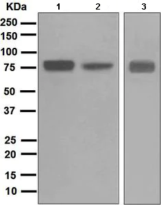
Western blot - Anti-Villin antibody [EPR3491(3)] (AB133510)
All lanes:
Western blot - Anti-Villin antibody [EPR3491(3)] (ab133510) at 1/10000 dilution
Lane 1:
Caco-2 cell lysate at 10 µg
Lane 2:
HT-29 cell lysate at 10 µg
Lane 3:
Human small intestine tissue lysate at 10 µg
Secondary
All lanes:
Goat anti-rabbit HRP conjugated at 1/2000 dilution
Predicted band size: 93 kDa
false
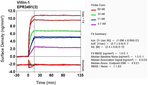
- OI-RD Scanning
Unknown
OI-RD Scanning - Anti-Villin antibody [EPR3491(3)] (AB133510)
We have systematically measured KD (the equilibrium dissociation constant between the antibody and its antigen), of more than 840 recombinant antibodies to assess not only their individual KD values but also to see the average affinity of antibody. Based on the comparison with published literature values for mouse monoclonal antibodies, Recombinant antibodies appear to be on average 1-2 order of magnitude higher affinity.
Related conjugates and formulations (1)
-
Anti-Villin antibody [EPR3491(3)] - BSA and Azide free
Alternative Products


Primary Antibodies
AB130751
BOND RX™ Validated|20ul selling size|RabMAb|Advanced Validation|Recombinant

- 5
- 5


Primary Antibodies
AB245749
Anti-Villin antibody [SP145] - BSA and Azide free
BOND RX™ Validated|RabMAb|Advanced Validation|Recombinant

- 0
- 0
Complementary Products
Primary Antibodies
AB272692
Anti-MUC2 antibody [EPR23479-47]
BOND RX™ Validated|20ul selling size|RabMAb|Advanced Validation|Recombinant|Trial Size

- 5
- 7
Primary Antibodies
AB108508
Anti-Lysozyme antibody [EPR2994(2)]
20ul selling size|RabMAb|Recombinant|KO Validated

- 5
- 4

Primary Antibodies
AB76126
Anti-Cytokeratin 20 antibody [EPR1622Y] - Cytoskeleton Marker
RabMAb|Recombinant|Lab Essentials|20ul selling size

- 5
- 17
Reactivity data
Product details
Species reactivity
Mouse, Rat: We have preliminary internal testing data to indicate this antibody may not react with these species.
Please contact us for more information.
Patented technology
Our RabMAb® technology is a patented hybridoma-based technology for making rabbit monoclonal antibodies. For details on our patents, please refer to RabMAb® patents.
What are the advantages of a recombinant monoclonal antibody?
This product is a recombinant monoclonal antibody, which offers several advantages including:
- - High batch-to-batch consistency and reproducibility
- - Improved sensitivity and specificity
- - Long-term security of supply
- - Animal-free batch production
For more information, read more on recombinant antibodies.
Properties and storage information
Form
Purification technique
Storage buffer
Shipped at conditions
Appropriate short-term storage conditions
Appropriate long-term storage conditions
Storage information
Product protocols
- Visit the General protocols
- Visit the Troubleshooting
Target data
Alternative Names
Publications (1)
Recent publications for all applications. Explore the full listand refine your search
Gut 64:1705-14 PubMed25753030
2015
Applications
Unspecified application
Species
Unspecified reactive species
Product promise
Please note: All products are 'FOR RESEARCH USE ONLY. NOT FOR USE IN DIAGNOSTIC OR THERAPEUTIC PROCEDURES'.
For licensing inquiries, please contact partnerships@abcam.com