Goat Anti-Rabbit IgG H&L (FITC) (ab97050)
 (ab97050)")
Key features and details
- Goat Anti-Rabbit IgG H&L (FITC)
- Conjugation: FITC. Ex: 493nm, Em: 528nm
- Host species: Goat
- Isotype: IgG
- Suitable for: IHC-P, ICC/IF, Flow Cyt
Overview
-
Product name
Goat Anti-Rabbit IgG H&L (FITC)
See all IgG secondary antibodies -
Host species
Goat -
Target species
Rabbit -
Specificity
By immunoelectrophoresis and ELISA this antibody reacts specifically with Rabbit IgG and with light chains common to other Rabbit immunoglobulins. No antibody was detected against non-immunoglobulin serum proteins. -
Tested applications
Suitable for: IHC-P, ICC/IF, Flow Cytmore details -
Immunogen
Full length protein. Rabbit IgG (containing the usual heavy and light chain)
-
Conjugation
FITC. Ex: 493nm, Em: 528nm
Properties
-
Form
Liquid -
Storage instructions
Shipped at 4°C. Store at +4°C. -
Storage buffer
pH: 6.8
Preservative: 0.09% Sodium azide
Constituents: 0.2% BSA, PBS -
 Concentration information loading...
 -
Purity
Immunogen affinity purified -
Purification notes
This antibody was isolated by affinity chromatography using antigen coupled to agarose beads and conjugated to FITC. -
Conjugation notes
F/P ratio is 4.85 -
Clonality
Polyclonal -
Isotype
IgG -
Research areas
Applications
The Abpromise guarantee
Our Abpromise guarantee covers the use of ab97050 in the following tested applications.
The application notes include recommended starting dilutions; optimal dilutions/concentrations should be determined by the end user.
| Application | Abreviews | Notes |
|---|---|---|
| IHC-P |
1/50 - 1/500.
|
|
| ICC/IF |
1/50 - 1/500.
|
|
| Flow Cyt |
1/50 - 1/200.
|
| Notes |
|---|
|
IHC-P
1/50 - 1/500. |
|
ICC/IF
1/50 - 1/500. |
|
Flow Cyt
1/50 - 1/200. |
Images
-
Immunocytochemistry/ Immunofluorescence - Goat Anti-Rabbit IgG H&L (FITC) (ab97050)Ren Y et al. Potential of adipose-derived mesenchymal stem cells and skeletal muscle-derived satellite cells for somatic cell nuclear transfer mediated transgenesis in Arbas Cashmere goats. PLoS One 9:e93583 (2014).
Immunocytochemical/immunofluorescent analysis of 4% paraformaldehyde-fixed Triton X-100 permeabilized Goat skeletal muscle-derived satellite cells stained for NSE using ab53025.
After washing in PBS, the cells were incubated with Goat Anti-Rabbit IgG H&L (FITC) (ab97050) (Green).
-
 Flow Cytometry - Goat Anti-Rabbit IgG H&L (FITC) (ab97050)This image is courtesy of an anonymous Abreview.
ab84235 staining melanoma inhibitory activity in a human melanoma cell line by Flow Cytometry. Cells were harvested with EDTA and washed in PBS. Cells were pemeabilized with saponine. The sample was incubated with the primary antibody (1/100 in PBS) for 15 minutes at 20°C. A FITC-conjugated goat anti-rabbit IgG H&L (ab97050) (1/100) was used as the secondary antibody.
-
 Immunohistochemistry (Frozen sections) - Goat Anti-Rabbit IgG H&L (FITC) (ab97050)Courtesy of Dr. Shaohua Li, UMDNJ-Robert Wood Johnson Medical School
Sample: mouse embryonic stem cell-differentiated embryoid bodies (EBs)
Preparation:- Fix in 3% PFA in PBS for 30 min at RT
- Incubate in 7.5% sucrose-PBS for 3h at RT
- Incubate in 15% sucrose-PBS at 4 degree Celsius overnight
- Embed the EBs in tissue-Tek OCT compound
- Cut frozen sections to 4-20 µm thickness
Primary antibodies:
Mouse anti-Ki67, 1:100
Rabbit anti-laminin alpha 1 (basement marker)
Secondary antibodies:
Goat polyclonal Secondary Antibody to Mouse IgG - H&L (AMCA) (ab47052), 1:100
Goat polyclonal Secondary Antibody to Rabbit IgG - H&L (FITC) (ab97050), 1:100 -
 Flow Cytometry - Goat Anti-Rabbit IgG H&L (FITC) (ab97050)
The FACS staining was perform on THP-1 cell lines with Rabbit polyclonal to CCR2 (ab21667) at a 1/100 dillution for 30 min at 4C. The secondary antibody was ab97050 used at 1/100 for 20min at 4C. The buffer used was PBS/BSA (0.5%)/Azide (0.05%). No fixation or permeabilization was performed and gating was done on alive cells.
This image is courtesy of an anonymous Abreview
-
 Immunohistochemistry (Frozen sections) - Goat Anti-Rabbit IgG H&L (FITC) (ab97050)Courtesy of Dr. Shaohua Li, UMDNJ-Robert Wood Johnson Medical School
Sample: mouse embryonic stem cell-differentiated embryoid bodies (EBs)
Preparation:- Fix in 3% PFA in PBS for 30 min at RT
- Incubate in 7.5% sucrose-PBS for 3h at RT
- Incubate in 15% sucrose-PBS at 4° overnight
- Embed the EBs in tissue-Tek OCT compound
- Cut frozen sections to 4-20 µm thickness
Primary antibody: Rabbit anti-laminin alpha 1, 1:400
Secondary antibody: Goat polyclonal Secondary Antibody to Rabbit IgG - H&L (FITC) (ab97050)
F-actin was stained with CytoPainter F-actin staining kit (blue) (ab112124), 1:1000
Nuclei were counterstained stained with DRAQ7™ (ab109202), 1:1000 -
 Flow Cytometry - Goat Anti-Rabbit IgG H&L (FITC) (ab97050)
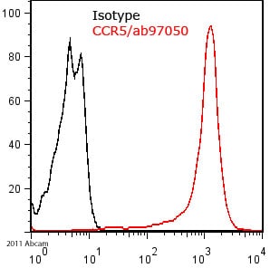
The FACS staining was performed on HEK cells expressing human CCR5 with PBS/BSA (0.5%)/Azide (0.05%) as FACS buffer. We used a primary polyclonal rabbit antibody against CCR5 at a 1/100 dillution, incubated for 1 hour at 4C. The secondary antibody was ab97050 used at a concentration of 1/100 for 30min at 4C. No fixation or permeabilization was performed and gaiting was done on alive cells.
This image is courtesy of an anonymous Abreview
Protocols
To our knowledge, customised protocols are not required for this product. Please try the standard protocols listed below and let us know how you get on.
Datasheets and documents
-
SDS download
-
Datasheet download
References (54)
ab97050 has been referenced in 54 publications.
- Enderami SE et al. Enhanced yield of cholinergic neurons from induced pluripotent stem cells (iPSC): A two-step induction protocol. Bratisl Lek Listy 124:267-272 (2023). PubMed: 36598319
- Adzraku SY et al. Robo4 inhibits gamma radiation-induced permeability of a murine microvascular endothelial cell by regulating the junctions. Cell Mol Biol Lett 28:2 (2023). PubMed: 36647012
- Segunda MN et al. Comparative Analysis of the Potential for Germ Cell (GC) Differentiation of Bovine Peripheral Blood Derived-Mesenchymal Stem Cells (PB-MSC) and Spermatogonial Stem Cells (SSC) in Co-Culture System with Sertoli Cells (SC). Animals (Basel) 13:N/A (2023). PubMed: 36670859
- Nushtaeva A et al. "Pulsed Hypoxia" Gradually Reprograms Breast Cancer Fibroblasts into Pro-Tumorigenic Cells via Mesenchymal-Epithelial Transition. Int J Mol Sci 24:N/A (2023). PubMed: 36768815
- Ohtsuki S et al. Deficiency of the CD155-CD96 immune checkpoint controls IL-9 production in giant cell arteritis. Cell Rep Med 4:101012 (2023). PubMed: 37075705