Human CDKN1C (p57 Kip2) knockout HeLa cell line (ab280061)
 knockout HeLa cell line (ab280061)")
Overview
-
Product name
Human CDKN1C (p57 Kip2) knockout HeLa cell line
See all p57 Kip2 lysates -
Parental Cell Line
HeLa -
Organism
Human -
Mutation description
Knockout achieved by using CRISPR/Cas9, Homozygous: 217 bp deletion in exon 1 -
Passage number
<20 -
Knockout validation
Sanger Sequencing, Western Blot (WB) -
Tested applications
Suitable for: WB, Sanger Sequencingmore details -
Biosafety level
2 -
General notes
Recommended control: Human wild-type HeLa cell line (ab275466). Please note a wild-type cell line is not automatically included with a knockout cell line order, if required please add recommended wild-type cell line at no additional cost using the code WILDTYPE-TMTK1.
Cryopreservation cell medium: Cell Freezing Medium-DMSO Serum free media, contains 8.7% DMSO in MEM supplemented with methyl cellulose.
Culture medium: DMEM (High Glucose) + 10% FBS
Initial handling guidelines: Upon arrival, the vial should be stored in liquid nitrogen vapor phase and not at -80°C. Storage at -80°C may result in loss of viability.
1. Thaw the vial in 37°C water bath for approximately 1-2 minutes.
2. Transfer the cell suspension (0.8 mL) to a 15 mL/50 mL conical sterile polypropylene centrifuge tube containing 8.4 mL pre-warmed culture medium, wash vial with an additional 0.8 mL culture medium (total volume 10 mL) to collect remaining cells, and centrifuge at 201 x g (rcf) for 5 minutes at room temperature. 10 mL represents minimum recommended dilution. 20 mL represents maximum recommended dilution.
3. Resuspend the cell pellet in 5 mL pre-warmed culture medium and count using a haemocytometer or alternative cell counting method. Based on cell count, seed cells in an appropriate cell culture flask at a density of 2x10⁴ cells/cm2. Seeding density is given as a guide only and should be scaled to align with individual lab schedules.
4. Incubate the culture at 37°C incubator with 5% CO2. Cultures should be monitored daily.Subculture guidelines:
- All seeding densities should be based on cell counts gained by established methods.
- A guide seeding density of 2x10⁴ cells/cm2 is recommended.
- A partial media change 24 hours prior to subculture may be helpful to encourage growth, if required.
- Cells should be passaged when they have achieved 80-90% confluence.
This product is subject to limited use licenses from The Broad Institute and ERS Genomics Limited, and is developed with patented technology. For full details of the limited use licenses and relevant patents please refer to our limited use license and patent pages.
We will provide viable cells that proliferate on revival.
Properties
-
Number of cells
1 x 10⁶ cells/vial, 1 mL -
Adherent /Suspension
Adherent -
Tissue
Cervix -
Cell type
epithelial -
Disease
Adenocarcinoma -
Gender
Female -
Mycoplasma free
Yes -
Storage instructions
Shipped on Dry Ice. Store in liquid nitrogen. -
Storage buffer
Constituents: 8.7% Dimethylsulfoxide, 2% Cellulose, methyl ether -
Research areas
Target
-
Function
Potent tight-binding inhibitor of several G1 cyclin/CDK complexes (cyclin E-CDK2, cyclin D2-CDK4, and cyclin A-CDK2) and, to lesser extent, of the mitotic cyclin B-CDC2. Negative regulator of cell proliferation. May play a role in maintenance of the non-proliferative state throughout life. -
Tissue specificity
Expressed in the heart, brain, lung, skeletal muscle, kidney, pancreas and testis. High levels are seen in the placenta while low levels are seen in the liver. -
Involvement in disease
Defects in CDKN1C are a cause of Beckwith-Wiedemann syndrome (BWS) [MIM:130650]. BWS is a genetically heterogeneous disorder characterized by anterior abdominal wall defects including exomphalos (omphalocele), pre- and postnatal overgrowth, and macroglossia. Additional less frequent complications include specific developmental defects and a predisposition to embryonal tumors.
Note=Defects in CDKN1C are involved in tumor formation. -
Sequence similarities
Belongs to the CDI family. -
Cellular localization
Nucleus. - Information by UniProt
Associated products
-
KO cell lysates
-
Related Products
Applications
The Abpromise guarantee
Our Abpromise guarantee covers the use of ab280061 in the following tested applications.
The application notes include recommended starting dilutions; optimal dilutions/concentrations should be determined by the end user.
| Application | Abreviews | Notes |
|---|---|---|
| WB |
Use at an assay dependent concentration.
|
|
| Sanger Sequencing |
Use at an assay dependent concentration.
|
| Notes |
|---|
|
WB
Use at an assay dependent concentration. |
|
Sanger Sequencing
Use at an assay dependent concentration. |
Images
-
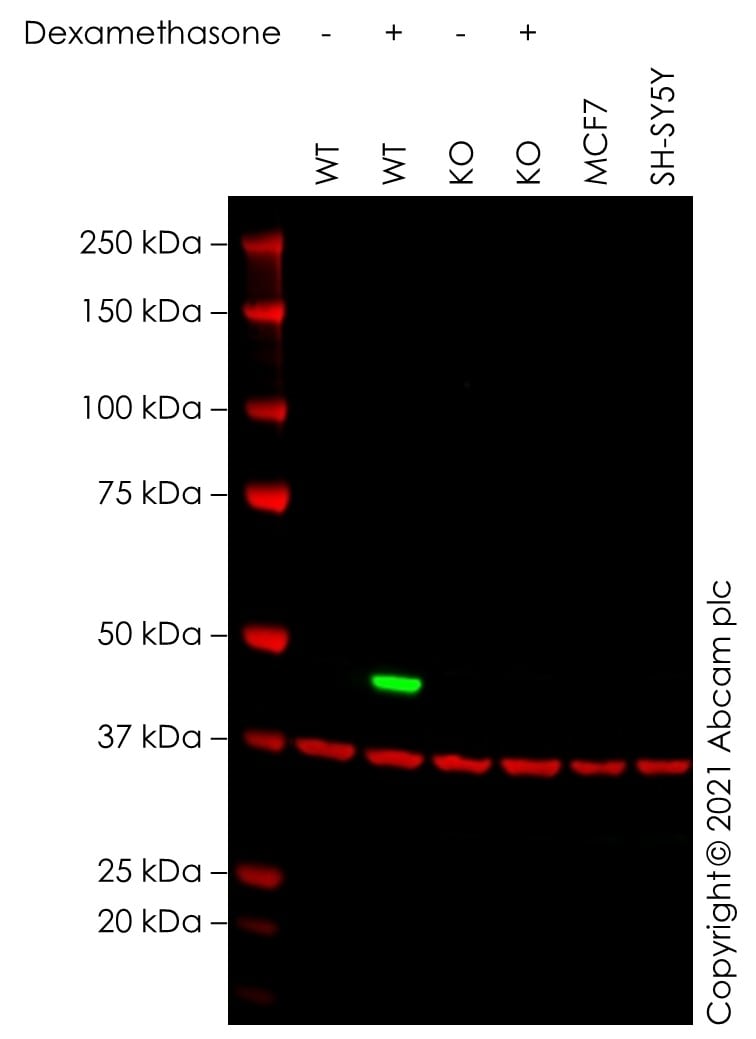
Western blot - Human CDKN1C (p57 Kip2) knockout HeLa cell line (ab280061)All lanes : Anti-p57 Kip2 antibody [EP2718(2)] (ab133531) at 1/1000 dilution
Lane 1 : Wild-type HeLa Vehicle Control Dexamethasone (0 nM, 16 h) ab277359 cell lysate
Lane 2 : Wild-type HeLa Treated Dexamethasone (50 nM, 16 h) ab287335 cell lysate
Lane 3 : CDKN1C knockout HeLa Vehicle Control Dexamethasone (0 nM, 16 h) ab277299 cell lysate
Lane 4 : CDKN1C knockout HeLa Treated Dexamethasone (50 nM, 16 h) ab281877 cell lysate
Lane 5 : MCF7 cell lysate
Lane 6 : SH-SY5Y cell lysate
Lysates/proteins at 20 µg per lane.
Performed under reducing conditions.
Observed band size: 50 kDa why is the actual band size different from the predicted?False colour image of Western blot: Anti-p57 Kip2 antibody [EP2718(2)] staining at 1/1000 dilution, shown in green; Mouse anti-GAPDH antibody [6C5] (ab8245) loading control staining at 1/20000 dilution, shown in red. In Western blot, ab133531 was shown to bind specifically to p57 Kip2. A band was observed at 50 kDa in wild-type HeLa cell lysates with no signal observed at this size in CDKN1C knockout cell line ab280061 (knockout cell lysate ab280120). To generate this image, wild-type and CDKN1C knockout HeLa cell lysates were analysed. First, samples were run on an SDS-PAGE gel then transferred onto a nitrocellulose membrane. Membranes were blocked in fluorescent western blot (TBS-based) blocking solution before incubation with primary antibodies overnight at 4°C. Blots were washed four times in TBS-T, incubated with secondary antibodies for 1 h at room temperature, washed again four times then imaged. Secondary antibodies used were Goat anti-Rabbit IgG H&L (IRDye® 800CW) preabsorbed (ab216773) and Goat anti-Mouse IgG H&L (IRDye® 800CW) preabsorbed (ab216772) at 1/20000 dilution.
-
 Western blot - Human CDKN1C (p57 Kip2) knockout HeLa cell line (ab280061)All lanes : Anti-p57 Kip2 antibody [EP2516] (ab119989) at 1/1000 dilution
Lane 1 : Wild-type HeLa Vehicle Control Dexamethasone (0 nM, 16 h) ab277359 cell lysate
Lane 2 : Wild-type HeLa Treated Dexamethasone (50 nM, 16 h) ab287335 cell lysate
Lane 3 : CDKN1C knockout HeLa Vehicle Control Dexamethasone (0 nM, 16 h) ab277299 cell lysate
Lane 4 : CDKN1C knockout HeLa Treated Dexamethasone (50 nM, 16 h) ab281877 cell lysate
Lane 5 : MCF7 cell lysate
Lane 6 : SH-SY5Y cell lysate
Lysates/proteins at 20 µg per lane.
Performed under reducing conditions.
Observed band size: 50 kDa why is the actual band size different from the predicted?False colour image of Western blot: Anti-p57 Kip2 antibody [EP2516] staining at 1/1000 dilution, shown in green; Mouse anti-GAPDH antibody [6C5] (ab8245) loading control staining at 1/20000 dilution, shown in red. In Western blot, ab119989 was shown to bind specifically to p57 Kip2. A band was observed at 50 kDa in wild-type HeLa cell lysates with no signal observed at this size in CDKN1C knockout cell line ab280061 (knockout cell lysate ab280120). To generate this image, wild-type and CDKN1C knockout HeLa cell lysates were analysed. First, samples were run on an SDS-PAGE gel then transferred onto a nitrocellulose membrane. Membranes were blocked in fluorescent western blot (TBS-based) blocking solution before incubation with primary antibodies overnight at 4°C. Blots were washed four times in TBS-T, incubated with secondary antibodies for 1 h at room temperature, washed again four times then imaged. Secondary antibodies used were Goat anti-Rabbit IgG H&L (IRDye® 800CW) preabsorbed (ab216773) and Goat anti-Mouse IgG H&L (IRDye® 800CW) preabsorbed (ab216772) at 1/20000 dilution.
-
 Sanger Sequencing - Human CDKN1C (p57 Kip2) knockout HeLa cell line (ab280061)
217 bp deletion in exon 1
Protocols
Datasheets and documents
-
SDS download
-
Datasheet download
References (0)
ab280061 has not yet been referenced specifically in any publications.