Human EGFR knockout HeLa cell line
- Advanced Validation
- 각 태그의 뜻
Be the first to review this product! 리뷰 제출
|
(2 제품이 사용된 논문 )

- WB
Lab
Western blot - Human EGFR knockout HeLa cell line (AB255385)
Western blot : Anti-EGFR antibody [E235] (ab32077) staining at 1/1000 dilution shown in green; Mouse anti-Alpha Tubulin [DM1A] (ab7291) loading control staining at 1/20000 dilution shown in red. In Western blot ab32077 was shown to bind specifically to EGFR. A band was observed at 160 kDa in wild-type cell lysates with no signal observed at this size in EGFR knockout cell lines. To generate this image wild-type and EGFR knockout A549 (ab286394) and HeLa (ab255385) cell lysates were analysed. First samples were run on an SDS-PAGE gel then transferred onto a nitrocellulose membrane. Membranes were blocked in 3% milk in TBS-0.1% Tween ® 20 (TBS-T) before incubation with primary antibodies overnight at 4°. Blots were washed four times in TBS-T incubated with secondary antibodies for 1 h at room temperature washed again four times then imaged. Secondary antibodies used were Goat anti-Rabbit IgG H&L 800CW and Goat anti-Mouse IgG H&L 680RD at 1/20000 dilution.
All lanes:
Western blot - Anti-EGFR antibody [E235] (<a href='/ko/products/primary-antibodies/egfr-antibody-e235-ab32077'>ab32077</a>) at 1/1000 dilution
Lane 1:
Wild-type A549 cell lysate at 20 µg
Lane 2:
EGFR knockout A549 cell lysate at 20 µg
Lane 3:
Wild-type HeLa cell lysate at 20 µg
Lane 4:
EGFR knockout HeLa cell lysate at 20 µg
Observed band size: 160 kDa
false

- WB
Lab
Western blot - Human EGFR knockout HeLa cell line (AB255385)
All lanes:
Western blot - Anti-EGFR antibody [E234] (<a href='/ko/products/primary-antibodies/egfr-antibody-e234-ab32198'>ab32198</a>) at 1/1000 dilution
Lane 1:
Wild-type A549 cell lysate at 20 µg
Lane 2:
EGFR knockout A549 cell lysate at 20 µg
Lane 3:
Wild-type HeLa cell lysate at 20 µg
Lane 4:
EGFR knockout HeLa cell lysate at 20 µg
Observed band size: 160 kDa
false

- Sanger seq
Unknown
Sanger Sequencing - Human EGFR knockout HeLa cell line (AB255385)
Allele-1 : 1 bp deletion in exon 1.

- Sanger seq
Unknown
Sanger Sequencing - Human EGFR knockout HeLa cell line (AB255385)
Allele-2 : 1 bp insertion in exon 1.

- Sanger seq
Lab
Sanger Sequencing - Human EGFR knockout HeLa cell line (AB255385)
Sequencing chromatogram displaying sequence edit in exon 1
- WB
Lab
Western blot - Human EGFR knockout HeLa cell line (AB255385)
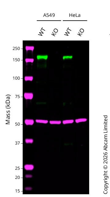
Western blot : Anti-EGFR antibody [SP84] - C-terminal ab227642 staining at 1/200 dilution, shown in green; Mouse anti alpha Tubulin ab7291 loading control staining at 1/20000 dilution, shown in magenta. A band was observed at 160 kDa in Wild-type A549 cell lysates with no signal observed at this size in EGFR knockout A549 cell line. To generate this image, samples were run on an SDS-PAGE gel then transferred onto a nitrocellulose membrane. Membranes were blocked in 3% Milk in TBS-0.1 % Tween® 20 (TBS-T) before incubation with primary antibodies overnight at 4 °C. Blots were washed four times in TBS-T, incubated with secondary antibodies for 1 h at room temperature, washed again four times then imaged. Secondary antibodies used were Goat anti-Rabbit 800CW and Goat anti-Mouse 680RD at 1/20,000 dilution.
All lanes:
Western blot - Anti-EGFR antibody [SP84] - C-terminal (<a href='/ko/products/primary-antibodies/egfr-antibody-sp84-c-terminal-ab227642'>ab227642</a>) at 1/200 dilution
Lane 1:
Wild-type A549 cell lysates at 20 µg
Lane 2:
EGFR knockout A549 cell lysates at 20 µg
Lane 3:
Wild-type HeLa ab255928 cell lysates at 20 µg
Lane 4:
Western blot - Human EGFR knockout HeLa cell line (ab255385) at 20 µg
Secondary
All lanes:
Goat anti-Rabbit 800CW & Goat anti-Mouse 680RD at 1/20000 dilution
Predicted band size: 134 kDa
Observed band size: 160 kDa
false
- WB
Lab
Western blot - Human EGFR knockout HeLa cell line (AB255385)
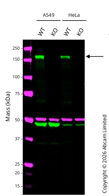
Western blot : Anti-EGFR antibody [EPR39Y] ab76153 staining at 1/1000 dilution, shown in green; Mouse anti alpha Tubulin ab7291 loading control staining at 1/20000 dilution, shown in magenta. A band was observed at 160 kDa in Wild-type A549 cell lysates with no signal observed at this size in EGFR knockout A549 cell line. To generate this image, samples were run on an SDS-PAGE gel then transferred onto a nitrocellulose membrane. Membranes were blocked in 3% Milk in TBS-0.1 % Tween® 20 (TBS-T) before incubation with primary antibodies overnight at 4 °C. Blots were washed four times in TBS-T, incubated with secondary antibodies for 1 h at room temperature, washed again four times then imaged. Secondary antibodies used were Goat anti-Rabbit 800CW and Goat anti-Mouse 680RD at 1/20,000 dilution.
All lanes:
Western blot - Anti-EGFR antibody [EPR39Y] (<a href='/ko/products/primary-antibodies/egfr-antibody-epr39y-ab76153'>ab76153</a>) at 1/1000 dilution
Lane 1:
Wild-type A549 cell lysates at 20 µg
Lane 2:
EGFR knockout A549 cell lysates at 20 µg
Lane 3:
Wild-type HeLa ab255928 cell lysates at 20 µg
Lane 4:
Western blot - Human EGFR knockout HeLa cell line (ab255385) at 20 µg
Secondary
All lanes:
Goat anti-Rabbit 800CW & Goat anti-Mouse 680RD at 1/20000 dilution
Predicted band size: 134 kDa
Observed band size: 160 kDa
false
보완 제품

Cell Lines & Lysates
AB265961
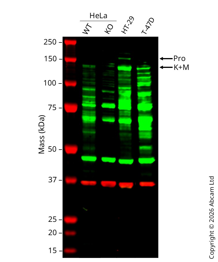
Human MET (Met (c-Met)) knockout HeLa cell line
Advanced Validation
- 0
- 0
Cell Lines & Lysates
AB264921
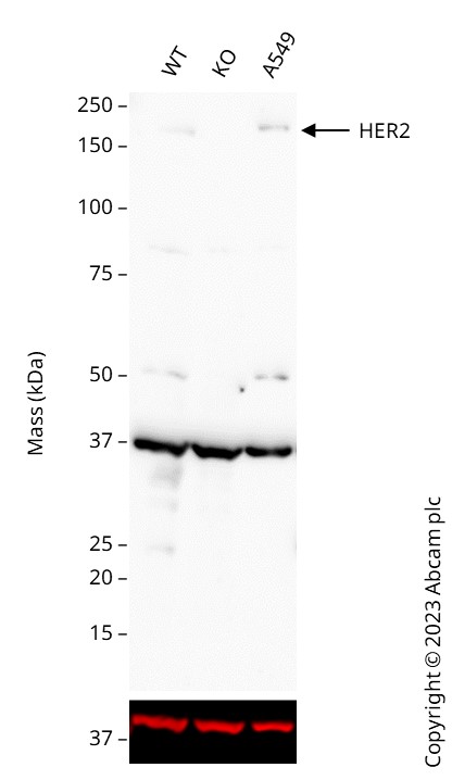
Human FOLR1 (Folate Binding Protein/FBP) knockout HeLa cell line
Advanced Validation

- 0
- 0
Reactivity 정보
제품 세부 정보
We will provide viable cells that proliferate on revival.
This product is subject to limited use licenses from The Broad Institute, ERS Genomics Limited and Sigma-Aldrich Co. LLC, and is developed with patented technology. For full details of the licenses and patents please refer to our limited use license and patent pages.
제품 구성
특성 및 보관 정보
유전자명
유전자 편집 종류
유전자 편집 방식
Knockout 검증
품질 관리 - STR analysis
배송 시 보관 조건
적절한 단기 보관 조건
적절한 장기 보관 조건
추가 정보
This supplementary information is collated from multiple sources and compiled automatically.
Biological function summary
The EGFR protein plays an important role in cellular communication and signaling processes. EGFR pairs with other receptor family members to form active dimers or even higher-order complexes which in turn initiate intracellular signaling cascades. Through these complexes EGFR influences many processes including cell differentiation and repair. This function of EGFR makes it an integral part of mammalian biology affecting how cells respond to their environment by mediating changes in gene expression.
Pathways
EGFR is a central player in the MAPK and PI3K/Akt signaling pathways. Alongside other protein partners like KRAS and PI3 kinase it contributes to transmitting signals from the cell surface to the nucleus affecting gene transcription and cell behavior. These pathways are important for normal cell growth and division and aberrations in these pathways can lead to excessive or insufficient cell proliferation.
Cell culture
Biosafety level
EU: 2 US: 2
Adherent/suspension
Adherent
Gender
Female
초기 취급 지침
Upon arrival, the vial should be stored in liquid nitrogen vapor phase and not at -80°C. Storage at -80°C may result in loss of viability. <p>1. Thaw the vial in 37°C water bath for approximately 1-2 minutes.<br>2. Transfer the cell suspension (0.8 mL) to a 15 mL/50 mL conical sterile polypropylene centrifuge tube containing 8.4 mL pre-warmed culture medium, wash vial with an additional 0.8 mL culture medium (total volume 10 mL) to collect remaining cells, and centrifuge at 201 x g (rcf) for 5 minutes at room temperature. 10 mL represents minimum recommended dilution. 20 mL represents maximum recommended dilution.<br>3. Resuspend the cell pellet in 5 mL pre-warmed culture medium and count using a haemocytometer or alternative cell counting method seed all remaining cells into a T25.<br>4. Incubate the culture at 37°C incubator with 5% CO<sub>2</sub>. Check the culture one day after revival and continue to check until 80% confluent. Media change can be given if needed.<br>5. Once confluent passage into an appropriate flask at a density of 2x10<sup>4</sup> cells/cm<sup>2</sup>. Seeding density is given as a guide only and should be scaled to align with individual lab schedules. Cultures should be monitored daily.</p>
Subculture 가이드라인
- All seeding densities should be based on cell counts gained by established methods.
- A guide seeding density of 2x10⁴ cells/cm2 is recommended.
- Cells should be passaged when they have achieved 80-90% confluence.
배양 배지
DMEM (High Glucose) + 10% FBS
동결보존 배지
Cell Freezing Medium-DMSO Serum free media, contains 8.7% DMSO in MEM supplemented with methyl cellulose.
타겟 정보
대체 명칭 보기
제품이 사용된 논문 (2)
Recent publications for all applications. Explore the 전체 목록and refine your search
Cell reports. Medicine 6:101874 PubMed39694038
2024
Applications
Unspecified application
Species
Unspecified reactive species
Nature structural & molecular biology 31:1404-1412 PubMed38724718
2024
Applications
Unspecified application
Species
Unspecified reactive species
Product promise
Please note: All products are 'FOR RESEARCH USE ONLY. NOT FOR USE IN DIAGNOSTIC OR THERAPEUTIC PROCEDURES'.
For licensing inquiries, please contact partnerships@abcam.com