Human FANCD2 knockout HeLa cell line
- Advanced Validation
- 각 태그의 뜻
Be the first to review this product! 리뷰 제출
|
(0 출판물)
- WB
Lab
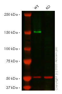
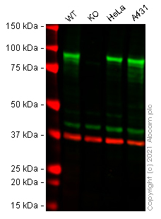
Western blot - Human FANCD2 knockout HeLa cell line (AB261743)
Lanes 1- 2 : Merged signal (red and green). Green - ab178705 observed at 166 kDa. Red - Anti-alpha Tubulin antibody [DM1A] - Loading Control (ab7291) observed at 50 kDa.
ab178705 was shown to react with FANCD2 in wild-type HeLa cells in western blot. Loss of signal was observed when knockout cell line ab261743 (knockout cell lysate ab257173) was used. Wild-type HeLa and FANCD2 knockout HeLa cell lysates were subjected to SDS-PAGE. Membrane was blocked for 1 hour at room temperature in 0.1% TBST with 3% non-fat dried milk. ab178705 and Anti-alpha Tubulin antibody [DM1A] - Loading Control (ab7291) overnight at 4°C at a 1 in 1000 dilution and a 1 in 20000 dilution respectively. Blots were developed with Goat anti-Rabbit IgG H&L (IRDye®800CW) preadsorbed (ab216773) and Goat anti-Mouse IgG H&L (IRDye®680RD) preadsorbed (ab216776) secondary antibodies at 1 in 20000 dilution for 1 hour at room temperature before imaging.
All lanes:
Western blot - Anti-FANCD2 antibody [EPR2279(3)] (<a href='/ko/products/primary-antibodies/fancd2-antibody-epr22793-ab178705'>ab178705</a>) at 1/1000 dilution
Lane 1:
Wild-type HeLa cell lysate at 20 µg
Lane 2:
FANCD2 knockout HeLa cell lysate at 20 µg
Lane 2:
Western blot - Human FANCD2 knockout HeLa cell line (ab261743)
Predicted band size: 166 kDa
Observed band size: 166 kDa
false
- WB
Lab
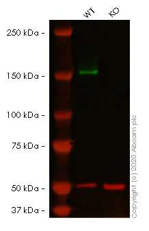
Western blot - Human FANCD2 knockout HeLa cell line (AB261743)
Lanes 1- 2 : Merged signal (red and green). Green - ab108928 observed at 166 kDa. Red - Anti-alpha Tubulin antibody [DM1A] - Loading Control (ab7291) observed at 50 kDa.
ab108928 was shown to react with FANCD2 in wild-type HeLa cells in western blot. Loss of signal was observed when knockout cell line ab261743 (knockout cell lysate ab257173) was used. Wild-type HeLa and FANCD2 knockout HeLa cell lysates were subjected to SDS-PAGE. Membrane was blocked for 1 hour at room temperature in 0.1% TBST with 3% non-fat dried milk. ab108928 and Anti-alpha Tubulin antibody [DM1A] - Loading Control (ab7291) overnight at 4°C at a 1 in 1000 dilution and a 1 in 20000 dilution respectively. Blots were developed with Goat anti-Rabbit IgG H&L (IRDye®800CW) preadsorbed (ab216773) and Goat anti-Mouse IgG H&L (IRDye®680RD) preadsorbed (ab216776) secondary antibodies at 1 in 20000 dilution for 1 hour at room temperature before imaging.
All lanes:
Western blot - Anti-FANCD2 antibody [EPR2302] (<a href='/ko/products/primary-antibodies/fancd2-antibody-epr2302-ab108928'>ab108928</a>) at 1/1000 dilution
Lane 1:
Wild-type HeLa cell lysate at 20 µg
Lane 2:
FANCD2 knockout HeLa cell lysate at 20 µg
Lane 2:
Western blot - Human FANCD2 knockout HeLa cell line (ab261743)
Predicted band size: 166 kDa
Observed band size: 166 kDa
false

- Sanger seq
Unknown
Sanger Sequencing - Human FANCD2 knockout HeLa cell line (AB261743)
Homozygous : 4 bp deletion in exon 13.
보완 제품

Cell Lines & Lysates
AB277911
Human CTNNB1 (beta Catenin) knockout Hep G2 cell line
Advanced Validation
- 0
- 0
Reactivity 정보
제품 세부 정보
We will provide viable cells that proliferate on revival.
This product is subject to limited use licenses from The Broad Institute and ERS Genomics Limited, and is developed with patented technology. For full details of the limited use licenses and relevant patents please refer to our limited use license and patent pages.
제품 구성
특성 및 보관 정보
유전자명
유전자 편집 종류
유전자 편집 방식
Knockout 검증
Zygosity
배송 시 보관 조건
적절한 단기 보관 조건
적절한 장기 보관 조건
추가 정보
This supplementary information is collated from multiple sources and compiled automatically.
Biological function summary
FANCD2 collaborates with multiple proteins as part of the Fanconi anemia (FA) complex to ensure genomic stability. The FA complex operates by facilitating the repair of DNA interstrand crosslinks that impede replication. FANCD2 becomes activated through monoubiquitination which serves as a signal for recruitment to DNA damage sites. It essentially functions as a coordinator that brings together important components for repair.
Pathways
FANCD2 integrates into the Fanconi anemia pathway and the homologous recombination repair pathway. These pathways are critical for maintaining DNA integrity and preventing chromosomal instability. Within these pathways interactions with proteins such as BRCA1 and BRCA2 reinforce the repair processes. FANCD2's connection to these proteins exemplifies its role in complex mechanisms that preserve genomic fidelity.
품질 관리
STR analysis
CSF1PO, D13S317, D7S820, D5S818, TH01, D16S539, TPOX
Cell culture
Biosafety level
EU: 2 US: 2
Adherent/suspension
Adherent
Gender
Female
초기 취급 지침
Upon arrival, the vial should be stored in liquid nitrogen vapor phase and not at -80°C. Storage at -80°C may result in loss of viability. <p>1. Thaw the vial in 37°C water bath for approximately 1-2 minutes.<br>2. Transfer the cell suspension (0.8 mL) to a 15 mL/50 mL conical sterile polypropylene centrifuge tube containing 8.4 mL pre-warmed culture medium, wash vial with an additional 0.8 mL culture medium (total volume 10 mL) to collect remaining cells, and centrifuge at 201 x g (rcf) for 5 minutes at room temperature. 10 mL represents minimum recommended dilution. 20 mL represents maximum recommended dilution.<br>3. Resuspend the cell pellet in 5 mL pre-warmed culture medium and count using a haemocytometer or alternative cell counting method seed all remaining cells into a T25.<br>4. Incubate the culture at 37°C incubator with 5% CO<sub>2</sub>. Check the culture one day after revival and continue to check until 80% confluent. Media change can be given if needed.<br>5. Once confluent passage into an appropriate flask at a density of 2x10<sup>4</sup> cells/cm<sup>2</sup>. Seeding density is given as a guide only and should be scaled to align with individual lab schedules. Cultures should be monitored daily.</p>
Subculture 가이드라인
- All seeding densities should be based on cell counts gained by established methods.
- A guide seeding density of 2x10⁴ cells/cm2 is recommended.
- Cells should be passaged when they have achieved 80-90% confluence.
배양 배지
DMEM (High Glucose) + 10% FBS
동결보존 배지
Cell Freezing Medium-DMSO Serum free media, contains 8.7% DMSO in MEM supplemented with methyl cellulose.
Product promise
Please note: All products are 'FOR RESEARCH USE ONLY. NOT FOR USE IN DIAGNOSTIC OR THERAPEUTIC PROCEDURES'.
For licensing inquiries, please contact partnerships@abcam.com