Human RIPK3 knockout THP-1 cell line
- Advanced Validation
- 각 태그의 뜻
Be the first to review this product! 리뷰 제출
|
(0 출판물)

- WB
Lab
Western blot - Human RIPK3 knockout THP-1 cell line (AB280065)
Blocking and diluting buffer and concentration : 5% NFDM/TBST.
Performed under reducing conditions.
In Western blot, ab325636 was shown to bind specifically to RIPK3. Target of interest was observed at 37-50 kDa in wild-type THP-1 cell lysates (lane 1) with no signal observed at this size in RIPK3 knockout cell line (lane 2)(knockout cell line ab280065).
In Western blot, Anti-Histone H3 antibody [EPR16987] - Nuclear Marker and ChIP Grade (ab176842) (1 : 100000) (17KDa).
All lanes:
Western blot - Anti-RIP3 antibody [RM1355] (<a href='/ko/products/primary-antibodies/rip3-antibody-rm1355-ab325636'>ab325636</a>) at 1/1000 dilution
Lane 1:
Wilde-type THP-1 (human monocytic leukemia monocyte) whole cell lysate at 20 µg
Lane 2:
Western blot - Human RIPK3 knockout THP-1 cell line (ab280065) at 20 µg
Secondary
All lanes:
Western blot - Goat Anti-Rabbit IgG H&L (HRP) (<a href='/ko/products/secondary-antibodies/goat-rabbit-igg-h-l-hrp-ab97051'>ab97051</a>) at 1/20000 dilution
Observed band size: 37-56 kDa,17 kDa
false
Exposure time: 180s
- WB
Lab
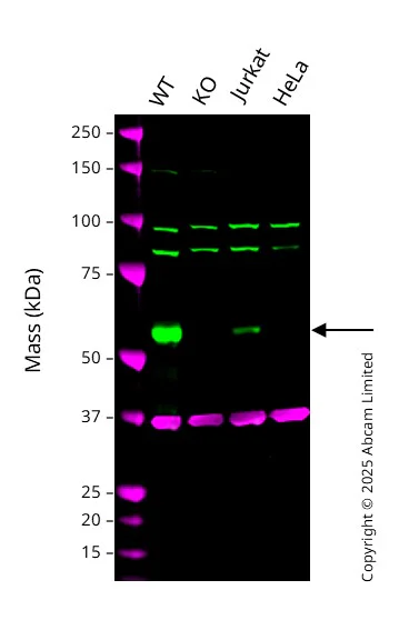
Western blot - Human RIPK3 knockout THP-1 cell line (AB280065)
Western blot : Anti-RIP3 antibody [EPR24374-135] ab305054 staining at 1/1000 dilution, shown in green; Mouse anti GAPDH ab8245 loading control staining at 1/20000 dilution, shown in magenta. A band was observed at 57 kDa in Wild-type THP-1 cell lysates with no signal observed at this size in RIPK3 knockout THP-1 cell line. To generate this image, samples were run on an SDS-PAGE gel then transferred onto a nitrocellulose membrane. Membranes were blocked in 3pc Milk in TBS-0.1 % Tween® 20 (TBS-T) before incubation with primary antibodies overnight at 4 °C. Blots were washed four times in TBS-T, incubated with secondary antibodies for 1 h at room temperature, washed again four times then imaged. Secondary antibodies used were Goat anti-Rabbit 800CW and Goat anti-Mouse 680RD at 1/20,000 dilution.
All lanes:
Western blot - Anti-RIP3 antibody [EPR24374-135] (<a href='/ko/products/primary-antibodies/rip3-antibody-epr24374-135-ab305054'>ab305054</a>) at 1/1000 dilution
Lane 1:
Wild-type THP-1 whole cell lysate at 20 µg
Lane 2:
Western blot - Human RIPK3 knockout THP-1 cell line (ab280065) at 20 µg
Lane 3:
Jurkat whole cell lysate at 20 µg
Lane 4:
HeLa whole cell lysate at 20 µg
Secondary
All lanes:
Goat anti-Rabbit 800CW & Goat anti-Mouse 680RD at 1/20000 dilution
Predicted band size: 57 kDa
Observed band size: 57 kDa
false
Reactivity 정보
제품 세부 정보
Recommended control: Human wild-type THP-1 cell line (ab281894). Please note a wild-type cell line is not automatically included with a knockout cell line order, if required please add recommended wild-type cell line at no additional cost using the code WILDTYPE-TMTK1.
- Upon thawing make banks as soon as possible and use each bank within 10-20 passages
- As cell line growth can vary please attempt culture for 2 weeks from revival of initial bank before contacting the technical team.
제품 구성
특성 및 보관 정보
유전자명
유전자 편집 종류
유전자 편집 방식
Knockout 검증
품질 관리 - STR analysis
배송 시 보관 조건
적절한 단기 보관 조건
적절한 장기 보관 조건
Cell culture
Biosafety level
EU: 1 US: 1
Adherent/suspension
Suspension
Gender
Male
Viability
~ 60%
Subculture 가이드라인
- All seeding densities should be based on cell counts gained by established methods.
- Cells should be seeded at 2x10⁵ - 3x10⁵ cells/mL and subcultured when they have reached 8x10⁵ cells/mL.
- It is not recommended to allow the cell density to exceed 1x10⁶ cells/mL.
배양 배지
RPMI + 10% FBS + 0.05 mM beta-mercaptoethanol
동결보존 배지
Cell Freezing Medium-DMSO Serum free media, contains 8.7% DMSO in MEM supplemented with methyl cellulose.
Product promise
Please note: All products are 'FOR RESEARCH USE ONLY. NOT FOR USE IN DIAGNOSTIC OR THERAPEUTIC PROCEDURES'.
For licensing inquiries, please contact partnerships@abcam.com