Human PXN (Paxillin) knockout A-431 cell lysate
Be the first to review this product! 리뷰 제출
|
(0 출판물)
- WB
Lab
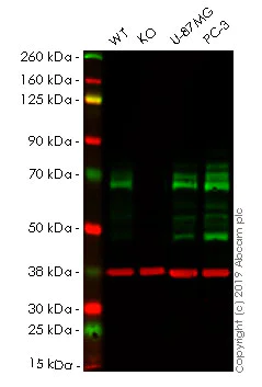
Western blot - Human PXN (Paxillin) knockout A-431 cell lysate (AB261701)
Lane 1 : Wild-type A-431 (Human epidermoid carcinoma cell line) whole cell lysate 20 ug
Lane 2 : PXN knockout A-431 (Human epidermoid carcinoma cell line) whole cell lysate 20 ug
Lane 3 : U-87 MG (Human glioblastoma-astrocytoma epithelial cell line) whole cell lysate 20 ug
Lane 4 : PC3 (Human prostate adenocarcinoma cell line) whole cell lysate 20 ug
Lanes 1 - 4 : Merged signal (red and green). Green - ab23510 observed at 65 kDa. Red - loading control, ab181602, observed at 37 kDa.
ab23510 was shown to recognize PXN in wild-type A-431 cells as signal was lost at the expected MW in PXN knockout cell line ab261892 (knockout cell lysate ab261701). Additional cross-reactive bands were observed in the wild-type and knockout samples. Wild-type and PXN knockout samples were subjected to SDS-PAGE. ab23510 and ab181602 (Rabbit anti-GAPDH loading control) were incubated overnight at 4°C at 1/1000 dilution and 1/20000 dilution respectively. Blots were developed with Goat anti-Mouse IgG H&L (IRDye® 800CW) preabsorbed ab216772 and Goat anti-Rabbit IgG H&L (IRDye® 680RD) preabsorbed ab216777 secondary antibodies at 1/20000 dilution for 1 hour at room temperature before imaging.
All lanes:
Western blot - Anti-Paxillin antibody [M107] (<a href='/ko/products/primary-antibodies/paxillin-antibody-m107-ab23510'>ab23510</a>) at 1/1000 dilution
Lane 1:
Wild-type A-431 (Human epidermoid carcinoma cell line) whole cell lysate at 20 µg
Lane 2:
PXN knockout A-431 (Human epidermoid carcinoma cell line) whole cell lysate at 20 µg
Lane 2:
Western blot - Human PXN (Paxillin) knockout A-431 cell line (<a href='/ko/products/cell-lines/human-pxn-paxillin-knockout-a-431-cell-line-ab261892'>ab261892</a>)
Lane 3:
U-87 MG (Human glioblastoma-astrocytoma epithelial cell line) whole cell lysate at 20 µg
Lane 4:
PC3 (Human prostate adenocarcinoma cell line) whole cell lysate at 20 µg
Predicted band size: 65 kDa
false
- WB
Lab
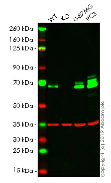
Western blot - Human PXN (Paxillin) knockout A-431 cell lysate (AB261701)
Lane 1 : Wild-type A-431 (Human epidermoid carcinoma cell line) whole cell lysate 20 ug
Lane 2 : PXN knockout A-431 (Human epidermoid carcinoma cell line) whole cell lysate 20 ug
Lane 3 : U-87 MG (Human glioblastoma-astrocytoma epithelial cell line) whole cell lysate 20 ug
Lane 4 : PC3 (Human prostate adenocarcinoma cell line) whole cell lysate 20 ug
Lanes 1 - 4 : Merged signal (red and green). Green - ab32115 observed at 65 kDa. Red - loading control, ab8245, observed at 37 kDa.
ab32115 was shown to specifically react with PXN in wild-type A-431 cells as signal was lost in PXN knockout cell line ab261892 (knockout cell lysate ab261701). Wild-type and PXN knockout samples were subjected to SDS-PAGE. The membrane was blocked with 3% milk. ab32115 and ab8245 (Mouse anti GAPDH loading control) were incubated overnight at 4°C at 1/10000 dilution and 1/20000 dilution respectively. Blots were developed with Goat anti-Rabbit IgG H&L (IRDye® 800CW) preabsorbed ab216773 and Goat anti-Mouse IgG H&L (IRDye® 680RD) preabsorbed ab216776 secondary antibodies at 1/20000 dilution for 1 hour at room temperature before imaging.
All lanes:
Western blot - Anti-Paxillin antibody [E228] (<a href='/ko/products/primary-antibodies/paxillin-antibody-e228-ab32115'>ab32115</a>) at 1/10000 dilution
Lane 1:
Wild-type A431 whole cell lysate at 20 µg
Lane 2:
PXN knockout A431 whole cell lysate at 20 µg
Lane 2:
Western blot - Human PXN (Paxillin) knockout A-431 cell line (<a href='/ko/products/cell-lines/human-pxn-paxillin-knockout-a-431-cell-line-ab261892'>ab261892</a>)
Lane 3:
U-87 MG whole cell lysate at 20 µg
Lane 4:
PC-3 whole cell lysate at 20 µg
Predicted band size: 65 kDa
Observed band size: 65 kDa
false
Exposure time: 10s
- WB
Lab
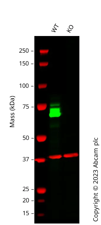
Western blot - Human PXN (Paxillin) knockout A-431 cell lysate (AB261701)
Lane 1 : Wild-type A431 cell lysate 20 ug Lane 2 : PXN knockout A431 cell lysate 20 ug Anti-PXN antibody [Y113] (ab32084) staining at 1/1000 dilution, shown in green; Mouse anti-GAPDH antibody [6C5] (ab8245) loading control staining at 1/20000 dilution, shown in red. In Western blot, ab32084 was shown to bind specifically to PXN. A band was observed at 70 kDa in wild-type A431 cell lysates with no signal observed at this size in PXN knockout cell line ab261892 (knockout cell lysate ab261701). To generate this image, wild-type and PXN knockout A431 cell lysates were analysed. First, samples were run on an SDS-PAGE gel then transferred onto a nitrocellulose membrane. Membranes were blocked in 3 % milk in TBS-0.1 % Tween$®$ 20 (TBS-T) before incubation with primary antibodies overnight at 4 °C. Blots were washed four times in TBS-T, incubated with secondary antibodies for 1 h at room temperature, washed again four times then imaged. Secondary antibodies used were Goat anti-Rabbit IgG H&L 800CW and Goat anti-Mouse IgG H&L 680RD at 1/20000 dilution.
All lanes:
Western blot - Anti-Paxillin antibody [Y113] (<a href='/ko/products/primary-antibodies/paxillin-antibody-y113-ab32084'>ab32084</a>) at 1/1000 dilution
Lane 2:
Western blot - Human PXN (Paxillin) knockout A-431 cell lysate (ab261701)
Lane 2:
Western blot - Human PXN (Paxillin) knockout A-431 cell line (<a href='/ko/products/cell-lines/human-pxn-paxillin-knockout-a-431-cell-line-ab261892'>ab261892</a>)
Predicted band size: 65 kDa
Observed band size: 70 kDa
false

- NGS
Lab
Next Generation Sequencing - Human PXN (Paxillin) knockout A-431 cell lysate (AB261701)
X = 7 bp deletion, 1 bp insertion
보완 제품
Primary Antibodies
AB32084
BOND RX™ Validated|RabMAb|Recombinant|KO Validated|20ul selling size

- 5
- 21
Reactivity 정보
제품 세부 정보
Knockout cell lysate achieved by CRISPR/Cas9.
REACH authorisation
Abcam has not and does not intend to apply for the REACH Authorisation of customers' uses of products that contain European Authorisation list (Annex XIV) substances.
It is the responsibility of our customers to check the necessity of application of REACH Authorisation, and any other relevant authorisations, for their intended uses.
Lysate preparation: Our lysates are made using RIPA buffer to which we add a protease inhibitor cocktail and phosphatase inhibitor cocktail (ratio: 300:100:10). This means that the protein of interest is denatured. If you require a native form of the protein please use the live cell version. Please refer to our lysis protocol for further details on how our lysates are prepared.
User storage instructions: Lyophilizate may be stored at 4°C. After reconstitution, store at -20°C for short-term storage or -80°C for long-term storage.
This product is subject to limited use licenses from The Broad Institute, ERS Genomics Limited and Sigma-Aldrich Co. LLC, and is developed with patented technology. For full details of the licenses and patents please refer to our limited use license and patent pages.
제품 구성
특성 및 보관 정보
유전자명
유전자 편집 종류
유전자 편집 방식
Knockout 검증
배송 시 보관 조건
적절한 단기 보관 조건
적절한 장기 보관 조건
추가 정보
This supplementary information is collated from multiple sources and compiled automatically.
Biological function summary
Paxillin serves key functions in cellular processes such as migration proliferation and survival. It interacts with different proteins including vinculin and talin forming part of a complex at the focal adhesion sites. The phosphorylation state of Paxillin can modulate its interactions and in this way influence the assembly of signaling complexes that control dynamic cellular processes.
Pathways
Paxillin participates in the integrin signaling and MAPK pathways. It operates by linking integrin receptors to the actin cytoskeleton facilitating signal transduction. Paxillin phosphorylation is an important regulatory mechanism within these pathways. In particular its interaction with focal adhesion kinase (FAK) and Src family kinases signifies its role in transmitting signals from the extracellular matrix to the cellular interior which impacts cell behavior and response.
품질 관리
STR analysis
CSF1PO, D13S317, D7S820, D5S818, TH01, D16S539, TPOX
Cell culture
Biosafety level
EU: 1 US: 1
Adherent/suspension
Adherent
Gender
Female
Product promise
Please note: All products are 'FOR RESEARCH USE ONLY. NOT FOR USE IN DIAGNOSTIC OR THERAPEUTIC PROCEDURES'.
For licensing inquiries, please contact partnerships@abcam.com