Alexa Fluor® 488 Anti-Transferrin Receptor antibody [EPR20584]
- BOND RX™ Validated
- Recombinant
- RabMAb
- 각 태그의 뜻
Be the first to review this product! 리뷰 제출
|
(0 출판물)

- IHC-P
Supplier Data
Immunohistochemistry (Formalin/PFA-fixed paraffin-embedded sections) - Alexa Fluor® 488 Anti-Transferrin Receptor antibody [EPR20584] (AB317529)
Immunohistochemical analysis of paraffin-embedded Human placenta tissue labeling Transferrin Receptor with ab317529 at 1/50 (10.0 ug/ml) dilution.
Positive staining on human placental trophoblast.The section was incubated with ab317529 for 60 mins at room temperature (shown in green). Nuclear DNA was labeled with DAPI (shown in blue). The section was then mounted using Fluoromount®.The immunostaining was performed on a Leica Biosystems BOND RX instrument.Image was taken with a confocal microscope (Leica-Microsystems, TCS SP8).
Counterstained with DAPI.
Heat mediated antigen retrieval was performed with Tris-EDTA buffer (pH 9.0, Epitope Retrieval Solution2) for 40 mins

- IHC-P
Supplier Data
Immunohistochemistry (Formalin/PFA-fixed paraffin-embedded sections) - Alexa Fluor® 488 Anti-Transferrin Receptor antibody [EPR20584] (AB317529)
Immunohistochemical analysis of paraffin-embedded Human cerebrum tissue labeling Transferrin Receptor with ab317529 at 1/50 (10.0 ug/ml) dilution.
Positive staining on capillaries of human cerebrum.The section was incubated with ab317529 for 60 mins at room temperature (shown in green). Nuclear DNA was labeled with DAPI (shown in blue). The section was then mounted using Fluoromount®.The immunostaining was performed on a Leica Biosystems BOND RX instrument.Image was taken with a confocal microscope (Leica-Microsystems, TCS SP8).
Counterstained with DAPI.
Heat mediated antigen retrieval was performed with Tris-EDTA buffer (pH 9.0, Epitope Retrieval Solution2) for 40 mins
- IHC-P
Supplier Data
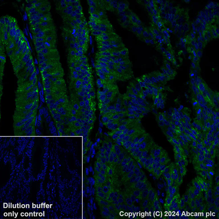
Immunohistochemistry (Formalin/PFA-fixed paraffin-embedded sections) - Alexa Fluor® 488 Anti-Transferrin Receptor antibody [EPR20584] (AB317529)
Immunohistochemical analysis of paraffin-embedded Human stomach cancer tissue labeling Transferrin Receptor with ab317529 at 1/50 (10.0 ug/ml) dilution.
Cytoplasmic staining on human stomach cancer.The section was incubated with ab317529 for 60 mins at room temperature (shown in green). Nuclear DNA was labeled with DAPI (shown in blue). The section was then mounted using Fluoromount®.The immunostaining was performed on a Leica Biosystems BOND RX instrument. Image was taken with a confocal microscope (Leica-Microsystems, TCS SP8).
Counterstained with DAPI.
Heat mediated antigen retrieval was performed with Tris-EDTA buffer (pH 9.0, Epitope Retrieval Solution2) for 40 mins
- ICC/IF
Supplier Data
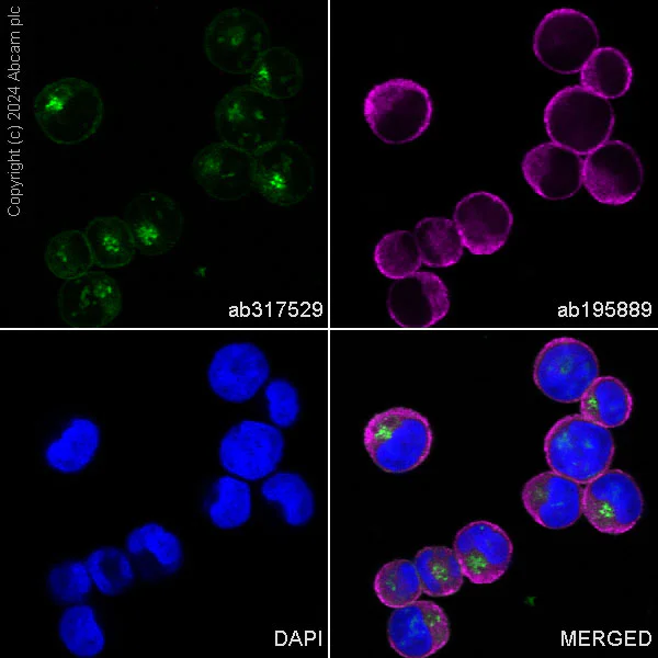
Immunocytochemistry/ Immunofluorescence - Alexa Fluor® 488 Anti-Transferrin Receptor antibody [EPR20584] (AB317529)
Immunofluorescent analysis of 80% Methanol-fixed, 0.1% TritonX-100 permeabilized K-562 (human chronic myelogenous leukemia lymphoblast) cells labelling Transferrin Receptor with ab317529 at 1/50 (10.0 ug/ml) dilution (Green).
Confocal image showing cytoplasmic and membranous staining in K-562 cell line (shown in green). The counterstain was observed in magenta. Nuclear DNA was labeled with DAPI (shown in blue). Image was taken with a confocal microscope(Leica-Microsystems, TCS SP8).
ab195889 Anti-alpha Tubulin mouse monoclonal antibody - Microtubule Marker (Alexa Fluor® 594) was used to counterstain tubulin at 1/200 2.5ug/ml dilution (Magenta). The Nuclear counterstain was DAPI (Blue).

- IHC-P
Supplier Data
Immunohistochemistry (Formalin/PFA-fixed paraffin-embedded sections) - Alexa Fluor® 488 Anti-Transferrin Receptor antibody [EPR20584] (AB317529)
Immunohistochemical analysis of paraffin-embedded Mouse cerebrum tissue labeling Transferrin Receptor with ab317529 at 1/50 (10.0 ug/ml) dilution.
Positive staining on capillaries of mouse cerebrum. The section was incubated with ab317529 for 60 mins at room temperature (shown in green). Nuclear DNA was labeled with DAPI (shown in blue). The section was then mounted using Fluoromount®. The immunostaining was performed on a Leica Biosystems BOND RX instrument.Image was taken with a confocal microscope (Leica-Microsystems, TCS SP8).
Counterstained with DAPI.
Heat mediated antigen retrieval was performed with Tris-EDTA buffer (pH 9.0, Epitope Retrieval Solution2) for 40 mins
- ICC/IF
Supplier Data
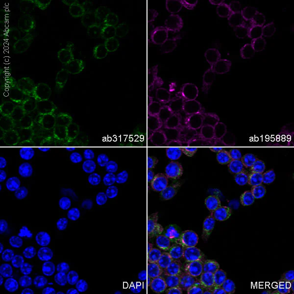
Immunocytochemistry/ Immunofluorescence - Alexa Fluor® 488 Anti-Transferrin Receptor antibody [EPR20584] (AB317529)
Immunofluorescent analysis of 100% methanol-fixed, 0.1% TritonX-100 permeabilized Raw 264.7 (mouse Abelson murine leukemia virus-induced tumor macrophage) cells labelling Transferrin Receptor with ab317529 at 1/50 (10.0 ug/ml) dilution (Green).
Confocal image showing cytoplasmic and membranous staining in Raw 264.7 cell line (shown in green). The counterstain was observed in magenta. Nuclear DNA was labeled with DAPI (shown in blue). Image was taken with a confocal microscope(Leica-Microsystems, TCS SP8).
ab195889 Anti-alpha Tubulin mouse monoclonal antibody - Microtubule Marker (Alexa Fluor® 594) was used to counterstain tubulin at 1/200 2.5ug/ml dilution (Magenta). The Nuclear counterstain was DAPI (Blue).
관련 conjugated 항체와 다양한 조성의 항체 (2)
-
Anti-Transferrin Receptor antibody [EPR20584]
-
Anti-Transferrin Receptor antibody [EPR20584] - BSA and Azide free
보완 제품

Primary Antibodies
AB185033
Alexa Fluor® 488 Anti-Desmin antibody [Y66] - Cytoskeleton Marker
RabMAb|Recombinant

- 0
- 0
Primary Antibodies
AB197640
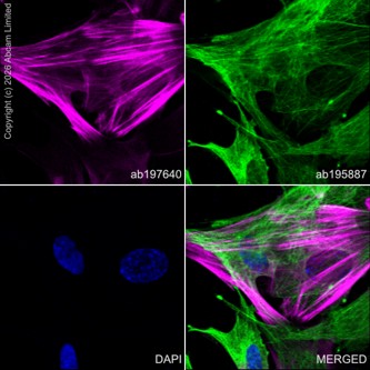
Alexa Fluor® 647 Anti-Calponin 1 antibody [EP798Y]
RabMAb|Recombinant
- 0
- 0
Primary Antibodies
AB311126
Alexa Fluor® 647 Anti-Claudin 5 antibody [EPR7583]
RabMAb|Recombinant
- 0
- 0
Primary Antibodies
AB215695
Alexa Fluor® 555 Anti-Lactate Dehydrogenase antibody [EP1563Y]
RabMAb|Recombinant

- 0
- 0
Reactivity 정보
제품 세부 정보
Patented technology
Our RabMAb® technology is a patented hybridoma-based technology for making rabbit monoclonal antibodies. For details on our patents, please refer to RabMAb® patents.
What are the advantages of a recombinant monoclonal antibody?
This product is a recombinant monoclonal antibody, which offers several advantages including:
- - High batch-to-batch consistency and reproducibility
- - Improved sensitivity and specificity
- - Long-term security of supply
- - Animal-free batch production
For more information, read more on recombinant antibodies.
Alexa Fluor® is a registered trademark of Molecular Probes, Inc, a Thermo Fisher Scientific Company. The Alexa Fluor® dye included in this product is provided under an intellectual property license from Life Technologies Corporation. As this product contains the Alexa Fluor® dye, the purchase of this product conveys to the buyer the non-transferable right to use the purchased product and components of the product only in research conducted by the buyer (whether the buyer is an academic or for-profit entity). As this product contains the Alexa Fluor® dye the sale of this product is expressly conditioned on the buyer not using the product or its components, or any materials made using the product or its components, in any activity to generate revenue, which may include, but is not limited to use of the product or its components: in manufacturing; (ii) to provide a service, information, or data in return for payment (iii) for therapeutic, diagnostic or prophylactic purposes; or (iv) for resale, regardless of whether they are sold for use in research. For information on purchasing a license to this product for purposes other than research, contact Life Technologies Corporation, 5781 Van Allen Way, Carlsbad, CA 92008 USA or outlicensing@thermofisher.com.
특성 및 보관 정보
제형
Purification 테크닉
보관 버퍼
배송 시 보관 조건
적절한 단기 보관 기간
적절한 단기 보관 조건
적절한 장기 보관 조건
분주 정보
보관 정보
추가 정보
This supplementary information is collated from multiple sources and compiled automatically.
Biological function summary
TfR plays a critical role in iron homeostasis by mediating the internalization of transferrin and release of iron in the endosomes. It operates as part of the transferrin-transferrin receptor complex facilitating iron assimilation necessary for DNA synthesis and cell growth. Iron release involves acidifying endosomes allowing transferrin to bind with specific cellular receptors including alternate forms like beta 2 transferrin. The process subsequently contributes to erythropoiesis and various metabolic processes by regulating essential cellular iron levels.
Pathways
The transferrin receptor is central to iron metabolism and the receptor-mediated endocytosis pathway. It tightly interacts with transferrin and intracellular pathways process the iron released from transferrin within endosomes. The receptor's role in this pathway involves a dynamic with other proteins such as HFE and hepcidin. These interactions help control systemic iron levels linking closely to the maintenance of erythroid cell health and proliferation.
제품 프로토콜
- Visit the General protocols
- Visit the Troubleshooting
타겟 정보
대체 명칭 보기
Product promise
Please note: All products are 'FOR RESEARCH USE ONLY. NOT FOR USE IN DIAGNOSTIC OR THERAPEUTIC PROCEDURES'.
For licensing inquiries, please contact partnerships@abcam.com