Anti-ATAD2 antibody [EPR30238-508]
- Advanced Validation
- BOND RX™ Validated
- RabMAb
- Recombinant
- 20ul selling size
- KO Validated
- 각 태그의 뜻
Be the first to review this product! 리뷰 제출
|
(0 출판물)

- IHC-P
Supplier Data
Immunohistochemistry (Formalin/PFA-fixed paraffin-embedded sections) - Anti-ATAD2 antibody [EPR30238-508] (AB324560)
Immunohistochemical analysis of paraffin-embedded Human testis tissue labeling ATAD2 with ab324560 at 1/100 (4.88 ug/ml) dilution, followed by a ready to use LeicaDS9800 (Bond™ Polymer Refine Detection).
Positive staining on human testis. The primary antibody was incubated for 30 mins at room temperature.
The immunostaining was performed on a Leica Biosystems BOND® RX instrument
Counterstained with Hematoxylin.
Secondary antibody only control : Secondary antibody is a ready to use LeicaDS9800 (Bond™ Polymer Refine Detection).
Heat mediated antigen retrieval was performed with Tris-EDTA buffer (pH 9.0, Epitope Retrieval Solution2) for 20 mins

- IHC-P
Supplier Data
Immunohistochemistry (Formalin/PFA-fixed paraffin-embedded sections) - Anti-ATAD2 antibody [EPR30238-508] (AB324560)
Immunohistochemical analysis of paraffin-embedded Human tonsil tissue labeling ATAD2 with ab324560 at 1/100 (4.88 ug/ml) dilution, followed by a ready to use LeicaDS9800 (Bond™ Polymer Refine Detection).
Positive staining on human tonsil. The primary antibody was incubated for 30 mins at room temperature.
The immunostaining was performed on a Leica Biosystems BOND® RX instrument
Counterstained with Hematoxylin.
Secondary antibody only control : Secondary antibody is a ready to use LeicaDS9800 (Bond™ Polymer Refine Detection).
Heat mediated antigen retrieval was performed with Tris-EDTA buffer (pH 9.0, Epitope Retrieval Solution2) for 20 mins

- IHC-P
Supplier Data
Immunohistochemistry (Formalin/PFA-fixed paraffin-embedded sections) - Anti-ATAD2 antibody [EPR30238-508] (AB324560)
Immunohistochemical analysis of paraffin-embedded (A) Wild-type HAP1 (human chronic myelogenous leukemia near-haploid cell) cell pellet (B) ATAD2 knockout HAP1 cell pellet tissue labeling ATAD2 with ab324560 at 1/100 (4.88 ug/ml) dilution, followed by a ready to use LeicaDS9800 (Bond™ Polymer Refine Detection).
Positive staining on (A) Wild-type HAP1 (human chronic myelogenous leukemia near-haploid cell) cell pellet, no staining on (B) ATAD2 knockout HAP1 cell pellet. The primary antibody was incubated for 30 mins at room temperature.
The immunostaining was performed on a Leica Biosystems BOND® RX instrument
Counterstained with Hematoxylin.
Secondary antibody only control : Secondary antibody is a ready to use LeicaDS9800 (Bond™ Polymer Refine Detection).
Heat mediated antigen retrieval was performed with Tris-EDTA buffer (pH 9.0, Epitope Retrieval Solution2) for 20 mins

- ICC/IF
Supplier Data
Immunocytochemistry/ Immunofluorescence - Anti-ATAD2 antibody [EPR30238-508] (AB324560)
Immunofluorescent analysis of 4% Paraformaldehyde-fixed, 0.1% TritonX-100 permeabilized ATAD2 KO HAP1 (ATAD2 knockout human chronic myelogenous leukemia near-haploid cell) cells labelling ATAD2 with ab324560 at 1/50 (9.76 ug/ml) dilution, followed by ab150081 Goat Anti-Rabbit IgG H&L (Alexa Fluor® 488) preadsorbed antibody at 1/1000 (2ug/ml) dilution (Green).
Confocal image showing nuclear staining in wildtype HAP1 cells(shown in green) with no staining in ATAD2 knockout HAP1 cells. The counterstain was observed in magenta. Nuclear DNA was labelled with DAPI (shown in blue). Image was taken with a confocal microscope (Leica-Microsystems, TCS SP8).
ab7291 Anti-alpha Tubulin mouse monoclonal antibody was used to counterstain tubulin at 1/1000 (1ug/ml) dilution, followed by ab150120 Goat Anti-Mouse IgG H&L (Alexa Fluor® 594) antibody at 1/1000 (2ug/ml) dilution (Magenta). The Nuclear counterstain was DAPI (Blue).
Secondary antibody only control : Secondary antibody is ab150081 Goat Anti-Rabbit IgG H&L (Alexa Fluor® 488) preadsorbed at 1/1000 (2ug/ml) dilution.
- mIHC
Lab
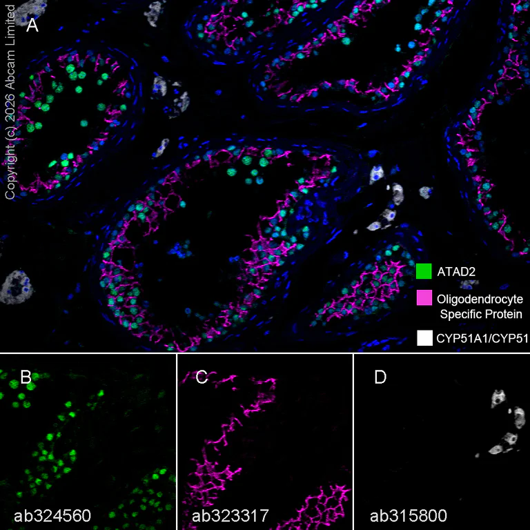
Multiplex immunohistochemistry - Anti-ATAD2 antibody [EPR30238-508] (AB324560)
Multiplex immunohistochemistry analysis of formalin/PFA-fixed paraffin-embedded human testis tissue staining ATAD2 with ab324560 at a 1/100 dilution, ab323317 anti-Oligodendrocyte Specific Protein used at 1/500 dilution and ab315800 anti-CYP51A1/CYP51 used at a 1/2000 dilution.
Panel A : anti-ATAD2 (green; Opal™570), anti-Oligodendrocyte Specific Protein (magenta; Opal™690) and anti-CYP51A1/CYP51 (gray; Opal™520) on human testis.
Panel B : anti-ATAD2 staining nucleus of proliferating cells in human testis.
Panel C : anti-Oligodendrocyte Specific Protein staining apical Sertoli cell crypts in human testis.
Panel D : anti-CYP51A1/CYP51 staining Leydig cells in human testis.
Nuclear DNA was labeled with DAPI (shown in blue).
The section was incubated in three rounds of staining : in the order of ab324560, ab323317 and ab315800 for 30 mins at room temperature. Each round was followed by a separate fluorescent tyramide signal amplification system.
The immunostaining was performed on a Leica Biosystems BOND® RX instrument with an Opal™ 4-color kit. Image acquisition was performed with Leica SP8 confocal microscope.
Heat mediated antigen retrieval was performed with Tris-EDTA buffer (pH 9.0, Epitope Retrieval Solution2) for 20 mins.

- IHC-P
Supplier Data
Immunohistochemistry (Formalin/PFA-fixed paraffin-embedded sections) - Anti-ATAD2 antibody [EPR30238-508] (AB324560)
Immunohistochemical analysis of paraffin-embedded Human skeletal muscle tissue labeling ATAD2 with ab324560 at 1/100 (4.88 ug/ml) dilution, followed by a ready to use LeicaDS9800 (Bond™ Polymer Refine Detection).
Low expression : no staining on human skeletal muscle. The primary antibody was incubated for 30 mins at room temperature.
The immunostaining was performed on a Leica Biosystems BOND® RX instrument
Counterstained with Hematoxylin.
Secondary antibody only control : Secondary antibody is a ready to use LeicaDS9800 (Bond™ Polymer Refine Detection).
Heat mediated antigen retrieval was performed with Tris-EDTA buffer (pH 9.0, Epitope Retrieval Solution2) for 20 mins

- IHC-P
Supplier Data
Immunohistochemistry (Formalin/PFA-fixed paraffin-embedded sections) - Anti-ATAD2 antibody [EPR30238-508] (AB324560)
Immunohistochemical analysis of paraffin-embedded Human endometrial cancer tissue labeling ATAD2 with ab324560 at 1/100 (4.88 ug/ml) dilution, followed by a ready to use LeicaDS9800 (Bond™ Polymer Refine Detection).
Positive staining on human endometrial cancer. The primary antibody was incubated for 30 mins at room temperature.
The immunostaining was performed on a Leica Biosystems BOND® RX instrument
Counterstained with Hematoxylin.
Secondary antibody only control : Secondary antibody is a ready to use LeicaDS9800 (Bond™ Polymer Refine Detection).
Heat mediated antigen retrieval was performed with Tris-EDTA buffer (pH 9.0, Epitope Retrieval Solution2) for 20 mins

- ICC/IF
Supplier Data
Immunocytochemistry/ Immunofluorescence - Anti-ATAD2 antibody [EPR30238-508] (AB324560)
Immunofluorescent analysis of 4% Paraformaldehyde-fixed, 0.1% TritonX-100 permeabilized HeLa (human cervical adenocarcinoma epithelial cell) cells labelling ATAD2 with ab324560 at 1/50 (9.76 ug/ml) dilution, followed by ab150081 Goat Anti-Rabbit IgG H&L (Alexa Fluor® 488) preadsorbed antibody at 1/1000 (2ug/ml) dilution (Green).
Confocal image showing nuclear staining in HeLa cell line (shown in green). The counterstain was observed in magenta. Nuclear DNA was labelled with DAPI (shown in blue). Image was taken with a confocal microscope (Leica-Microsystems, TCS SP8).
ab7291 Anti-alpha Tubulin mouse monoclonal antibody was used to counterstain tubulin at 1/1000 (1ug/ml) dilution, followed by ab150120 Goat Anti-Mouse IgG H&L (Alexa Fluor® 594) antibody at 1/1000 (2ug/ml) dilution (Magenta). The Nuclear counterstain was DAPI (Blue).
-ve control 1 : ab324560 at 1/50 dilution, followed by ab150120 Goat Anti-Mouse IgG H&L (Alexa Fluor® 594) antibody at 1/1000 dilution.
-ve control 2 : ab7291 at 1/1000 dilution, followed by ab150081 Goat Anti-Rabbit IgG H&L (Alexa Fluor® 488) preadsorbed antibody at 1/1000 dilution.

- mIHC
Lab
Multiplex immunohistochemistry - Anti-ATAD2 antibody [EPR30238-508] (AB324560)
Multiplex immunohistochemistry analysis of formalin/PFA-fixed paraffin-embedded mouse testis tissue staining ATAD2 with ab324560 at a 1/500 dilution, ab323317 anti-Oligodendrocyte Specific Protein used at 1/500 dilution and ab315800 anti-CYP51A1/CYP51 used at a 1/2000 dilution.
Panel A : anti-ATAD2 (green; Opal™570), anti-Oligodendrocyte Specific Protein (magenta; Opal™690) and anti-CYP51A1/CYP51 (gray; Opal™520) on mouse testis.
Panel B : anti-ATAD2 staining nucleus of proliferating cells in mouse testis.
Panel C : anti-Oligodendrocyte Specific Protein staining apical Sertoli cell crypts in mouse testis.
Panel D : anti-CYP51A1/CYP51 staining Leydig cells in mouse testis.
Nuclear DNA was labeled with DAPI (shown in blue).
The section was incubated in three rounds of staining : in the order of ab324560, ab323317 and ab315800 for 30 mins at room temperature. Each round was followed by a separate fluorescent tyramide signal amplification system.
The immunostaining was performed on a Leica Biosystems BOND® RX instrument with an Opal™ 4-color kit. Image acquisition was performed with Leica SP8 confocal microscope.
Heat mediated antigen retrieval was performed with Tris-EDTA buffer (pH 9.0, Epitope Retrieval Solution2) for 20 mins.
- mIHC
Lab
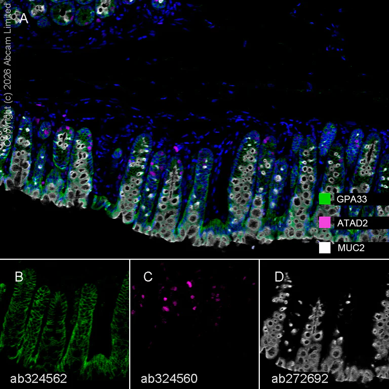
Multiplex immunohistochemistry - Anti-ATAD2 antibody [EPR30238-508] (AB324560)
Multiplex immunohistochemistry analysis of formalin/PFA-fixed paraffin-embedded mouse colon tissue staining GPA33 with ab324562 at a 1/100 dilution, ab324560 anti-ATAD2 used at 1/500 dilution and ab272692 anti-MUC2 used at a 1/2000 dilution.
Panel A : anti-GPA33 (green; Opal™690), anti-ATAD2 (magenta; Opal™520) and anti-MUC2 (gray; Opal™570) on mouse colon.
Panel B : anti-GPA33 staining membrane of epithelium in mouse colon.
Panel C : anti-ATAD2 staining nucleus of proliferating cells in mouse colon.
Panel D : anti-MUC2 staining goblet cells in mouse colon.
Nuclear DNA was labeled with DAPI (shown in blue).
The section was incubated in three rounds of staining : in the order of ab324562, ab324560 and ab272692 for 30 mins at room temperature. Each round was followed by a separate fluorescent tyramide signal amplification system.
The immunostaining was performed on a Leica Biosystems BOND® RX instrument with an Opal™ 4-color kit. Image acquisition was performed with Leica SP8 confocal microscope.
Heat mediated antigen retrieval was performed with Tris-EDTA buffer (pH 9.0, Epitope Retrieval Solution2) for 20 mins.

- IHC-P
Supplier Data
Immunohistochemistry (Formalin/PFA-fixed paraffin-embedded sections) - Anti-ATAD2 antibody [EPR30238-508] (AB324560)
Immunohistochemical analysis of paraffin-embedded Mouse skeletal muscle tissue labeling ATAD2 with ab324560 at 1/500 (0.976 ug/ml) dilution, followed by a ready to use LeicaDS9800 (Bond™ Polymer Refine Detection).
Low expression : no staining on mouse skeletal muscle (PMID : 20581866). The primary antibody was incubated for 30 mins at room temperature.
The immunostaining was performed on a Leica Biosystems BOND® RX instrument
Counterstained with Hematoxylin.
Secondary antibody only control : Secondary antibody is a ready to use LeicaDS9800 (Bond™ Polymer Refine Detection).
Heat mediated antigen retrieval was performed with Tris-EDTA buffer (pH 9.0, Epitope Retrieval Solution2) for 20 mins

- IHC-P
Supplier Data
Immunohistochemistry (Formalin/PFA-fixed paraffin-embedded sections) - Anti-ATAD2 antibody [EPR30238-508] (AB324560)
Immunohistochemical analysis of paraffin-embedded Mouse testis tissue labeling ATAD2 with ab324560 at 1/500 (0.976 ug/ml) dilution, followed by a ready to use LeicaDS9800 (Bond™ Polymer Refine Detection).
Positive staining on mouse testis (PMID : 20581866). The primary antibody was incubated for 30 mins at room temperature.
The immunostaining was performed on a Leica Biosystems BOND® RX instrument
Counterstained with Hematoxylin.
Secondary antibody only control : Secondary antibody is a ready to use LeicaDS9800 (Bond™ Polymer Refine Detection).
Heat mediated antigen retrieval was performed with Tris-EDTA buffer (pH 9.0, Epitope Retrieval Solution2) for 20 mins

- IHC-P
Supplier Data
Immunohistochemistry (Formalin/PFA-fixed paraffin-embedded sections) - Anti-ATAD2 antibody [EPR30238-508] (AB324560)
Immunohistochemical analysis of paraffin-embedded Mouse lung cancer tissue labeling ATAD2 with ab324560 at 1/500 (0.976 ug/ml) dilution, followed by a ready to use LeicaDS9800 (Bond™ Polymer Refine Detection).
Positive staining on mouse lung cancer. The primary antibody was incubated for 30 mins at room temperature.
The immunostaining was performed on a Leica Biosystems BOND® RX instrument
Counterstained with Hematoxylin.
Secondary antibody only control : Secondary antibody is a ready to use LeicaDS9800 (Bond™ Polymer Refine Detection).
Heat mediated antigen retrieval was performed with Tris-EDTA buffer (pH 9.0, Epitope Retrieval Solution2) for 20 mins

- ICC/IF
Supplier Data
Immunocytochemistry/ Immunofluorescence - Anti-ATAD2 antibody [EPR30238-508] (AB324560)
Immunofluorescent analysis of 4% Paraformaldehyde-fixed, 0.1% TritonX-100 permeabilized Neuro-2a (mouse neuroblastoma neuroblast) cells labelling ATAD2 with ab324560 at 1/50 (9.76 ug/ml) dilution, followed by ab150081 Goat Anti-Rabbit IgG H&L (Alexa Fluor® 488) preadsorbed antibody at 1/1000 (2ug/ml) dilution (Green).
Confocal image showing nuclear staining in Neuro-2a cell line (shown in green). The counterstain was observed in magenta. Nuclear DNA was labelled with DAPI (shown in blue). Image was taken with a confocal microscope (Leica-Microsystems, TCS SP8).
ab7291 Anti-alpha Tubulin mouse monoclonal antibody was used to counterstain tubulin at 1/1000 (1ug/ml) dilution, followed by ab150120 Goat Anti-Mouse IgG H&L (Alexa Fluor® 594) antibody at 1/1000 (2µg/ml) (Magenta). The Nuclear counterstain was DAPI (Blue).
-ve control 1 : ab324560 at 1/50 dilution, followed by ab150120 Goat Anti-Mouse IgG H&L (Alexa Fluor® 594) antibody at 1/1000 dilution.
-ve control 2 : ab7291 at 1/1000 dilution, followed by ab150081 Goat Anti-Rabbit IgG H&L (Alexa Fluor® 488) preadsorbed antibody at 1/1000 dilution.

- WB
Supplier Data
Western blot - Anti-ATAD2 antibody [EPR30238-508] (AB324560)
Blocking and diluting buffer and concentration : 5% NFDM/TBST.
In Western blot, ab324560 was shown to bind specifically to ATAD2. Target of interest was observed at 200 kDa in wild-type HAP1 cell lysates (lane 1) with no signal observed at this size in ATAD2 knockout cell line (lane 2).
In Western blot, Anti-Vinculin antibody [EPR8185] (ab129002) staining at 1/10000 dilution.
All lanes:
Western blot - Anti-ATAD2 antibody [EPR30238-508] (ab324560) at 1/1000 dilution
Lane 1:
Wild-type HAP1 (human chronic myelogenous leukemia near-haploid cell) whole cell lysate at 20 µg
Lane 2:
ATAD2 knockout HAP1 whole cell lysate at 20 µg
Secondary
All lanes:
Western blot - Goat Anti-Rabbit IgG H&L (HRP) (<a href='/ko/products/secondary-antibodies/goat-rabbit-igg-h-l-hrp-ab97051'>ab97051</a>) at 1/20000 dilution
Observed band size: 200 kDa,124 kDa
false
Exposure time: 15s

- WB
Supplier Data
Western blot - Anti-ATAD2 antibody [EPR30238-508] (AB324560)
Blocking and diluting buffer and concentration : 5% NFDM/TBST.
To minimize protein degradation, cells were lysed immediately after harvest and then applied to a gel and transfer membrane for Western blotting as soon as possible.
In Western blot, Anti-Vinculin antibody [EPR8185] (ab129002) staining at 1/10000 dilution.
Exposure time : Lane 1 : 8 seconds; Lanes 2-3 : 37 seconds.
All lanes:
Western blot - Anti-ATAD2 antibody [EPR30238-508] (ab324560) at 1/1000 dilution
Lane 1:
Neuro-2a (mouse neuroblastoma neuroblast) whole cell lysate at 20 µg
Lane 2:
U-87 MG (human glioblastoma-astrocytoma epithelial cell) whole cell lysate at 20 µg
Lane 3:
SH-SY5Y (human neuroblastoma epithelial cell) whole cell lysate at 20 µg
Secondary
All lanes:
Western blot - Goat Anti-Rabbit IgG H&L (HRP) (<a href='/ko/products/secondary-antibodies/goat-rabbit-igg-h-l-hrp-ab97051'>ab97051</a>) at 1/20000 dilution
Observed band size: 200 kDa,124 kDa
false

- WB
Supplier Data
Western blot - Anti-ATAD2 antibody [EPR30238-508] (AB324560)
Blocking and diluting buffer and concentration : 5% NFDM/TBST.
To minimize protein degradation, the fresh lysates were lysed immediately after harvest and then applied to a gel and transfer membrane for Western blotting as soon as possible.
In Western blot, Anti-Vinculin antibody [EPR8185] (ab129002) staining at 1/10000 dilution.
All lanes:
Western blot - Anti-ATAD2 antibody [EPR30238-508] (ab324560) at 1/1000 dilution
Lane 1:
MCF7 (human breast adenocarcinoma epithelial cell) whole cell lysate (Frozen lysate) at 20 µg
Lane 2:
HeLa (human cervical adenocarcinoma epithelial cell) whole cell lysate (Frozen lysate) at 20 µg
Lane 3:
HeLa (human cervical adenocarcinoma epithelial cell) whole cell lysate (Fresh lysate) at 20 µg
Lane 4:
F9 (mouse embryonal carcinoma epithelial cell) whole cell lysate (Fresh lysate) at 20 µg
Lane 5:
F9 (mouse embryonal carcinoma epithelial cell) whole cell lysate (Frozen lysate) at 20 µg
Lane 6:
Mouse testis tissue lysate (Frozen lysate) at 20 µg
Secondary
All lanes:
Western blot - Goat Anti-Rabbit IgG H&L (HRP) (<a href='/ko/products/secondary-antibodies/goat-rabbit-igg-h-l-hrp-ab97051'>ab97051</a>) at 1/20000 dilution
Observed band size: 200 kDa,124 kDa
false
Exposure time: 37s
보완 제품

Primary Antibodies
AB181602
Anti-GAPDH antibody [EPR16891] - Loading Control
RabMAb|Recombinant|Lab Essentials|20ul selling size

- 5
- 23
Primary Antibodies
AB155977
Anti-Nucleolin (phospho T84) antibody [EPR8080]
20ul selling size|BOND RX™ Validated|RabMAb|Recombinant

- 0
- 0
Primary Antibodies
AB33911
Anti-Cyclin E1 antibody [EP435E]
20ul selling size|BOND RX™ Validated|KO Validated|RabMAb|Recombinant

- 4
- 8
Reactivity 정보
제품 세부 정보
Patented technology
Our RabMAb® technology is a patented hybridoma-based technology for making rabbit monoclonal antibodies. For details on our patents, please refer to RabMAb® patents.
What are the advantages of a recombinant monoclonal antibody?
This product is a recombinant monoclonal antibody, which offers several advantages including:
- - High batch-to-batch consistency and reproducibility
- - Improved sensitivity and specificity
- - Long-term security of supply
- - Animal-free batch production
For more information, read more on recombinant antibodies.
특성 및 보관 정보
제형
Purification 테크닉
보관 버퍼
배송 시 보관 조건
적절한 단기 보관 기간
적절한 단기 보관 조건
적절한 장기 보관 조건
분주 정보
보관 정보
제품 프로토콜
- Visit the General protocols
- Visit the Troubleshooting
타겟 정보
대체 명칭 보기
Product promise
Please note: All products are 'FOR RESEARCH USE ONLY. NOT FOR USE IN DIAGNOSTIC OR THERAPEUTIC PROCEDURES'.
For licensing inquiries, please contact partnerships@abcam.com