Anti-c-Myc (phospho S62) antibody [33A12E10]
- KD Validated
- BOND RX™ Validated
- Lab Essentials
- 각 태그의 뜻
Be the first to review this product! 리뷰 제출
|
(34 제품이 사용된 논문 )

- ICC/IF
Unknown
Immunocytochemistry/ Immunofluorescence - Anti-c-Myc (phospho S62) antibody [33A12E10] (AB78318)
ab78318 staining (green) and DAPH staining (blue) of c-Myc (phospho S62) in HeLa cells by ICC/IF.

- ICC/IF
Supplier Data
Immunocytochemistry/ Immunofluorescence - Anti-c-Myc (phospho S62) antibody [33A12E10] (AB78318)
Proximity Ligation Analysis with anti-cMyc pS62 and CIP2A antibodies, association of cMyc pS62 with CIP2A (red) in nuclei (DAPI, blue) of HeLa cells by ICC/IF.

- Flow Cyt (Intra)
Unknown
Flow Cytometry (Intracellular) - Anti-c-Myc (phospho S62) antibody [33A12E10] (AB78318)
Overlay histogram showing HL60 cells stained with ab78318 (red line). The cells were fixed with 80% methanol (5 min) and then permeabilized with 0.1% PBS-Tween for 20 min. The cells were then incubated in 1x PBS / 10% normal goat serum / 0.3M glycine to block non-specific protein-protein interactions followed by the antibody (ab78318, 1μg/1x106 cells) for 30 min at 22°C. The secondary antibody used was a Goat Anti-Mouse DyLight® 488 (IgG; H+L) (ab96879) at 1/500 dilution for 30 min at 22°C. Isotype control antibody (black line) was mouse IgG2b [PLPV219] (ab91366, 2μg/1x106 cells) used under the same conditions. Acquisition of >5,000 events was performed. This antibody gave a positive signal in HL60 cells fixed in 4% paraformaldehyde (10 min)/permeabilized with 0.1% PBS-Tween for 20 min used under the same conditions.

- IHC-P
Unknown
Immunohistochemistry (Formalin/PFA-fixed paraffin-embedded sections) - Anti-c-Myc (phospho S62) antibody [33A12E10] (AB78318)
IHC image of ab78318 staining in human normal cervical carcinoma formalin fixed paraffin embedded tissue section, performed on a Leica BondTM system using the standard protocol F. The section was pre-treated using heat mediated antigen retrieval with sodium citrate buffer (pH6, epitope retrieval solution 1) for 20 mins. The section was then incubated with ab78318, 1µg/ml, for 15 mins at room temperature and detected using an HRP conjugated compact polymer system. DAB was used as the chromogen. The section was then counterstained with haematoxylin and mounted with DPX.
For other IHC staining systems (automated and non-automated) customers should optimize variable parameters such as antigen retrieval conditions, primary antibody concentration and antibody incubation times.

- WB
Unknown
Western blot - Anti-c-Myc (phospho S62) antibody [33A12E10] (AB78318)
All lanes:
Western blot - Anti-c-Myc (phospho S62) antibody [33A12E10] (ab78318) at 1 µg/mL
Lane 1:
Crude cell extracts of AGS (gastric adenocarcinoma) cells with Scr (scrambled) siRNA introduced into the cells as a negative control
Lane 2:
Crude cell extracts of AGS (gastric adenocarcinoma) cells transfected with a negative control siRNA
Lane 3:
Crude cell extracts of AGS (gastric adenocarcinoma) cells transfected with siRNA for c-Myc
Predicted band size: 48 kDa
Observed band size: 60 kDa
false
- WB
CiteAb
Western blot - Anti-c-Myc (phospho S62) antibody [33A12E10] (AB78318)
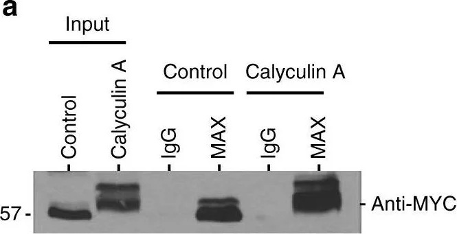
Western Blotting using Anti-c-Myc (phospho S62) antibody [33A12E10]Anti-c-Myc (phospho S62) antibody [33A12E10], ab78318. Publication image from Dingar, D. et al., 2018, Nat Commun, 30158517. Legend direct from paper.
Hyperphosphorylated MYC retains interaction with MAX but is compromised for chromatin binding. a MCF10A cells were treated with Calyculin A (50 nM) or vehicle control for 30 min, lysed, and immunoprecipitated with MAX rabbit or IgG rabbit antibody. The input and IP was resolved by SDS-PAGE and immunoblotted with MYC mouse antibody. Shown is a representative image of n = 3. b For ChIP analysis, MCF10A cells were treated with MG132 (10 µM) for 4 h ± Calyculin A (50 nM) for 30 min, and lysate incubated with MYC rabbit antibody along with IgG antibody as a control. ChIP’d DNA was quantified by qPCR for MYC target genes promoters of CAD, CDK4, LDHA, and NCL, and chr6 (negative control). c For nuclear and cytoplasmic fractionation, MCF10A cells were treated with MG132 (10 µM) for 4 h ± Calyculin A (50 nM) for 30 min (left panel), or transfected with 10 nM siRNA directed against PP1α, β, and γ or universal scramble negative control by calcium phosphate for 44 h and then treated with MG132 (10 µM) for 4 h (right panel). Lysates were used for fractionation, and immunoblotted with indicated antibodies. d MCF10A cells carrying inducible FLAG-tagged EV, MYC wt, MYC12A, or MYC12D were treated with 4-OHT (24 h) to induce expression, treated with MG132 (10 µM) for 4 h, and then lysed for immunoblot analysis (top) or for ChIP-qPCR with FLAG or IgG antibody (bottom) as outlined in b above. For b, d : shown are % input mean (MYC and FLAG ChIP : n = 3). *p < 0.05, **p < 0.01, ***p < 0.001, ****p < 0.0001, two-way ANOVA with Bonferroni test. Not statistically significant = ns. Error bars represent s.d. e Proposed model of PP1/PNUTS regulation of MYC hyperphosphorylation in the control of chromatin binding and degradation
false
- WB
CiteAb
Western blot - Anti-c-Myc (phospho S62) antibody [33A12E10] (AB78318)
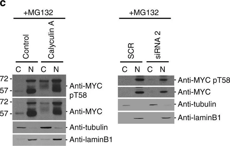
Western Blotting using Anti-c-Myc (phospho S62) antibody [33A12E10]Anti-c-Myc (phospho S62) antibody [33A12E10], ab78318. Publication image from Dingar, D. et al., 2018, Nat Commun, 30158517. Legend direct from paper.
Hyperphosphorylated MYC retains interaction with MAX but is compromised for chromatin binding. a MCF10A cells were treated with Calyculin A (50 nM) or vehicle control for 30 min, lysed, and immunoprecipitated with MAX rabbit or IgG rabbit antibody. The input and IP was resolved by SDS-PAGE and immunoblotted with MYC mouse antibody. Shown is a representative image of n = 3. b For ChIP analysis, MCF10A cells were treated with MG132 (10 µM) for 4 h ± Calyculin A (50 nM) for 30 min, and lysate incubated with MYC rabbit antibody along with IgG antibody as a control. ChIP’d DNA was quantified by qPCR for MYC target genes promoters of CAD, CDK4, LDHA, and NCL, and chr6 (negative control). c For nuclear and cytoplasmic fractionation, MCF10A cells were treated with MG132 (10 µM) for 4 h ± Calyculin A (50 nM) for 30 min (left panel), or transfected with 10 nM siRNA directed against PP1α, β, and γ or universal scramble negative control by calcium phosphate for 44 h and then treated with MG132 (10 µM) for 4 h (right panel). Lysates were used for fractionation, and immunoblotted with indicated antibodies. d MCF10A cells carrying inducible FLAG-tagged EV, MYC wt, MYC12A, or MYC12D were treated with 4-OHT (24 h) to induce expression, treated with MG132 (10 µM) for 4 h, and then lysed for immunoblot analysis (top) or for ChIP-qPCR with FLAG or IgG antibody (bottom) as outlined in b above. For b, d : shown are % input mean (MYC and FLAG ChIP : n = 3). *p < 0.05, **p < 0.01, ***p < 0.001, ****p < 0.0001, two-way ANOVA with Bonferroni test. Not statistically significant = ns. Error bars represent s.d. e Proposed model of PP1/PNUTS regulation of MYC hyperphosphorylation in the control of chromatin binding and degradation
false
- WB
CiteAb
Western blot - Anti-c-Myc (phospho S62) antibody [33A12E10] (AB78318)
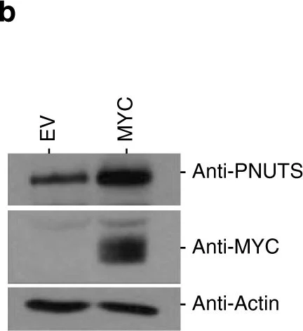
Western Blotting using Anti-c-Myc (phospho S62) antibody [33A12E10]Anti-c-Myc (phospho S62) antibody [33A12E10], ab78318. Publication image from Dingar, D. et al., 2018, Nat Commun, 30158517. Legend direct from paper.
MYC and PP1/PNUTS are co-amplified in cancer and co-bind to promoters of MYC target genes. aMYC and PPP1R10, PPP1CA, PPP1CB, or PPP1CC gene amplification is evident and demonstrates significant co-occurrence in breast invasive carcinoma. b HO15.19 MYC knockout cells (EV) or cells ectopically expressing MYC were lysed and immunoblotted with antibodies to PNUTS, MYC, and actin. Shown are representative images of n = 3. c MCF10A cells were analyzed by chromatin immunoprecipitation (ChIP) using MYC or IgG antibody. ChIP’d DNA was quantified by qPCR for MYC or IgG binding to CAD and PPP1R10 promoters or chr6 (negative control). d MCF10A cells probed with MYC rabbit antibody and/or a PNUTS mouse antibody. The proximity of MYC and PNUTS was assayed using the Duolink Proximity Ligation Assay (PLA) In Situ Red Starter kit (Sigma) as per manufacturer’s instructions. Representative PLA signal (red or white) and nuclear staining (DAPI; blue) are shown for cells probed with anti-MYC alone (top row), anti-PNUTS alone (middle row), or anti-MYC and anti-PNUTS (bottom row). PLA signal was quantified and shown as median number of foci per cell with range (n = 3). *p < 0.05, **p < 0.01, one-way ANOVA with Bonferroni test. Scale bars represent 20 µm. e MCF10A cells analyzed by ChIP with MYC, PNUTS or IgG antibody. ChIP’d DNA was quantified using qPCR for MYC, PNUTS or IgG binding to MYC target genes promoters, CDK4, NCL, and HES1, or chr6 (negative control). f For ChIP-re-ChIP analysis, MCF10A cells lysate incubated with MYC rabbit antibody followed by PNUTS (top panel), or MAX (bottom panel) antibody along with IgG antibody as a control. ChIP’d DNA was quantified similarly as above for ChIP experiment. For c, e, f : shown are % input mean (n = 3). *p < 0.05, **p < 0.01, ***p < 0.001, ****p < 0.0001, two-way ANOVA with Bonferroni test. Error bars represent s.d.
false
대체품
Primary Antibodies
AB190560
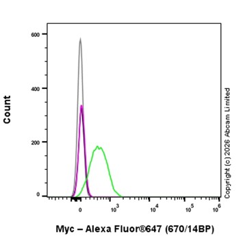
Alexa Fluor® 647 Anti-c-Myc antibody [Y69]
RabMAb|Recombinant|KO Validated
- 5
- 1
Primary Antibodies
AB201780
Alexa Fluor® 555 Anti-c-Myc antibody [Y69]
RabMAb|Recombinant|KO Validated

- 1
- 1
Primary Antibodies
AB190026
Alexa Fluor® 488 Anti-c-Myc antibody [Y69]
RabMAb|Recombinant|KO Validated

- 0
- 0

Primary Antibodies
AB185655
Anti-c-Myc (phospho T58) antibody [EPR17923]
RabMAb|Recombinant|Lab Essentials|20ul selling size

- 4
- 1
Primary Antibodies
AB185656
Anti-c-Myc (phospho S62) antibody [EPR17924]
RabMAb|Recombinant|Lab Essentials|20ul selling size|Advanced Validation

- 0
- 0
Primary Antibodies
AB32072
Anti-c-Myc antibody [Y69] - ChIP Grade
BOND RX™ Validated|KO Validated|RabMAb|Recombinant|Lab Essentials|20ul selling size|Advanced Validation

- 4
- 56
Primary Antibodies
AB168727
Anti-c-Myc antibody [Y69] - ChIP Grade - BSA and Azide free
BOND RX™ Validated|RabMAb|KO Validated|Advanced Validation|Recombinant

- 4
- 11
보완 제품
Primary Antibodies
AB185655
Anti-c-Myc (phospho T58) antibody [EPR17923]
RabMAb|Recombinant|Lab Essentials|20ul selling size
- 4
- 1
Primary Antibodies
AB32072
Anti-c-Myc antibody [Y69] - ChIP Grade
BOND RX™ Validated|KO Validated|RabMAb|Recombinant|Lab Essentials|20ul selling size|Advanced Validation
- 4
- 56
Primary Antibodies
AB16667
BOND RX™ Validated|KO Validated|RabMAb|Advanced Validation|Recombinant|20ul selling size

- 5
- 133
Primary Antibodies
AB184787
Anti-Caspase-3 antibody [EPR18297]
20ul selling size|RabMAb|Recombinant|KO Validated

- 3
- 3
Reactivity 정보
특성 및 보관 정보
제형
Purification 테크닉
보관 버퍼
배송 시 보관 조건
적절한 단기 보관 조건
적절한 장기 보관 조건
분주 정보
보관 정보
추가 정보
This supplementary information is collated from multiple sources and compiled automatically.
Biological function summary
C-Myc is involved in regulating cell growth and proliferation. It forms part of the Myc/Max complex which binds to DNA to regulate gene expression. This activity affects cellular metabolism ribosome biogenesis and cell cycle entry emphasizing its regulation of cellular energy and stress response. Its expression levels critically govern normal cellular functions and homeostasis.
Pathways
C-Myc plays an important role in the cell cycle pathway and apoptosis regulation. Specifically c-Myc is associated with the Wnt signaling pathway which influences cellular proliferation and differentiation. It also interacts with other proteins like Cyclin D1 influencing cell cycle control. These interactions ensure c-Myc's involvement in regulating key processes related to cell proliferation and stability.
제품 프로토콜
- Visit the General protocols
- Visit the Troubleshooting
타겟 정보
대체 명칭 보기
제품이 사용된 논문 (34)
Recent publications for all applications. Explore the 전체 목록and refine your search
Cell communication and signaling : CCS 23:390 PubMed40898325
2025
Applications
Unspecified application
Species
Unspecified reactive species
CytoJournal 21:49 PubMed39737117
2024
Applications
Unspecified application
Species
Unspecified reactive species
Pharmaceutics 15: PubMed38139993
2023
Applications
Unspecified application
Species
Unspecified reactive species
Clinical and translational medicine 13:e1153 PubMed36639831
2023
Applications
Unspecified application
Species
Unspecified reactive species
Molecular oncology 17:1007-1023 PubMed36461911
2022
Applications
Unspecified application
Species
Unspecified reactive species
Molecular cancer research : MCR 20:1151-1165 PubMed35380701
2022
Applications
Unspecified application
Species
Unspecified reactive species
Nucleic acids research 50:3505-3522 PubMed35244724
2022
Applications
Unspecified application
Species
Unspecified reactive species
Cell chemical biology 29:358-372.e5 PubMed34525344
2021
Applications
Unspecified application
Species
Unspecified reactive species
Molecular cancer therapeutics 20:676-690 PubMed33568357
2021
Applications
Unspecified application
Species
Unspecified reactive species
Phytomedicine : international journal of phytotherapy and phytopharmacology 80:153383 PubMed33091855
2020
Applications
Unspecified application
Species
Unspecified reactive species
Product promise
Please note: All products are 'FOR RESEARCH USE ONLY. NOT FOR USE IN DIAGNOSTIC OR THERAPEUTIC PROCEDURES'.
For licensing inquiries, please contact partnerships@abcam.com