- IHC-P
Supplier Data
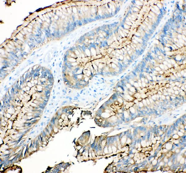
Immunohistochemistry (Formalin/PFA-fixed paraffin-embedded sections) - Anti-CEACAM5 antibody (AB131070)
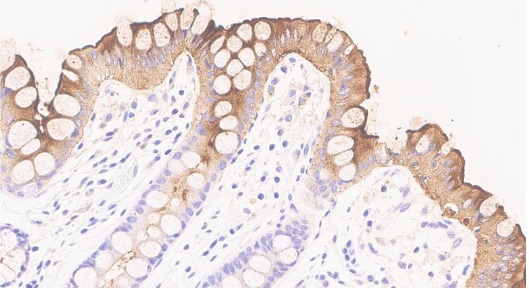
Immunohistochemical analysis of paraffin-embedded Human colon cancer tissue labeling Carcino Embryonic Antigen CEA with ab131070 at 1 ug/ml. Peroxidase Conjugated Goat Anti-rabbit IgG was used as secondary antibody and incubated for 30 minutes at 37°C.
The section was blocked with 10% goat serum then incubated with ab131070 overnight at 4°C.
The tissue section was developed using HRP Conjugated Rabbit IgG Super Vision Assay Kit with DAB as the chromogen.
Heat mediated antigen retrieval was performed in EDTA buffer (pH 8.0, epitope retrieval solution).
- WB
Lab
Western blot - Anti-CEACAM5 antibody (AB131070)
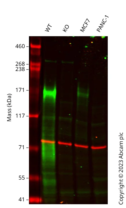
Western blot : Anti-CEACAM5 antibody (ab131070) staining at 0.1 ug/ml, shown in green; Mouse anti-CANX [CANX/1543] (ab238078) loading control staining at 1/20000 dilution, shown in red. In Western blot, ab131070 was shown to bind specifically to CEACAM5. A band was observed at 170 kDa in wild-type A549 cell lysates with no signal observed at this size in CEACAM5 knockout cell line. To generate this image, wild-type and CEACAM5 knockout A549 cell lysates were analysed. First, samples were run on an SDS-PAGE gel then transferred onto a nitrocellulose membrane. Membranes were blocked in 3 % milk in TBS-0.1 % Tween® 20 (TBS-T) before incubation with primary antibodies overnight at 4 °C. Blots were washed four times in TBS-T, incubated with secondary antibodies for 1 h at room temperature, washed again four times then imaged. Secondary antibodies used were Goat anti-Rabbit IgG H&L 800CW and Goat anti-Mouse IgG H&L 680RD at 1/20000 dilution.
All lanes:
Western blot - Anti-CEACAM5 antibody (ab131070) at 0.1 µg/mL
Lane 1:
Wild-type A549 cell lysate at 20 µg
Lane 2:
CEACAM5 knockout A549 cell lysate at 20 µg
Lane 3:
MCF7 cell lysate at 20 µg
Lane 4:
PANC-1 cell lysate at 20 µg
Predicted band size: 76 kDa
false
대체품
Primary Antibodies
AB207718
Anti-CEACAM5 + CD66b + CEACAM1 + CEACAM6 antibody [EPR20721]
BOND RX™ Validated|RabMAb|Recombinant|20ul selling size
- 0
- 0
Primary Antibodies
AB133633
Anti-CEACAM5 antibody [EPCEAR7]
BOND RX™ Validated|KO Validated|RabMAb|Recombinant|20ul selling size
- 0
- 0
Primary Antibodies
AB229074
Anti-CEACAM5 + CD66b + CEACAM1 + CEACAM6 antibody [EPR20721] - BSA and Azide free
BOND RX™ Validated|RabMAb|Recombinant
- 0
- 0
Primary Antibodies
AB4451
Anti-CEACAM5 antibody [26/3/13] - BSA and Azide free
BOND RX™ Validated|Recombinant
- 1
- 2
보완 제품
Primary Antibodies
AB305048
Alexa Fluor® 647 Anti-CD8 alpha antibody [CAL66]
BOND RX™ Validated|RabMAb|Recombinant

- 5
- 1
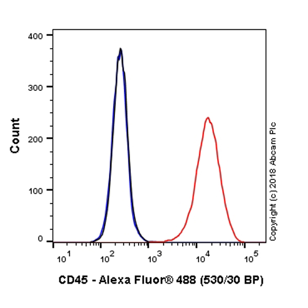
Primary Antibodies
AB68459
Anti-Cytokeratin 7 antibody [EPR1619Y] - Cytoskeleton Marker
KO Validated|RabMAb|Recombinant|Lab Essentials|20ul selling size

- 5
- 1
Primary Antibodies
AB16667
BOND RX™ Validated|KO Validated|RabMAb|Advanced Validation|Recombinant|20ul selling size

- 5
- 133
Reactivity 정보
특성 및 보관 정보
제형
Purification 테크닉
보관 버퍼
배송 시 보관 조건
적절한 단기 보관 기간
적절한 단기 보관 조건
적절한 장기 보관 조건
분주 정보
추가 정보
This supplementary information is collated from multiple sources and compiled automatically.
Biological function summary
CEACAM5 contributes to cellular processes involving cell adhesion and immune response modulation. This protein operates as part of a larger group of cell adhesion molecules and does not function as part of a complex. CEACAM5 can affect the interactions between cells and the extracellular matrix which has implications for tissue structuring and integrity. It helps the immune system recognize and respond to infectious agents or cellular anomalies.
Pathways
CEACAM5 involvement extends to immune response regulation and cell adhesion pathways. It interacts functionally with proteins like E-cadherin in these pathways which contribute to maintaining epithelial integrity. These pathways are critical for normal tissue maintenance and immune system function especially in mucosal surfaces where pathogen entry occurs. Understanding CEACAM5 interactions provides insight into cellular communication and adhesion mechanisms.
제품 프로토콜
- Visit the General protocols
- Visit the Troubleshooting
타겟 정보
대체 명칭 보기
제품이 사용된 논문 (4)
Recent publications for all applications. Explore the 전체 목록 and refine your search
Sensors (Basel, Switzerland) 20: PubMed32131502
2020
Applications
Unspecified application
Species
Unspecified reactive species
Science advances 5:eaaw3413 PubMed31844660
2019
Applications
Unspecified application
Species
Unspecified reactive species
Journal of cellular biochemistry 120:105-114 PubMed30216488
2018
Applications
Unspecified application
Species
Unspecified reactive species
The Journal of biological chemistry 293:11709-11726 PubMed29887526
2018
Applications
Unspecified application
Species
Unspecified reactive species
Product promise
Please note: All products are 'FOR RESEARCH USE ONLY. NOT FOR USE IN DIAGNOSTIC OR THERAPEUTIC PROCEDURES'.
For licensing inquiries, please contact partnerships@abcam.com